. 2019 Sep 1;28(17):2920-2936.
doi: 10.1093/hmg/ddz116.

A mouse model of Proteus syndrome

Affiliations

A mouse model of Proteus syndrome

Marjorie J Lindhurst et al. Hum Mol Genet. .

Abstract

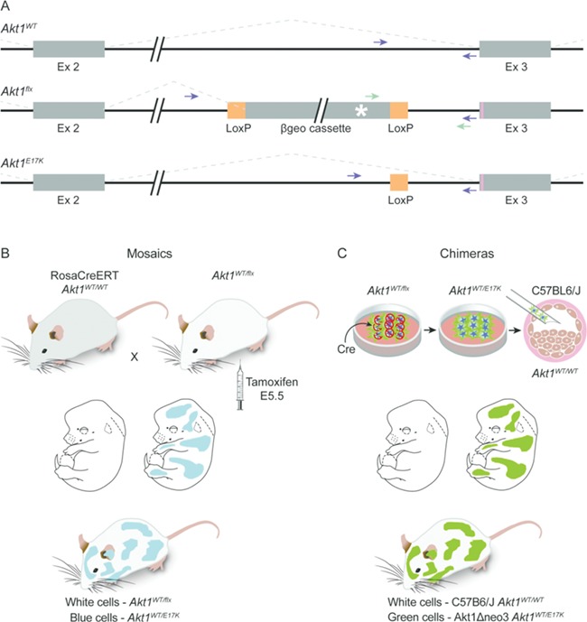
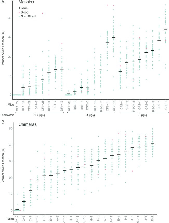
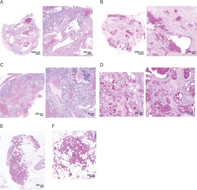
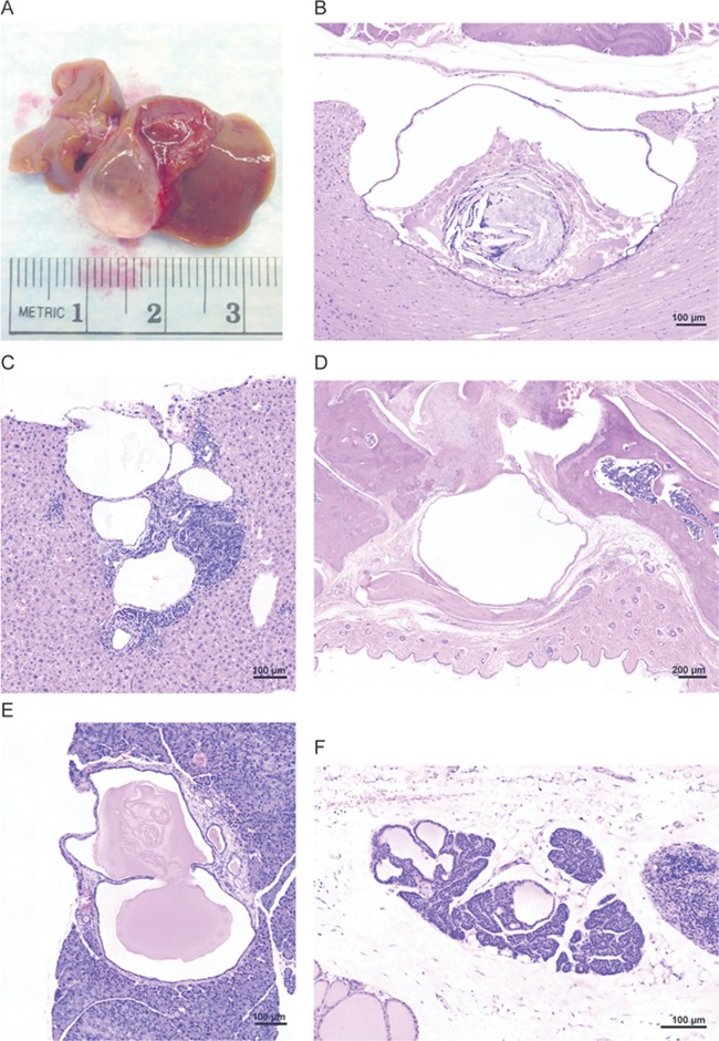
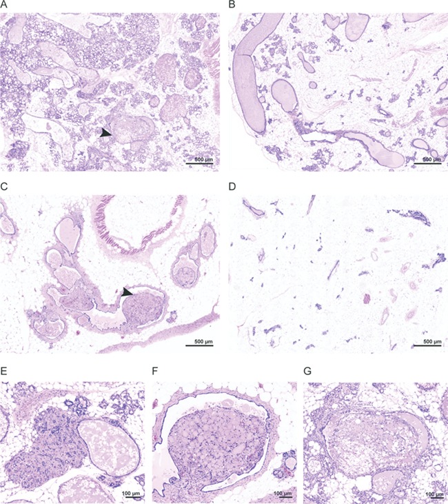
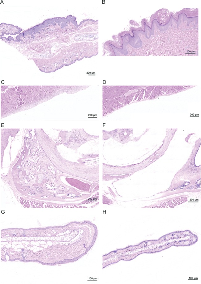
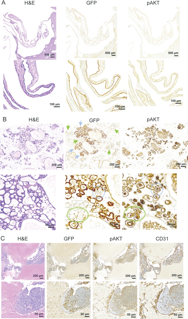
Proteus syndrome is a mosaic, progressive overgrowth disorder caused by a somatic activating variant c.49G > A p.(E17K) in AKT1. The presentation in affected individuals is variable, with a diversity of tissues demonstrating abnormalities. Common manifestations include skin and bony overgrowth, vascular malformations (VMs), cysts and benign tumors. We used two methods to create mouse models that had endogenously-regulated mosaic expression of the Proteus syndrome variant. Variant allele fractions (VAFs) ranged from 0% to 50% across numerous tissues in 44 Proteus syndrome mice. Mice were phenotypically heterogeneous with lesions rarely observed before 12 months of age. VMs were the most frequent finding with a total of 69 found in 29 of 44 Proteus syndrome mice. Twenty-eight cysts and ectasia, frequently biliary, were seen in 22 of 44 Proteus syndrome mice. Varying levels of mammary hyperplasia were seen in 10 of 16 female Proteus syndrome mice with other localized regions of hyperplasia and stromal expansion noted in several additional animals. Interestingly, 27 of 31 Proteus syndrome animals had non-zero blood VAF that is in contrast to the human disorder where it is rarely seen in peripheral blood. Identification of variant-positive cells by green fluorescent protein (GFP) staining in chimeric Proteus syndrome mice showed that in some lesions, hyperplastic cells were predominantly GFP/Akt1E17K-positive and showed increased pAKT signal compared to GFP-negative cells. However, hyperplastic mammary epithelium was a mixture of GFP/Akt1E17K-positive and negative cells with some GFP/Akt1E17K-negative cells also having increased pAKT signal suggesting that the variant-positive cells can induce lesion formation in a non-cell autonomous manner.


Figures

Figure 1
Figure 1
Strategy for making mosaic Proteus syndrome mice. (A) Diagrams of the wild type (top), conditional (middle) and p.(E17K)-expressing (bottom) Akt1 alleles. Exons are represented by the thick boxes; introns are shown by thin lines. The Akt1flx allele contains the 5 Kb floxed βgeo cassette inserted 124 bp upstream of the start of exon 3. Orange boxes represent LoxP sites. Splicing patterns are indicated by the dotted lines. The p.(E17K) variant is indicated by the pink line in exon 3 and the poly A signal that terminates transcription in the βgeo cassette is indicated by the white asterisk. After CRE-mediated recombination of Akt1flx, one LoxP site remains in intron 2 in the Akt1E17K allele as depicted in the bottom drawing. Green arrows mark the location of primers used for identifying Akt1flx. Purple arrows mark the location of primers used to identify Akt1WT and Akt1E17K, which can be distinguished by their size difference. Note, because of the βgeo cassette, Akt1flx does not amplify with primers denoted by the purple arrows using conditions to amplify the other alleles. (B) Diagram of the mosaic strategy. Pregnant females from timed matings between RosaCreERT-expressing and Akt1WT/flx animals were dosed with tamoxifen at E5.5 to activate CRE and the Akt1 conditional allele. Offspring are mosaic for Akt1E17K. (C) Diagram of the chimeric strategy. Akt1WT/flx embryonic stem (ES) cells were transfected with Cre to activate the conditional allele. One to three Akt1WT/E17K ES cells were injected into blastocysts, which were then transferred to pseudo-pregnant mothers for gestation, resulting in animals that were chimeric for Akt1E17K.
Figure 2
Figure 2
VAF of tissues collected from Proteus syndrome animals. The VAFs of all tissues except blood from the tamoxifen mosaics (A) and chimeras (B) are indicated by the blue dots. Red dots represent the VAF in blood DNA. The horizontal line in each column marks the average VAF across all tissues tested for each mouse and represents the overall average VAF for that animal. The tamoxifen dose per gram of body weight used to induce CRE activation is shown below the graph in (A).
Figure 3
Figure 3
Examples of VMs in mice and patients with Proteus syndrome. (A and B) VMs found in the abdominal connective tissue of chimera E-2 (A) and a patient with Proteus syndrome (B). (C and D) VMs found in the spleen of mosaic DF1–18 (C) and a patient with Proteus syndrome (D). All showed variably-sized thin-walled ectatic vessels separated by increased stroma. (E and F) VMs found in adipose tissue of chimera A-9 (E) and a patient with Proteus syndrome (F) that were comprised of numerous, small, thin-walled, ectatic channels.
Figure 4
Figure 4
Examples of cysts and ductal ectasia seen in Proteus syndrome mice. (A) A large biliary cyst seen in chimera E-3. (B) A cyst found in the spinal cord of mosaic animal CF1–28. (C) Biliary ductule ectasia seen in mosaic animal DF1–8. (D) A synovial cyst found in the ankle of mosaic animal CF2–15. (E) A pancreatic cyst seen in mosaic animal DF1–13. (F) A parathyroid cyst seen in chimera A-5.
Figure 5
Figure 5
Range of mammary overgrowth seen in Proteus syndrome mice. (A) An area in chimera A-9 with multiple areas of alveolar hyperplasia and moderate ductal ectasia. (B) An area of milder alveolar hyperplasia and ductal ectasia in mosaic animal CF1–28. (C) An area of moderate ductal ectasia and stromal expansion in chimeric animal H-1. (D) An area in control animal DF1–6 with mild focal hyperplasia and ectasia shown for comparison. (E) An area of stromal expansion in chimera A-9. (FG) Higher magnification of polypoid projections seen in chimeric animals H-1 and A-9 marked by arrows in (A) and (C).
Figure 6
Figure 6
Hyperplasia and stromal expansion in Proteus syndrome mice. (A) A region of epidermal hyperplasia on the ear of chimera E-2. The upper edge showed expanded epidermis whereas the lower edge epidermis was normal thickness. (B) A linear epidermal nevus from a patient with Proteus syndrome. Note the similarity to the epidermal hyperplasia shown in chimera E-2 shown in panel A. (C) Expansion of the stroma lining the peritoneal wall in chimera E-2. (D) Normal thickness of stroma in an adjacent region of the peritoneal wall of this animal. (E) Overgrowth of multiple tissues in the left middle ear of chimera A-9. Expanded stroma, osseous metaplasia, cholesterol clefts, and several small cysts and areas of mineralization were seen. (F) Normal right middle ear of that same animal for comparison. (G) Increased collagen in the dermis at the tip of the ear in chimera E-4. (H) A normal ear from control T2C-81 for comparison.
Figure 7
Figure 7
Identification of Akt1E17K-positive cells and increased AKT signaling in lesional tissue of chimeric Proteus syndrome mice. Hematoxylin and eosin staining and immunostaining with antibodies to green fluorescent protein (GFP), pAKT, and CD31 are shown. (A) A collapsed biliary cyst from chimera E-3 showed that the epithelial cells lining the cysts were predominantly GFP (Akt1E17K)-positive. Phospho-AKT staining correlated with the GFP staining in the cyst. (B) Immunostaining showed hyperplastic mammary alveolar epithelia were comprised of both GFP-positive (blue arrows) and GFP-negative (green arrows) cells. In the higher magnification images, hyperplastic GFP-negative alveolar epithelia that stained positive with the pAKT antibody can be seen (compare color-matched circles). (C) CD31-positive endothelial cells lining the ectatic channels of a VM found in a peri-ovarian lymph node in A-9 were largely GFP-positive. Importantly, substantial numbers of GFP-negative endothelial cells were found that stained positive for pAKT.

Similar articles

  • Allelic heterogeneity of Proteus syndrome.
    Buser A, Lindhurst MJ, Kondolf HC, Yourick MR, Keppler-Noreuil KM, Sapp JC, Biesecker LG. Buser A, et al. Cold Spring Harb Mol Case Stud. 2020 Jun 12;6(3):a005181. doi: 10.1101/mcs.a005181. Print 2020 Jun. Cold Spring Harb Mol Case Stud. 2020. PMID: 32327430 Free PMC article.
  • A mosaic activating mutation in AKT1 associated with the Proteus syndrome.
    Lindhurst MJ, Sapp JC, Teer JK, Johnston JJ, Finn EM, Peters K, Turner J, Cannons JL, Bick D, Blakemore L, Blumhorst C, Brockmann K, Calder P, Cherman N, Deardorff MA, Everman DB, Golas G, Greenstein RM, Kato BM, Keppler-Noreuil KM, Kuznetsov SA, Miyamoto RT, Newman K, Ng D, O'Brien K, Rothenberg S, Schwartzentruber DJ, Singhal V, Tirabosco R, Upton J, Wientroub S, Zackai EH, Hoag K, Whitewood-Neal T, Robey PG, Schwartzberg PL, Darling TN, Tosi LL, Mullikin JC, Biesecker LG. Lindhurst MJ, et al. N Engl J Med. 2011 Aug 18;365(7):611-9. doi: 10.1056/NEJMoa1104017. Epub 2011 Jul 27. N Engl J Med. 2011. PMID: 21793738 Free PMC article.
  • A dyadic genotype-phenotype approach to diagnostic criteria for Proteus syndrome.
    Sapp JC, Buser A, Burton-Akright J, Keppler-Noreuil KM, Biesecker LG. Sapp JC, et al. Am J Med Genet C Semin Med Genet. 2019 Dec;181(4):565-570. doi: 10.1002/ajmg.c.31744. Epub 2019 Nov 6. Am J Med Genet C Semin Med Genet. 2019. PMID: 31692258 Free PMC article.
  • [A clinical study of Proteus syndrome caused by a mosaic somatic mutation in AKT1 gene].
    Xu Y, Fu WZ, He JW, Yue H, Zhang ZL. Xu Y, et al. Zhonghua Nei Ke Za Zhi. 2019 Jul 1;58(7):508-513. doi: 10.3760/cma.j.issn.0578-1426.2019.07.005. Zhonghua Nei Ke Za Zhi. 2019. PMID: 31269567 Review. Chinese.
  • Proteus syndrome review: molecular, clinical, and pathologic features.
    Cohen MM Jr. Cohen MM Jr. Clin Genet. 2014 Feb;85(2):111-9. doi: 10.1111/cge.12266. Epub 2013 Oct 23. Clin Genet. 2014. PMID: 23992099 Review.

Cited by

References

    1. Lindhurst M.J., Sapp J.C., Teer J.K., Johnston J.J., Finn E.M., Peters K., Turner J., Cannons J.L., Bick D., Blakemore L. et al. (2011) A mosaic activating mutation in AKT1 associated with the Proteus syndrome. N. Engl. J. Med., 365, 611–619. - PMC - PubMed
    1. Sapp J.C., Hu L., Zhao J., Gruber A., Schwartz B., Ferrari D. and Biesecker L.G. (2017) Quantifying survival in patients with Proteus syndrome. Genet. Med., 19, 1376–1379. - PMC - PubMed
    1. Doucet M.E., Bloomhardt H.M., Moroz K., Lindhurst M.J. and Biesecker L.G. (2016) Lack of mutation-histopathology correlation in a patient with Proteus syndrome. Am. J. Med. Genet. A, 170, 1422–1432. - PMC - PubMed
    1. Biesecker L. (2006) The challenges of Proteus syndrome: diagnosis and management. Eur. J. Hum. Genet., 14, 1151–1157. - PubMed
    1. Biesecker L.G., Happle R., Mulliken J.B., Weksberg R., Graham J.M. Jr., Viljoen D.L. and Cohen M.M. Jr. (1999) Proteus syndrome: diagnostic criteria, differential diagnosis, and patient evaluation. Am. J. Med. Genet., 84, 389–395. - PubMed

Publication types

Substances