. 2019 Jun;9(2):e63.
doi: 10.1002/cpmo.63. Epub 2019 Jun 13.

High Resolution Imaging of Mouse Embryos and Neonates with X-Ray Micro-Computed Tomography

Affiliations

High Resolution Imaging of Mouse Embryos and Neonates with X-Ray Micro-Computed Tomography

Chih-Wei Hsu et al. Curr Protoc Mouse Biol. 2019 Jun.

Abstract

Iodine-contrast micro-computed tomography (microCT) 3D imaging provides a non-destructive and high-throughput platform for studying mouse embryo and neonate development. Here we provide protocols on preparing mouse embryos and neonates between embryonic day 8.5 (E8.5) to postnatal day 4 (P4) for iodine-contrast microCT imaging. With the implementation of the STABILITY method to create a polymer-tissue hybrid structure, we have demonstrated that not only is soft tissue shrinkage minimized but also the minimum required time for soft tissue staining with iodine is decreased, especially for E18.5 to P4 samples. In addition, we also provide a protocol on using commercially available X-CLARITYTM hydrogel solution to create the similar polymer-tissue hybrid structure on delicate early post-implantation stage (E8.5 to E14.5) embryos. With its simple sample staining and mounting processes, this protocol is easy to adopt and implement for most of the commercially available, stand-alone microCT systems in order to study mouse development between early post-implantation to early postnatal stages. © 2019 by John Wiley & Sons, Inc.

Keywords: STABILITY; X-CLARITY; iodine contrasting; microCT.


Figures

Figure 1.
Figure 1.
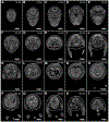
Virtual sectioning of a STABILITY processed, iodine contrasted E18.5 mouse embryo imaged on microCT with a 0.5 mm aluminum attenuation filter at 11 um voxel size from the anterior to the posterior. The step size is 1 mm from the anterior to the posterior of the embryo. The scale bar for each panel is 2 mm.
Figure 2.
Figure 2.
Example of a commercial stand along X-Ray microCT system (Skyscan 1272, Bruker).
Figure 3.
Figure 3.
Example of a commercial hydrogel polymerization system (Logos Biosystem). The system contains heating blocks that can accommodate either multi-well sample plates or 50 ml conical tubes. (A) A 12-well plate and three 50 ml conical tubes were setup in the polymerization system. (B) The setting panel on the system. The crosslinking condition is set at −90 kPa and 37 °C for 3 hours. (C) Alternative method with laboratory common equipment to perform the air removal process with a vacuum desiccator and benchtop built-in vacuum. (D - F) Example of a E18.5 mouse embryo after crosslinked in the STABILITY hydrogel (D-E) and removal from the embedded gel (F).
Figure 4.
Figure 4.
Comparison of E18.5 mouse embryos in (a) PBS, (b) STABILITY, and (c) X-CLARITY™ hydrogel solution after 3 hours crosslinking at 37 °C under −90 kPa vacuum (Logos Biosystem).
Figure 5.
Figure 5.
Volume difference between control and X-CLARITY™ processed mouse embryos after overnight iodine staining at E9.5 and E12.5.
Figure 6.
Figure 6.
Example of E18.5 mouse embryos mounted in (A) 1% agarose for immobilization and positioning of the sample. (B) Back projection image acquired from the detector of the iodine contrasted embryos. (C and D) Skyscan 1272 Control Software for oversize and batch scanning and imaging parameters setup.
Figure 7.
Figure 7.
Example of volume rendering of a E18.5 mouse embryo microCT dataset and virtual sections at sagittal, transverse, and coronal axis.

References

    1. Chung K, and Deisseroth K 2013. CLARITY for mapping the nervous system. Nature methods 10:508–513. - PubMed
    1. Degenhardt K, Wright AC, Horng D, Padmanabhan A, and Epstein JA 2010. Rapid 3D phenotyping of cardiovascular development in mouse embryos by micro-CT with iodine staining. Circulation. Cardiovascular imaging 3:314–322. - PMC - PubMed
    1. Dickinson ME, Flenniken AM, Ji X, Teboul L, Wong MD, White JK, Meehan TF, Weninger WJ, Westerberg H, Adissu H, et al. 2016. High-throughput discovery of novel developmental phenotypes. Nature 537:508–514. - PMC - PubMed
    1. Ermakova O, Orsini T, Gambadoro A, Chiani F, and Tocchini-Valentini GP 2018. Three-dimensional microCT imaging of murine embryonic development from immediate post-implantation to organogenesis: application for phenotyping analysis of early embryonic lethality in mutant animals. Mammalian genome : official journal of the International Mammalian Genome Society 29:245–259. - PMC - PubMed
    1. Hsu CW, Wong L, Rasmussen TL, Kalaga S, McElwee ML, Keith LC, Bohat R, Seavitt JR, Beaudet AL, and Dickinson ME 2016. Three-dimensional microCT imaging of mouse development from early post-implantation to early postnatal stages. Developmental biology 419:229–236. - PMC - PubMed

LinkOut - more resources