. 2019 Jun 5;8(6):802.
doi: 10.3390/jcm8060802.

Occipital Petalia and Albinism: A Study of Interhemispheric VEP Asymmetries in Albinism with No Nystagmus

Affiliations

Occipital Petalia and Albinism: A Study of Interhemispheric VEP Asymmetries in Albinism with No Nystagmus

Alkiviades Liasis et al. J Clin Med. .

Abstract

The purpose of this study was to assess chiasmal misrouting in a cohort of children with albinism with no nystagmus using hemifield visual evoked potentials (VEP) measures.

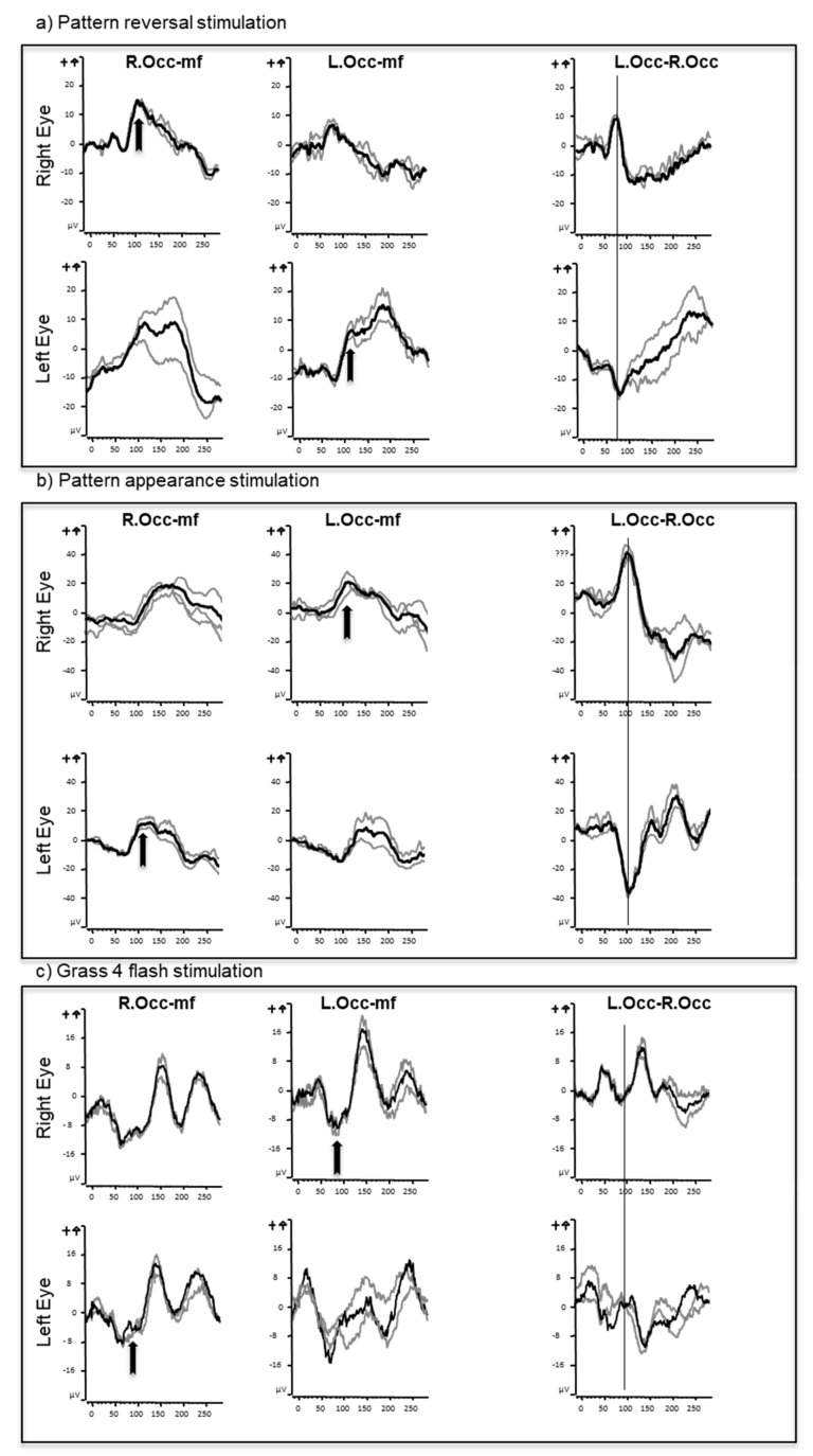
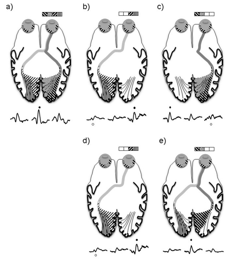
Methods: Monocular VEPs were recorded and analyzed from three electrodes (O1, Oz, and O2 referred to Fz) from 16 children with albinism without nystagmus. Pattern reversal (full field and hemifield stimulation), full field pattern appearance and flash stimuli were used to evoke VEPs for each eye.

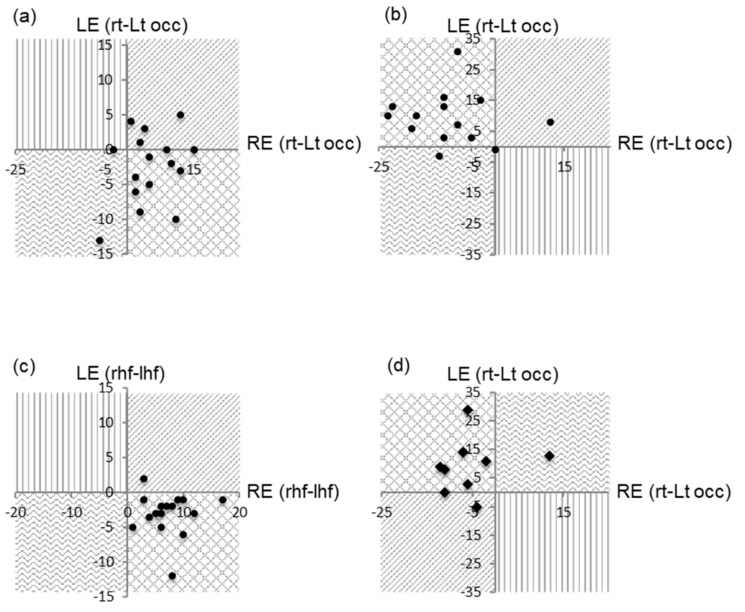
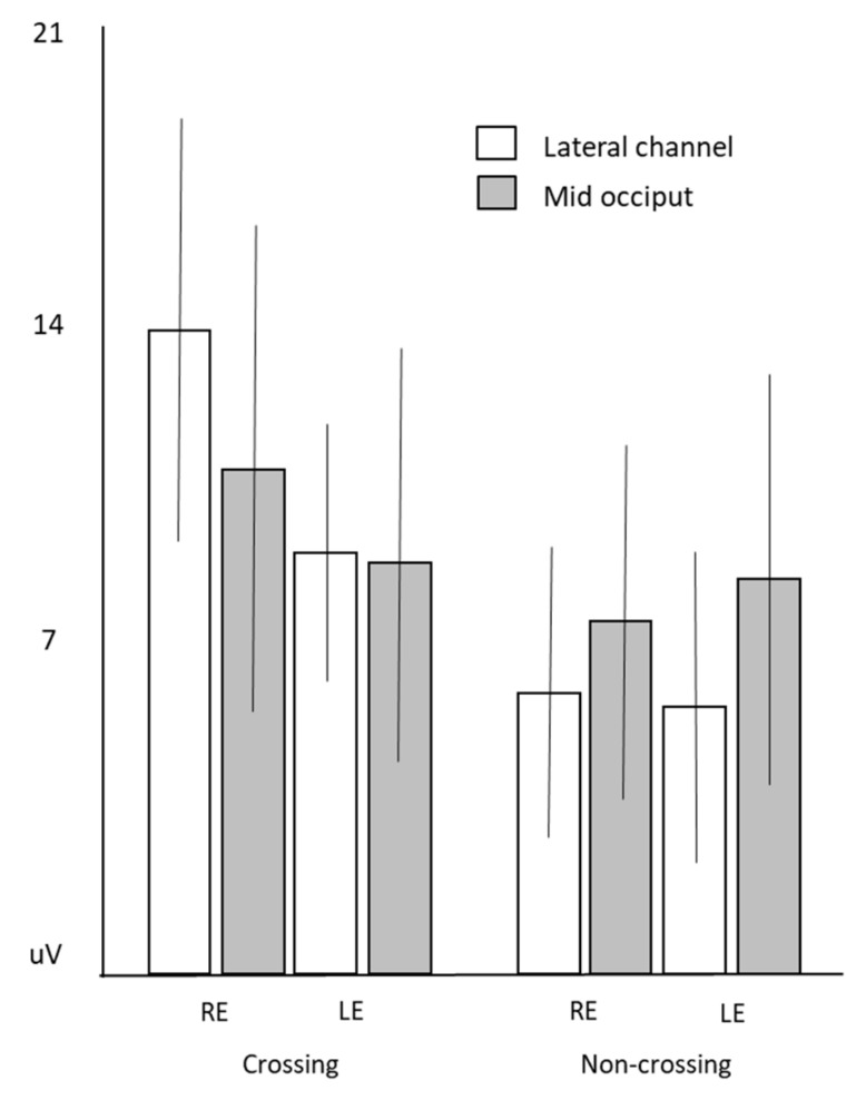
Results: The amplitude of the pattern reversal VEPs to stimulation of the hemifield corresponding to the crossing pathways were as expected significantly larger than those to the non-crossing in each eye ((right eye p = 0.000004), (left eye p = 0.001)). Pattern reversal VEPs recorded from the left hemisphere were also larger than those from the right and most evident when comparing the crossing pathways of each eye (p = 0.004).

Conclusions: This study has demonstrated electrophysiological differences in visual pathway function of the left and right hemisphere in subjects with albinism like that previously described in controls. Nasal field stimulation activated crossing and non-crossing pathways in patients with albinism and as a result, nasal hemifield VEPs in albinism are less lateralized compared to what is found in normal subjects.

Keywords: albinism; hemifield; misrouting; visual evoked potential.


Conflict of interest statement

The authors declare no conflict of interest.

Figures

Figure 1
Figure 1
Full field pattern reversal (a), appearance (b), and flash (c) visual evoked potentials from one subject. Responses recorded from the right (R.Occ) and left (L.Occ) occiput from a mid-frontal (mf) reference. Gray lines represent individual trials, and black the grand average. Note the different patterns of crossed asymmetry between different stimuli. These are best seen with the subtraction waveform where responses from the right occiput are subtracted from the left (L.Occ–R.Occ). The crossing components are shown with arrows.
Figure 2
Figure 2
Amplitude and latency of P100 during monocular full field stimulation to pattern reversal test checks subtending 50 min of arc presented in a 28 degree field. The white area represents the 1st and 99th centile of the laboratories normal limits, whilst gray represents outside of normal limits. Responses for all eyes except two were within normal limits. LE: left eye; RE: right eye.
Figure 3
Figure 3
Comparison of monocular trans occipital amplitude asymmetries of the major VEP components. (a) Monocular full field pattern reversal responses, (b) hemifield responses, (c) pattern appearance, and (d) monocular flash.
Figure 4
Figure 4
Mean amplitude of iP100 measured over the hemisphere ipsilateral to the field being stimulated, error bars are ± standard deviation. White bars measured over the ipsilateral channel and gray measured at mid occiput.
Figure 5
Figure 5
A schematic explaining the trans occipital distribution of a normal subject during monocular pattern reversal stimulation of the full field (a), right hemifield (b), and left hemifield (c). In comparison to in a subject with albinism when the right hemifield (d) and left hemifield (e) are stimulated. ● = P100, o = iP100.

References

    1. Kinnear P.E., Jay B., Witkop C.J. Albinism. Surv. Ophthalmol. 1985;30:75–101. doi: 10.1016/0039-6257(85)90077-3. - DOI - PubMed
    1. Creel D.J., Summers C.G., King R.A. Visual anomalies associated with albinism. Ophthalmic Paediatr. Genet. 1990;11:193–200. doi: 10.3109/13816819009020979. - DOI - PubMed
    1. Schmitz B., Schaefer T., Krick C.M., Reith W., Backens M., Käsmann-Kellner B. Configuration of the optic chiasm in humans with albinism as revealed by magnetic resonance imaging. Investig. Ophthalmol. Vis. Sci. 2003;44:16–21. doi: 10.1167/iovs.02-0156. - DOI - PubMed
    1. Bridge H., von dem Hagen E.A.H., Davies G., Chambers C., Gouws A., Hoffmann M., Morland A.B. Changes in brain morphology in albinism reflect reduced visual acuity. Cortex. 2014;56:64–72. doi: 10.1016/j.cortex.2012.08.010. - DOI - PubMed
    1. Hoffmann M.B., Lorenz B., Morland A.B., Schmidtborn L.C. Misrouting of the optic nerves in albinism: Estimation of the extent with visual evoked potentials. Investig. Ophthalmol. Vis. Sci. 2005;46:3892–3898. doi: 10.1167/iovs.05-0491. - DOI - PubMed

LinkOut - more resources