

Quantitative Studies of Diabetic Foot Ulcer Evolution Under Treatment by Digital Stereotactic Photography
- PMID: 31195816
- PMCID: PMC6955448
- DOI: 10.1177/1932296819853843
Quantitative Studies of Diabetic Foot Ulcer Evolution Under Treatment by Digital Stereotactic Photography
Abstract
Background: Imaging the lower extremity reproducibly and accurately remains an elusive goal. This is particularly true in the high risk diabetic foot, where tissue loss, edema, and color changes are often concomitant. The purpose of this study was to evaluate the reproducibility of a novel and inexpensive stereotaxic frame in assessment of wound healing.
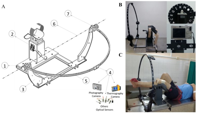
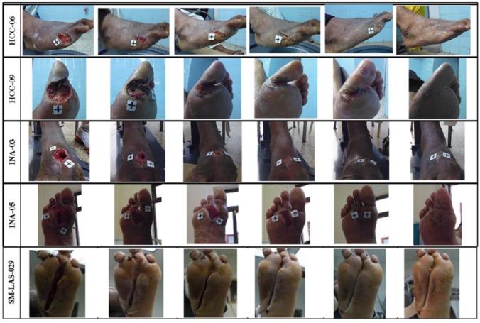
Methods: The main idea is to keep constant and reproducible the relative position of extremities related to the sensor used for the examination during a serial studies by stereotaxic digital photographic sequence. Ten healthy volunteers were evaluated at 10 different time moments to estimate the foot position variations in the stereotaxic frame. The evolution of 40 of DFU patients under treatment was evaluated before and during the epidemical grow factor intralesional treatment.
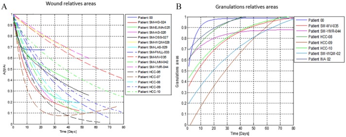
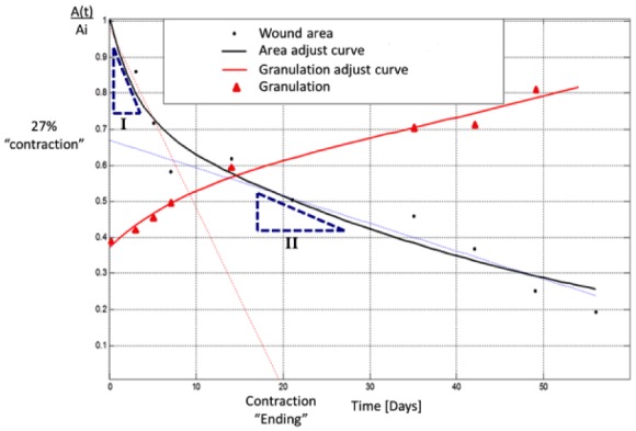
Results: The wound closing and granulation speeds, the relative contribution of the contraction and tissue restauration mechanism were evaluated by stereotaxic digital photography.
Conclusions: The results of this study suggest that the stereotaxic frame is a robust platform for serial study of the evolution of wound healing which allow to obtain consistent information from a variety of visible and hyperspectral measurement technologies. New stereotaxic digital photography evidences related to the diabetic foot ulcer healing process under treatment has been presented.
Keywords: diabetic foot ulcer; imaging; quantitative evolution; serial studies.
Conflict of interest statement
Figures
Similar articles
-
Diabetic foot ulcer photography study: a study within a trial to assess the reliability of two-dimensional (2D) photography for the assessment of ulcer healing in patients with diabetes-related foot ulcers-protocol paper.BMJ Open. 2025 Jan 9;15(1):e090299. doi: 10.1136/bmjopen-2024-090299. BMJ Open. 2025. PMID: 39788763 Free PMC article.
-
Area Determination of Diabetic Foot Ulcer Images Using a Cascaded Two-Stage SVM-Based Classification.IEEE Trans Biomed Eng. 2017 Sep;64(9):2098-2109. doi: 10.1109/TBME.2016.2632522. Epub 2016 Nov 23. IEEE Trans Biomed Eng. 2017. PMID: 27893380
-
Reliability and validity of the revised photographic wound assessment tool on digital images taken of various types of chronic wounds.Adv Skin Wound Care. 2013 Aug;26(8):360-73. doi: 10.1097/01.ASW.0000431329.50869.6f. Adv Skin Wound Care. 2013. PMID: 23860221 Clinical Trial.
-
Evidence-based protocol for diabetic foot ulcers.Plast Reconstr Surg. 2006 Jun;117(7 Suppl):193S-209S; discussion 210S-211S. doi: 10.1097/01.prs.0000225459.93750.29. Plast Reconstr Surg. 2006. PMID: 16799388 Review.
-
Wound nitric oxide bioactivity: a promising diagnostic indicator for diabetic foot ulcer management.J Wound Ostomy Continence Nurs. 2010 Jan-Feb;37(1):25-32; quiz 33-4. doi: 10.1097/WON.0b013e3181c68b61. J Wound Ostomy Continence Nurs. 2010. PMID: 20075688 Review.
Cited by
-
The role of artificial intelligence technology in the care of diabetic foot ulcers: the past, the present, and the future.World J Diabetes. 2022 Dec 15;13(12):1131-1139. doi: 10.4239/wjd.v13.i12.1131. World J Diabetes. 2022. PMID: 36578875 Free PMC article. Review.
References
-
- Cabal-Mirabal C, González Dalmau E, Berlanga Acosta J. et al. Quantitative studies of the evolution of diabetic foot lesions under EGF treatment by magnetic resonance imaging. J Radiol Res Pract. 2014;2014:783980.
-
- Frykberg RG, Zgonis T, Armstrong DG. et al. Diabetic foot disorders: a clinical practice guideline. American College of Foot and Ankle Surgeons. J Foot Ankle Surg. 2006;45:S1-S66. - PubMed
-
- Official Site of Image J Software. https://imagej.nih.gov/ij/.
Publication types
MeSH terms
LinkOut - more resources
Full Text Sources
Medical
