. 2019 Jun 13;20(1):120.
doi: 10.1186/s12931-019-1081-3.

Role of PM2.5 in the development and progression of COPD and its mechanisms

Affiliations

Role of PM2.5 in the development and progression of COPD and its mechanisms

Junling Zhao et al. Respir Res. .

Abstract

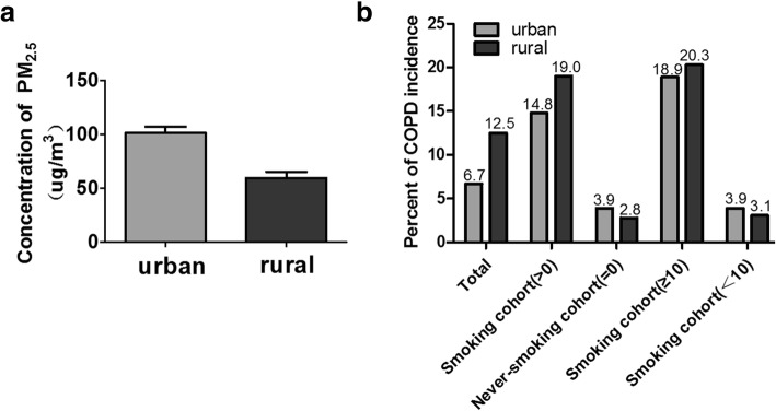
Background: A multitude of epidemiological studies have shown that ambient fine particulate matter 2.5 (diameter < 2.5um; PM2.5) was associated with increased morbidity and mortality of chronic obstructive pulmonary disease (COPD). However, the underlying associated mechanisms have not yet been elucidated. We conducted this study to investigate the role of PM2.5 in the development of COPD and associated mechanisms.

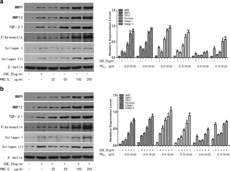
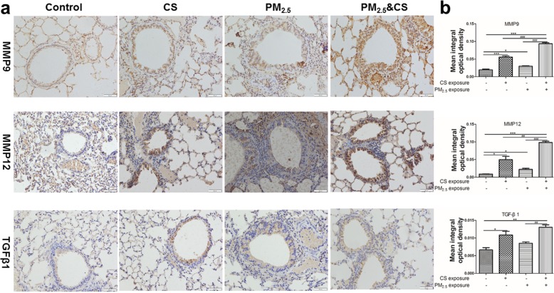
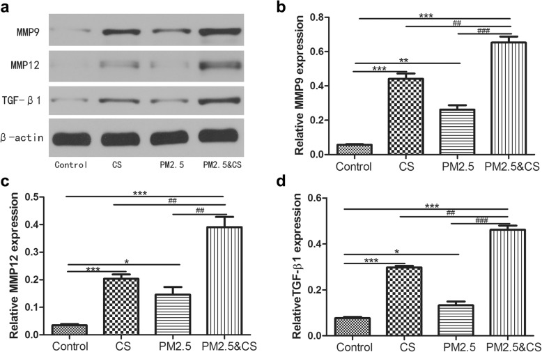
Methods: We firstly conducted a cross-sectional study in Chinese han population to observe PM2.5 effects on COPD morbidity. Then, in vitro, we incubated human bronchial epithelial cells to different concentrations of PM2.5 for 24 h. The expression levels of IL-6 and IL-8 were detected by ELISA and the levels of MMPs, TGF-β1, fibronectin and collagen was determined by immunoblotting. In vivo, we subjected C57BL/6 mice to chronic prolonged exposure to PM2.5 for 48 weeks to study the influence of PM2.5 exposure on lung function, pulmonary structure and inflammation.

Results: We found that the effect of PM2.5 on COPD morbidity was associated with its levels and that PM2.5 and cigarette smoke could have a synergistic impact on COPD development and progression. Both vitro and vivo studies demonstrated that PM2.5 exposure could induce pulmonary inflammation, decrease lung function, and cause emphysematous changes. Furthermore, PM2.5 could markedly aggravated cigarette smoke-induced changes.

Conclusions: In short, we found that prolonged chronic exposure to PM2.5 resulted in decreased lung function, emphysematous lesions and airway inflammation. Most importantly, long-term PM2.5 exposure exacerbateed cigarette smoke-induced changes in COPD.

Keywords: Airway inflammation; Ambient fine particulate matter; Chronic obstructive pulmonary disease; Emphysematous lesions; Lung function.


Conflict of interest statement

The authors declare that they have no competing interests.

Figures

Fig. 1
Fig. 1
The association of PM2.5 with COPD incidence in the population study. a The levels of PM2.5 in urban and rural settings; b COPD incidence among different groups based on smoking-pack-years
Fig. 2
Fig. 2
Effects of PM2.5 on the cytokines release of IL-6 and IL-8 in HBEs. HBEs were, respectively, incubated with CSE (25 μg/ml) or 25 μg/ml to 200 μg/ml PM2.5 for 24 h to examine the levels of pro-inflammatory cytokines by ELISA. a IL-6, b IL-8. HBEs were co-stimulated with CSE and different concentrations of PM2.5 for 24 h to examine the levels of proinflammatory cytokines by ELISA. c IL-6, d IL-8.The results are mean ± SD of three independent experiments. *P < 0.05, ** P < 0.01, *** P < 0.001 vs control group; #P < 0.05, ## P < 0.01, ### P < 0.001 vs CSE group
Fig. 3
Fig. 3
Effects of PM2.5 on the protein expression levels of MMPs, TGF-β1, fibronectin and collagen proteins. HBEs were incubated with PM2.5 with or without CSE for 24 h. The protein expression levels of MMPs, TGF-β1, fibronectin and collagens were detected by WB. a The protein expression of MMPs, TGF-β1, fibronectin and collagen induced by PM2.5 /CSE. b The protein expression levels of MMPs, TGF-β1, fibronectin and collagens induced by PM2.5 + CSE. * P < 0.05, ** P < 0.01, *** P < 0.001 vs control group; #P < 0.05, ## P < 0.01, ### P < 0.001 vs CSE group
Fig. 4
Fig. 4
Morphological and pathological changes of lung tissue in mouse model. The representative micrographs of H&E-stained lung tissue of long-term exposure to PM2.5 with or without CS for 48 weeks (a-d) (200x). a control group; b CS-exposed group; c PM2.5-exposed group; d PM2.5 + CS -exposed group. e Quantitative morphometric measurements of MLI. ** P < 0.01, *** P < 0.001 vs control group
Fig. 5
Fig. 5
Effect of PM2.5 and cigarette smoke exposure on pulmonary inflammation. The representative changes of pulmonary inflammation after long-term exposure to PM2.5 with or without CS for 48 weeks (a-d) (200x). a control group; b CS-exposed group; c PM2.5-exposed group; d PM2.5 + CS -exposed group. The levels of inflammatory cytokines IL-6 and KC (mouse IL-8) in BAL were detected by ELISA; e the level of IL-8 in BAL and f the level of IL-6 in BAL. Data are presented as mean ± SD (n = 6). * P < 0.05, ** P < 0.01 vs control group
Fig. 6
Fig. 6
Immunohistochemical staining of MMP9, MMP12, and TGF-β1 in lungs sections induced by PM2.5 exposure (400x) (a) and semiquantitative assessment of MMP9, MMP12, and TGF-β1 protein expression using Image-Pro Plus (b). Data are presented as mean ± SD (n = 6). *P < 0.05, ** P < 0.01, *** P < 0.001 vs control group; #P < 0.05, ## P < 0.01, ### P < 0.001 vs PM2.5 + CSE-exposed group
Fig. 7
Fig. 7
Western blot examination for MMP9, MMP12, and TGF-β1 protein levels expression in lungs sections induced by PM2.5/CS exposure. The expressions of MMP9, MMP12, and TGF-β1 were analyzed with western blotting (a) and (b-d) the proteins expression were evaluated. Data are expressed as mean ± SD (n = 6). *P < 0.05, ** P < 0.01, *** P < 0.001 vs control group; #P < 0.05, ## P < 0.01, ### P < 0.001 vs PM2.5 + CSE-exposed group
Fig. 8
Fig. 8
Effect of PM2.5 and cigarette smoke exposure on airway wall remodeling. a Photomicrographs of peribronchial collagen and fibronectin deposition in lung tissue induced by chronic exposure to PM2.5 with or without CS (200X). b The assessment of peribronchial collagen deposition in lung tissue induced by chronic exposure to PM2.5 with or without CS. c Semiquantitative assessment of fibronectin protein expression using Image-Pro Plus. Results were expressed as means± SD (n = 6). *P < 0.05, ** P < 0.01, *** P < 0.001 vs control group; #P < 0.05, ## P < 0.01, ### P < 0.001 vs PM2.5 + CSE-exposed group

References

    1. Vestbo J, Hurd SS, Agusti AG, Jones PW, Vogelmeier C, Anzueto A, Barnes PJ, Fabbri LM, Martinez FJ, Nishimura M, et al. Global strategy for the diagnosis, management, and prevention of chronic obstructive pulmonary disease: GOLD executive summary. Am J Respir Crit Care Med. 2013;187:347–365. doi: 10.1164/rccm.201204-0596PP. - DOI - PubMed
    1. Barnes PJ. Chronic obstructive pulmonary disease: a growing but neglected global epidemic. PLoS Med. 2007;4:e112. doi: 10.1371/journal.pmed.0040112. - DOI - PMC - PubMed
    1. Lamprecht B, McBurnie MA, Vollmer WM, Gudmundsson G, Welte T, Nizankowska-Mogilnicka E, Studnicka M, Bateman E, Anto JM, Burney P, et al. COPD in never smokers: results from the population-based burden of obstructive lung disease study. Chest. 2011;139:752–763. doi: 10.1378/chest.10-1253. - DOI - PMC - PubMed
    1. COPD Improving prevention and care. Lancet. 2015;385:830. - PubMed
    1. Salvi SS, Barnes PJ. Chronic obstructive pulmonary disease in non-smokers. Lancet. 2009;374:733–743. doi: 10.1016/S0140-6736(09)61303-9. - DOI - PubMed