. 2019 Jun 13;10(1):166.
doi: 10.1186/s13287-019-1255-4.

Examining the fundamental biology of a novel population of directly reprogrammed human neural precursor cells

Affiliations

Examining the fundamental biology of a novel population of directly reprogrammed human neural precursor cells

Jan-Eric Ahlfors et al. Stem Cell Res Ther. .

Abstract

Background: Cell reprogramming is a promising avenue for cell-based therapies as it allows for the generation of multipotent, unipotent, or mature somatic cells without going through a pluripotent state. While the use of autologous cells is considered ideal, key challenges for their clinical translation include the ability to reproducibly generate sufficient quantities of cells within a therapeutically relevant time window.

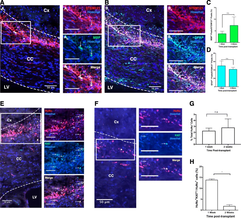
Methods: We performed transfection of three distinct human somatic starting populations of cells with a non-integrating synthetic plasmid expressing Musashi 1 (MSI1), Neurogenin 2 (NGN2), and Methyl-CpG-Binding Domain 2 (MBD2). The resulting directly reprogrammed neural precursor cells (drNPCs) were examined in vitro using RT-qPCR, karyotype analysis, immunohistochemistry, and FACS at early and late time post-transfection. Electrophysiology (patch clamp) was performed on drNPC-derived neurons to determine their capacity to generate action potentials. In vivo characterization was performed following transplantation of drNPCs into two animal models (Shiverer and SCID/Beige mice), and the numbers, location, and differentiation profile of the transplanted cells were examined using immunohistochemistry.

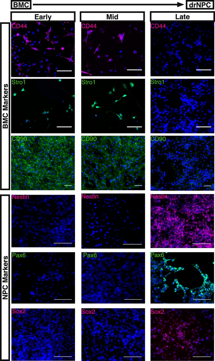
Results: Human somatic cells can be directly reprogrammed within two weeks to neural precursor cells (drNPCs) by transient exposure to Msi1, Ngn2, and MBD2 using non-viral constructs. The drNPCs generate all three neural cell types (astrocytes, oligodendrocytes, and neurons) and can be passaged in vitro to generate large numbers of cells within four weeks. drNPCs can respond to in vivo differentiation and migration cues as demonstrated by their migration to the olfactory bulb and contribution to neurogenesis in vivo. Differentiation profiles of transplanted cells onto the corpus callosum of myelin-deficient mice reveal the production of oligodendrocytes and astrocytes.

Conclusions: Human drNPCs can be efficiently and rapidly produced from donor somatic cells and possess all the important characteristics of native neural multipotent cells including differentiation into neurons, astrocytes, and oligodendrocytes, and in vivo neurogenesis and myelination.

Keywords: Direct reprogramming; In vivo neurogenesis; In vivo remyelination; Neural precursor cells; Neural stem cells; drNPC.


Conflict of interest statement

JEA is a shareholder of New World Laboratories. RE, CB, SM, and OM are employees of New World Laboratories. The other authors declare that they have no competing interests.

Figures

Fig. 1
Fig. 1
Generation and in vitro characterization of drNPCs. a Schematic of protocol for reprogramming cells to drNPCs. b At passage 7, drNPCs continue expressing high levels of Nestin, Sox2, GFAP, NG2, Ascl1, Tuj1, Dcx, and Pax6. Nuclei stained with Hoechst (blue). Scale bar = 100 μm. c drNPCs at passage 12 were analyzed for expression of multiple markers, graphed by intensity of expression to determine cell population purity, using a 12-channel Amnis FlowSight® Imaging flow cytometer. d Normal karyotype of drNPCs at passage 12
Fig. 2
Fig. 2
BMC to drNPC reprogramming. In vitro cultures of bone marrow cells (BMCs) during reprogramming to drNPCs. At early time-points (days 1–3 in vitro), BMC-specific markers CD44, Stro1, and CD90 are expressed and no NPC-specific markers Nestin, Pax6, or Sox2 are observed. By the mid time-points (days 6–7 in vitro), downregulation of BMC-specific markers occurs. At late time-points (days 14–16 in vitro), no BMC-specific markers are observed and NPC-specific markers Nestin, Pax6, and Sox2 are highly expressed. Nuclei stained with Hoechst (blue). Scale bar = 100 μm.
Fig. 3
Fig. 3
drNPC differentiation in vitro. In differentiation conditions, drNPC monolayers give rise to neurons (Tuj1 and Map 2b), oligodendrocytes (A2B5 and O1) and astrocytes (GFAP). Early = 1–3 in vitro; mid = days 6–7 in vitro; late = days 14–16 in vitro. Nuclei stained with Hoechst (blue). Scale bar = 100 μm.
Fig. 4
Fig. 4
Electrophysiological profiles of neurons derived from drNPCs. a, b Infrared DIC images of the drNPCs, with patch pipette attached. ch Whole cell recordings from cells shown in a and b. cf Current clamp recordings of action potentials evoked with 50 ms (c, d) and 250 ms (e, f) current pulses of varying intensities. The current pulses are shown in lower traces of cf. Note the differences between the sizes and shapes of abortive (c, e) and more advanced action potentials (d, f). g, h Voltage clamp recordings from these cells show smaller Na currents (downward deflections, shown with arrows) in the cell with an abortive action potential (g) compared to the cell with a more advanced action potential (h). The voltage steps from − 80 mV holding level, with 10 mV increments, are shown in lower traces. ik Statistical comparison of action potential parameters and Na and K currents in cells with abortive and more advanced action potentials
Fig. 5
Fig. 5
Transplanted drNPCs respond to migratory cues in vivo. A At 2 weeks post-transplant STEM121+ drNPCs are seen in both the anterior SVZ, near the site of transplant (Ai), and in the OB (Aii, Aiii). B At 1 month post-transplant HuNu+ drNPCs are found in both the anterior SVZ, near the site of transplant (BaiBaiv), and in the OB (BbiBbiv). Arrowheads indicate HuNu+/NSE+ cells; white boxes outline higher magnification insets; CC corpus callosum, LV lateral ventricle, GCL granule cell layer, MCL mitral cell layer, ONL olfactory nerve layer, GL glomerular layer; scale bars = 50 μm. n = 3 transplanted hemispheres
Fig. 6
Fig. 6
drNPC transplants in myelin-deficient Shiverer mice. drNPCs give rise to MBP and GFAP expressing cells post-transplant (A, B). STEM121 (red) expressing drNPCs express MBP (Aiii; green) and GFAP (Biii; turquoise) in the CC (outlined with dotted lines). C, D MBP and GFAP expression in the transplanted drNPCs between 1 week and 2 weeks post-transplant. E, F A subpopulation of transplanted drNPCs are HuNu+/Ki67+ on the corpus callosum of Shi−/− mice at 1 week (A) and 2 weeks (B) post-transplant. White arrows indicate Ki67+/HuNu+ cells. There is no significant difference between the total number of HuNu+ drNPCs at the site of the transplant between 1 and 2 weeks post-transplant (G). There is a significant decrease in the percentage of Ki67+/HuNu+ drNPCs between 1 and 2 weeks post-transplant (H). Dotted lines indicate the border of corpus callosum border. Cx cortex, CC corpus callosum, LV lateral ventricle; scale bars = 50 μm, n = 4 transplanted hemispheres per group, * = p < 0.05; n.s. not significant

References

    1. Sun D, Gugliotta M, Rolfe A, Reid W, McQuiston AR, Hu W, et al. Sustained survival and maturation of adult neural stem/progenitor cells after transplantation into the injured brain. J Neurotrauma. 2011;28:961–972. doi: 10.1089/neu.2010.1697. - DOI - PMC - PubMed
    1. Karimi-Abdolrezaee S, Eftekharpour E, Wang J, Morshead CM, Fehlings MG. Delayed transplantation of adult neural precursor cells promotes remyelination and functional neurological recovery after spinal cord injury. J Neurosci. 2006;26:3377–3389. doi: 10.1523/JNEUROSCI.4184-05.2006. - DOI - PMC - PubMed
    1. Tsukamoto A, Uchida N, Capela A, Gorba T, Huhn S. Clinical translation of human neural stem cells. Stem Cell Res Ther. 2013;4:102. doi: 10.1186/scrt313. - DOI - PMC - PubMed
    1. Kelly S, Bliss TM, Shah AK, Sun GH, Ma M, Foo WC, et al. Transplanted human fetal neural stem cells survive, migrate, and differentiate in ischemic rat cerebral cortex. Proc Natl Acad Sci U S A. 2004;101:11839–11844. doi: 10.1073/pnas.0404474101. - DOI - PMC - PubMed
    1. Kim H-S, Choi S-M, Yang W, Kim D-S, Lee DR, Cho S-R, et al. PSA-NCAM(+) neural precursor cells from human embryonic stem cells promote neural tissue integrity and behavioral performance in a rat stroke model. Stem Cell Rev. 2014;10:761–771. doi: 10.1007/s12015-014-9535-y. - DOI - PubMed

Publication types

MeSH terms

Grants and funding

LinkOut - more resources