

Small molecule inhibitor of TGF-β signaling enables robust osteogenesis of autologous GMSCs to successfully repair minipig severe maxillofacial bone defects
- PMID: 31196174
- PMCID: PMC6567469
- DOI: 10.1186/s13287-019-1281-2
Small molecule inhibitor of TGF-β signaling enables robust osteogenesis of autologous GMSCs to successfully repair minipig severe maxillofacial bone defects
Abstract
Background: Clinically, for stem cell-based therapy (SCBT), autologous stem cells are considered better than allogenic stem cells because of little immune rejection and no risk of communicable disease infection. However, severe maxillofacial bone defects restoration needs sufficient autologous stem cells, and this remains a challenge worldwide. Human gingival mesenchymal stem cells (hGMSCs) derived from clinically discarded, easily obtainable, and self-healing autologous gingival tissues, have higher proliferation rate compared with autologous bone marrow mesenchymal stem cells (hBMSCs). But for clinical bone regeneration purpose, GMSCs have inferior osteogenic differentiation capability. In this study, a TGF-β signaling inhibitor SB431542 was used to enhance GMSCs osteogenesis in vitro and to repair minipig severe maxillofacial bone defects.
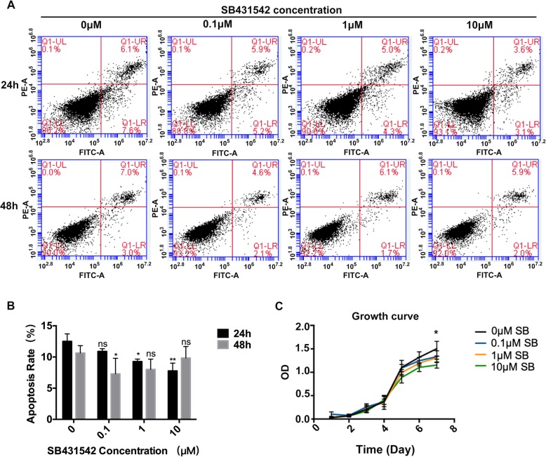
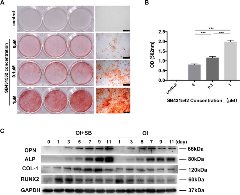
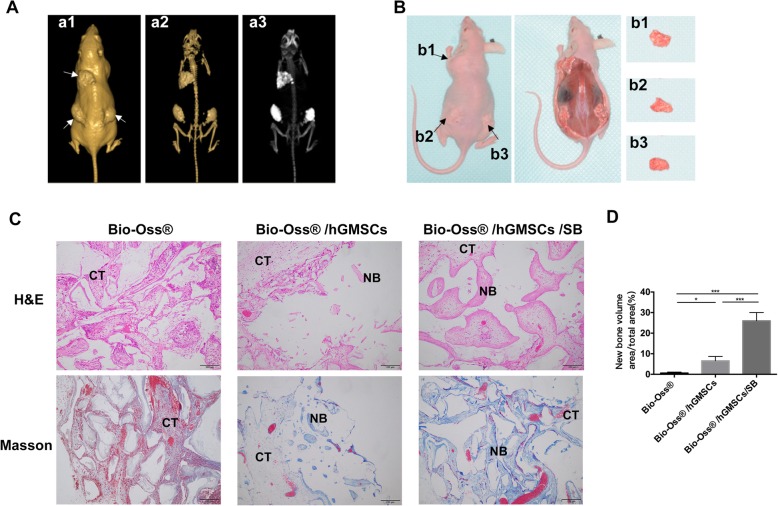
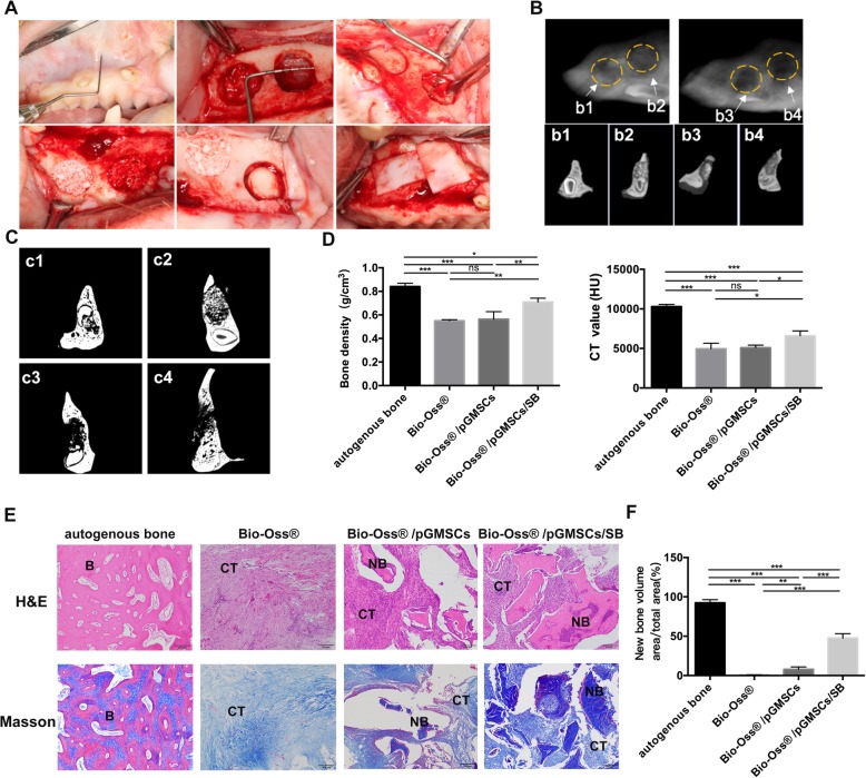
Methods: hGMSCs were isolated and cultured from clinically discarded gingival tissues. The effects of SB431542 on proliferation, apoptosis, and osteogenic differentiation of hGMSCs were analyzed in vitro, and then, SB431542-treated hGMSCs composited with Bio-Oss® were transplanted into immunocompromised mice subcutaneously to explore osteogenic differentiation in vivo. After that, SB431542-treated autologous pig GMSCs (pGMSCs) composited with Bio-Oss® were transplanted into circular confined defects (5 mm × 12 mm) in minipigs maxillary to investigate severe bone defect regeneration. Minipigs were sacrificed at 2 months and nude mice at 8 weeks to retrieve specimens for histological or micro-CT or CBCT analysis. Effects of SB431542 on TGF-β and BMP signaling in hGMSCs were investigated by Western Blot or qRT-PCR.
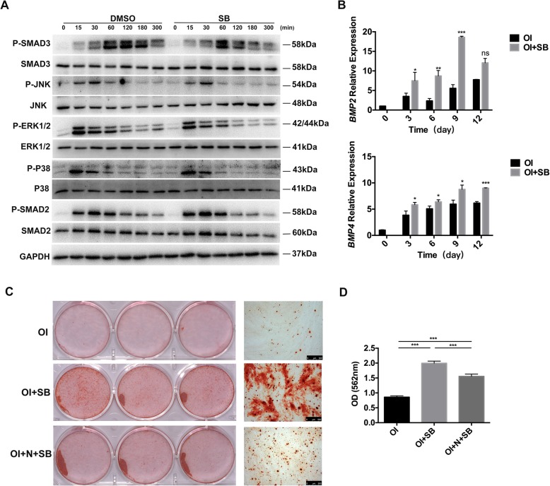
Results: One micromolar of SB431542 treatment induced a robust osteogenesis of hGMSCs in vitro, without adverse effect on apoptosis and growth. In vivo, 1 μM SB431542 treatment also enabled striking osteogenesis of hGMSCs subcutaneously in nude mice and advanced new bone formation of pGMSCs in minipig maxillary bone defect model. In addition, SB431542-treated hGMSCs markedly increased bone-related proteins expression, and BMP2 and BMP4 gene expression. Conversely, SMAD3 protein-dependent TGF-β signal pathway phosphorylation was decreased.
Conclusions: Our study show that osteogenic differentiation of GMSCs treated with TGF-β signaling inhibitor SB431542 was increased, and SB431542-treated autologous pig GMSCs could successfully repair minipig severe maxillofacial bone defects. This preclinical study brings about a promising large bone regeneration therapeutic potential of autologous GMSCs induced by SB431542 in clinic settings.
Keywords: BMP; Bone defect; GMSCs; Osteogenic differentiation; SB431542; TGF-β signaling.
Conflict of interest statement
The authors declare that they have no competing interests.
Figures

Similar articles
-
Rapid cell culture and pre-clinical screening of a transforming growth factor-beta (TGF-beta) inhibitor for orthopaedics.BMC Musculoskelet Disord. 2010 May 28;11:105. doi: 10.1186/1471-2474-11-105. BMC Musculoskelet Disord. 2010. PMID: 20509926 Free PMC article.
-
Inhibition of transforming growth factor-β signaling pathway enhances the osteogenic differentiation of unrestricted somatic stem cells.J Cell Biochem. 2018 Nov;119(11):9327-9333. doi: 10.1002/jcb.27209. Epub 2018 Aug 3. J Cell Biochem. 2018. PMID: 30074269
-
Bone regeneration potential of stem cells derived from periodontal ligament or gingival tissue sources encapsulated in RGD-modified alginate scaffold.Tissue Eng Part A. 2014 Feb;20(3-4):611-21. doi: 10.1089/ten.TEA.2013.0229. Epub 2013 Nov 6. Tissue Eng Part A. 2014. PMID: 24070211 Free PMC article.
-
Effects of nanofibers on mesenchymal stem cells: environmental factors affecting cell adhesion and osteogenic differentiation and their mechanisms.J Zhejiang Univ Sci B. 2020 Nov.;21(11):871-884. doi: 10.1631/jzus.B2000355. J Zhejiang Univ Sci B. 2020. PMID: 33150771 Free PMC article. Review.
-
Potential of Oral Cavity Stem Cells for Bone Regeneration: A Scoping Review.Cells. 2023 May 15;12(10):1392. doi: 10.3390/cells12101392. Cells. 2023. PMID: 37408226 Free PMC article.
Cited by
-
Time-Course Transcriptome Analysis of Gingiva-Derived Mesenchymal Stem Cells Reveals That Fusobacterium nucleatum Triggers Oncogene Expression in the Process of Cell Differentiation.Front Cell Dev Biol. 2020 Jan 14;7:359. doi: 10.3389/fcell.2019.00359. eCollection 2019. Front Cell Dev Biol. 2020. PMID: 31993418 Free PMC article.
-
Novel one-step lignin microsphere preparation for oral tissue regeneration applications.Front Bioeng Biotechnol. 2025 Jan 7;12:1521223. doi: 10.3389/fbioe.2024.1521223. eCollection 2024. Front Bioeng Biotechnol. 2025. PMID: 39840126 Free PMC article.
-
Botanicals and Oral Stem Cell Mediated Regeneration: A Paradigm Shift from Artificial to Biological Replacement.Cells. 2022 Sep 7;11(18):2792. doi: 10.3390/cells11182792. Cells. 2022. PMID: 36139367 Free PMC article. Review.
-
Human cells with osteogenic potential in bone tissue research.Biomed Eng Online. 2023 Apr 3;22(1):33. doi: 10.1186/s12938-023-01096-w. Biomed Eng Online. 2023. PMID: 37013601 Free PMC article. Review.
-
Glycogen synthase kinase-3β inhibitor promotes the migration and osteogenic differentiation of rat dental pulp stem cells via the β-catenin/PI3K/Akt signaling pathway.J Dent Sci. 2022 Apr;17(2):802-810. doi: 10.1016/j.jds.2021.09.035. Epub 2021 Oct 16. J Dent Sci. 2022. PMID: 35756816 Free PMC article.
References
-
- Senarath-Yapa K, Li S, Walmsley GG, Zielins E, Paik K, Britto JA, Grigoriadis AE, Wan DC, Liu KJ, Longaker MT, Quarto N. Small molecule inhibition of transforming growth factor Beta signaling enables the endogenous regenerative potential of the mammalian Calvarium. Eng Part A. 2016;22:707–720. doi: 10.1089/ten.tea.2015.0527. - DOI - PMC - PubMed
Publication types
MeSH terms
Substances
LinkOut - more resources
Full Text Sources
