Predifferentiated amniotic fluid mesenchymal stem cells enhance lung alveolar epithelium regeneration and reverse elastase-induced pulmonary emphysema
- PMID: 31196196
- PMCID: PMC6567664
- DOI: 10.1186/s13287-019-1282-1
Predifferentiated amniotic fluid mesenchymal stem cells enhance lung alveolar epithelium regeneration and reverse elastase-induced pulmonary emphysema
Abstract
Introduction: Pulmonary emphysema is a major component of chronic obstructive pulmonary disease (COPD). Emphysema progression attributed not only to alveolar structure loss and pulmonary regeneration impairment, but also to excessive inflammatory response, proteolytic and anti-proteolytic activity imbalance, lung epithelial cells apoptosis, and abnormal lung remodeling. To ameliorate lung damage with higher efficiency in lung tissue engineering and cell therapy, pre-differentiating graft cells into more restricted cell types before transplantation could enhance their ability to anatomically and functionally integrate into damaged lung. In this study, we aimed to evaluate the regenerative and repair ability of lung alveolar epithelium in emphysema model by using lung epithelial progenitors which pre-differentiated from amniotic fluid mesenchymal stem cells (AFMSCs).
Methods: Pre-differentiation of eGFP-expressing AFMSCs to lung epithelial progenitor-like cells (LEPLCs) was established under a modified small airway growth media (mSAGM) for 7-day induction. Pre-differentiated AFMSCs were intratracheally injected into porcine pancreatic elastase (PPE)-induced emphysema mice at day 14, and then inflammatory-, fibrotic-, and emphysema-related indices and pathological changes were assessed at 6 weeks after PPE administration.
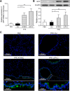
Results: An optimal LEPLCs pre-differentiation condition has been achieved, which resulted in a yield of approximately 20% lung epithelial progenitors-like cells from AFMSCs in a 7-day period. In PPE-induced emphysema mice, transplantation of LEPLCs significantly improved regeneration of lung tissues through integrating into the lung alveolar structure, relieved airway inflammation, increased expression of growth factors such as vascular endothelial growth factor (VEGF), and reduced matrix metalloproteinases and lung remodeling factors when compared with mice injected with AFMSCs. Histopathologic examination observed a significant amelioration in DNA damage in alveolar cells, detected by terminal deoxynucleotidyltransferase-mediated dUTP nick end labeling (TUNEL), the mean linear intercept, and the collagen deposition in the LEPLC-transplanted groups.
Conclusion: Transplantation of predifferentiated AFMSCs through intratracheal injection showed better alveolar regeneration and reverse elastase-induced pulmonary emphysema in PPE-induced pulmonary emphysema mice.
Keywords: Amniotic fluid mesenchymal stem cell; Elastase-induced pulmonary emphysema; Predifferentiation.
Conflict of interest statement
The authors declare that they have no competing interests.
Figures


Similar articles
-
Human Adipose-Derived Mesenchymal Stem Cells Ameliorate Elastase-Induced Emphysema in Mice by Mesenchymal-Epithelial Transition.Int J Chron Obstruct Pulmon Dis. 2021 Oct 8;16:2783-2793. doi: 10.2147/COPD.S324952. eCollection 2021. Int J Chron Obstruct Pulmon Dis. 2021. PMID: 34675503 Free PMC article.
-
Keratinocyte growth factor protects against elastase-induced pulmonary emphysema in mice.Am J Physiol Lung Cell Mol Physiol. 2007 Nov;293(5):L1230-9. doi: 10.1152/ajplung.00460.2006. Epub 2007 Aug 31. Am J Physiol Lung Cell Mol Physiol. 2007. PMID: 17766584
-
Therapeutic effects of amniotic fluid-derived mesenchymal stromal cells on lung injury in rats with emphysema.Respir Res. 2014 Oct 16;15(1):120. doi: 10.1186/s12931-014-0120-3. Respir Res. 2014. PMID: 25319435 Free PMC article.
-
The pulmonary matrix, glycosaminoglycans and pulmonary emphysema.Connect Tissue Res. 1999;40(2):97-104. doi: 10.3109/03008209909029105. Connect Tissue Res. 1999. PMID: 10761634 Review.
-
Lung regeneration using amniotic fluid mesenchymal stem cells.Artif Cells Nanomed Biotechnol. 2018 May;46(3):447-451. doi: 10.1080/21691401.2017.1337023. Epub 2017 Jul 4. Artif Cells Nanomed Biotechnol. 2018. PMID: 28675062 Review.
Cited by
-
Chronic Obstructive Pulmonary Disease and the Cardiovascular System: Vascular Repair and Regeneration as a Therapeutic Target.Front Cardiovasc Med. 2021 Apr 12;8:649512. doi: 10.3389/fcvm.2021.649512. eCollection 2021. Front Cardiovasc Med. 2021. PMID: 33912600 Free PMC article. Review.
-
Stem cell therapies for chronic obstructive pulmonary disease: mesenchymal stem cells as a promising treatment option.Stem Cell Res Ther. 2024 Sep 19;15(1):312. doi: 10.1186/s13287-024-03940-9. Stem Cell Res Ther. 2024. PMID: 39300523 Free PMC article. Review.
-
Can amniotic fluid protect developing fetal lungs against the harmful effects of oxidative stress?Turk J Med Sci. 2023 Feb;53(1):109-120. doi: 10.55730/1300-0144.5564. Epub 2023 Feb 22. Turk J Med Sci. 2023. PMID: 36945927 Free PMC article.
-
In Utero Cell Treatment of Hemophilia A Mice via Human Amniotic Fluid Mesenchymal Stromal Cell Engraftment.Int J Mol Sci. 2023 Nov 16;24(22):16411. doi: 10.3390/ijms242216411. Int J Mol Sci. 2023. PMID: 38003601 Free PMC article.
-
Aldo-keto reductase family 1 member A1 (AKR1A1) exerts a protective function in alcohol-associated liver disease by reducing 4-HNE accumulation and p53 activation.Cell Biosci. 2024 Feb 3;14(1):18. doi: 10.1186/s13578-024-01200-0. Cell Biosci. 2024. PMID: 38308335 Free PMC article.
References
Publication types
MeSH terms
Substances
LinkOut - more resources
Full Text Sources
Medical

