

Adipose-derived stem cells prevent the onset of bisphosphonate-related osteonecrosis of the jaw through transforming growth factor β-1-mediated gingival wound healing
- PMID: 31196208
- PMCID: PMC6567445
- DOI: 10.1186/s13287-019-1277-y
Adipose-derived stem cells prevent the onset of bisphosphonate-related osteonecrosis of the jaw through transforming growth factor β-1-mediated gingival wound healing
Abstract
Background: Due to its complex pathogenesis and low clinical cure rate, bisphosphonate-related osteonecrosis of the jaw (BRONJ) poses a substantial challenge for oral and maxillofacial surgeons. Therefore, the treatment of BRONJ should focus on prevention. In clinical studies, primary wound closure can significantly reduce the incidence of BRONJ. Whether local stem cell transplantation can promote primary gingival healing in patients with a medication history and prevent BRONJ has not been reported.
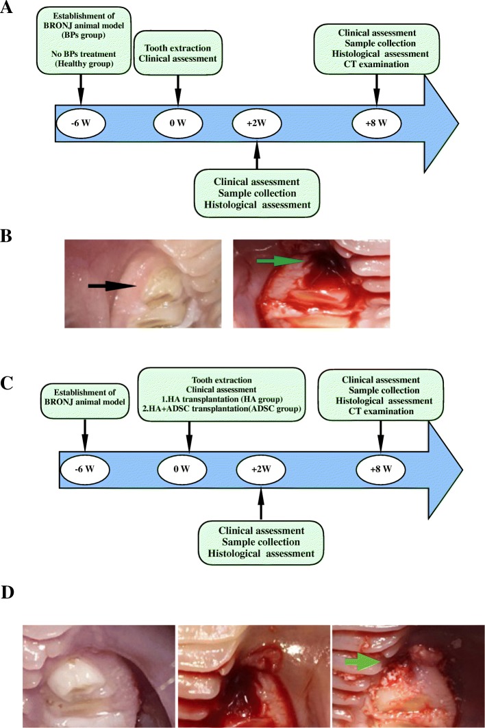
Methods: In this study, animals were divided into a healthy group (non-drug treatment), a BP group, a hydroxyapatite (HA) group, and an adipose-derived stem cell (ADSC) group. All groups except the healthy group were treated with BPs and immunosuppressive drugs once per week for 8 weeks, simulating clinical use for the treatment of cancer patients with bone metastasis, to induce BRONJ-like animals. After the sixth drug treatment, the bilateral premolars were extracted in all groups. In contrast to the healthy and BP groups, the extraction sockets in the HA and ADSC groups were filled with HA or HA + ADSCs simultaneously post extraction to observe the preventive effect of ADSCs on the occurrence of BRONJ. At 2 and 8 weeks post extraction, animals from all groups were sacrificed.
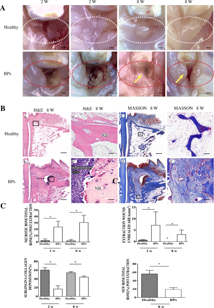
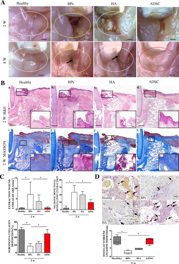
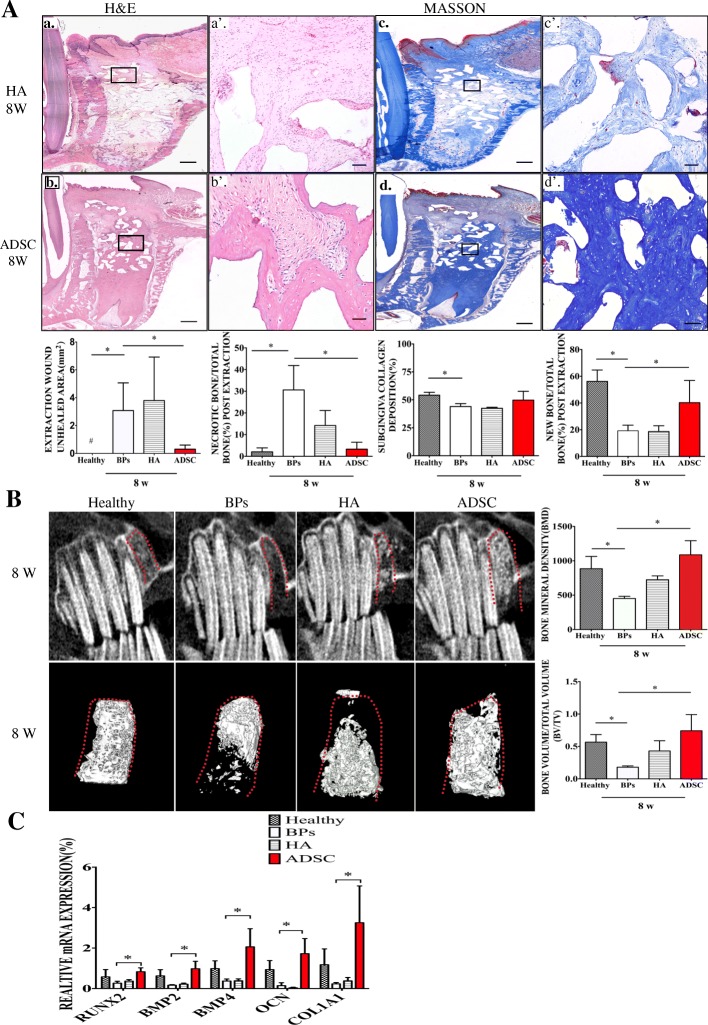
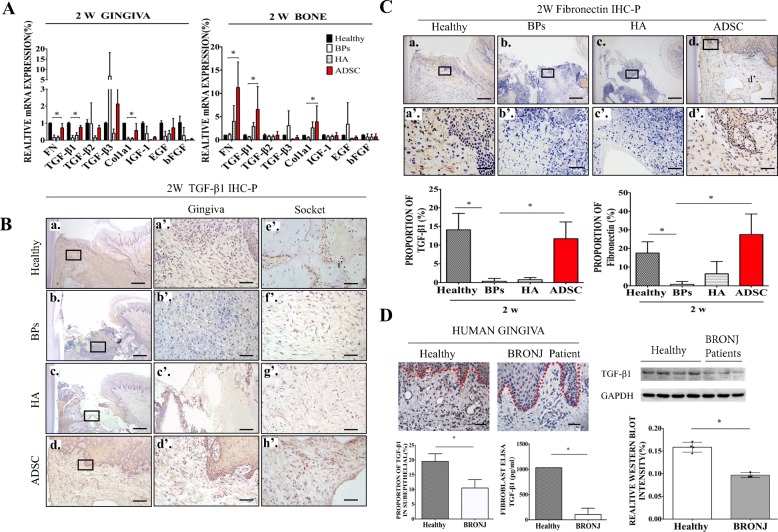
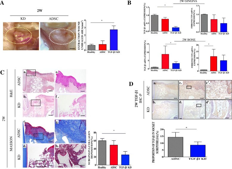
Results: At 8 weeks post transplantation, ADSCs prevented the occurrence of BRONJ, mainly through accelerating healing of the gingival epithelium at 2 weeks post extraction. We also found that ADSCs could upregulate the expression of transforming growth factor β1 (TGF-β1) and fibronectin in tissue from animals with a medication history by accelerating gingival healing of the extraction socket. A rescue assay further demonstrated that TGF-β1 and fibronectin expression decreased in TGF-β1-deficient ADSC-treated animals, which partially abolished the preventive effect of ADSCs on the onset of BRONJ.
Conclusion: ADSCs prevent the onset of BRONJ, mainly by upregulating the expression of TGF-β1 and fibronectin to promote primary gingival healing, ultimately leading to bone regeneration in the tooth extraction socket. Our new findings provide a novel stem cell treatment for the prevention of BRONJ.
Keywords: Adipose-derived mesenchymal stem cells; Bisphosphonate-related osteonecrosis of the jaw; Fibroblast; Fibronectin; Wound healing; Zoledronic acid.
Conflict of interest statement
The authors declare that they have no competing interests.
Figures
Similar articles
-
Characterization of Mesenchymal Stem Cells Derived from Bisphosphonate-Related Osteonecrosis of the Jaw Patients' Gingiva.Stem Cell Rev Rep. 2022 Jan;18(1):378-394. doi: 10.1007/s12015-021-10241-8. Epub 2021 Sep 22. Stem Cell Rev Rep. 2022. PMID: 34553308 Free PMC article.
-
Transplantation of Noncultured Stromal Vascular Fraction Cells of Adipose Tissue Ameliorates Osteonecrosis of the Jaw-Like Lesions in Mice.J Bone Miner Res. 2018 Jan;33(1):154-166. doi: 10.1002/jbmr.3292. Epub 2017 Oct 4. J Bone Miner Res. 2018. PMID: 28902422
-
Prevention of tooth extraction-triggered bisphosphonate-related osteonecrosis of the jaws with basic fibroblast growth factor: An experimental study in rats.PLoS One. 2019 Feb 8;14(2):e0211928. doi: 10.1371/journal.pone.0211928. eCollection 2019. PLoS One. 2019. PMID: 30735554 Free PMC article.
-
Bisphosphonate-related osteonecrosis of the jaws. A severe side effect of bisphosphonate therapy.Acta Medica (Hradec Kralove). 2012;55(3):111-5. doi: 10.14712/18059694.2015.47. Acta Medica (Hradec Kralove). 2012. PMID: 23297518 Review.
-
Current management concepts for bisphosphonate-related osteonecrosis of the jaw: a review.Gen Dent. 2018 Nov-Dec;66(6):e1-e5. Gen Dent. 2018. PMID: 30444713 Review.
Cited by
-
Calcium Phosphate Ceramics Can Prevent Bisphosphonate-Related Osteonecrosis of the Jaw.Materials (Basel). 2020 Apr 22;13(8):1955. doi: 10.3390/ma13081955. Materials (Basel). 2020. PMID: 32331240 Free PMC article.
-
Exosomes Derived from Adipose Tissue-Derived Mesenchymal Stromal Cells Prevent Medication-Related Osteonecrosis of the Jaw through IL-1RA.Int J Mol Sci. 2023 May 12;24(10):8694. doi: 10.3390/ijms24108694. Int J Mol Sci. 2023. PMID: 37240036 Free PMC article.
-
Recent Stem-Cell-Based and Stem-Cell-Free Possibilities for the Therapeutic Management of the Osteonecrosis of the Jaw.Biomolecules. 2025 Apr 16;15(4):595. doi: 10.3390/biom15040595. Biomolecules. 2025. PMID: 40305370 Free PMC article. Review.
-
Exosomes from Adipose-Derived Stem Cells Can Prevent Medication-Related Osteonecrosis of the Jaw.Med Sci Monit. 2021 Mar 10;27:e929684. doi: 10.12659/MSM.929684. Med Sci Monit. 2021. PMID: 33690263 Free PMC article.
-
Polymer-Bioactive Glass Composite Filaments for 3D Scaffold Manufacturing by Fused Deposition Modeling: Fabrication and Characterization.Front Bioeng Biotechnol. 2020 Jun 24;8:552. doi: 10.3389/fbioe.2020.00552. eCollection 2020. Front Bioeng Biotechnol. 2020. PMID: 32671025 Free PMC article.
References
-
- Ruggiero SL, Dodson TB, Fantasia J, Goodday R, Aghaloo T, Mehrotra B, O'Ryan F, O American Association of and S Maxillofacial American Association of Oral and Maxillofacial Surgeons position paper on medication-related osteonecrosis of the jaw--2014 update. J Oral Maxillofac Surg. 2014;72:1938–1956. doi: 10.1016/j.joms.2014.04.031. - DOI - PubMed
-
- Berenson JR, Rosen LS, Howell A, Porter L, Coleman RE, Morley W, Dreicer R, Kuross SA, Lipton A, Seaman JJ. Zoledronic acid reduces skeletal-related events in patients with osteolytic metastases. Cancer. 2001;91:1191–1200. doi: 10.1002/1097-0142(20010401)91:7<1191::AID-CNCR1119>3.0.CO;2-0. - DOI - PubMed
-
- Ogata K, Katagiri W, Osugi M, Kawai T, Sugimura Y, Hibi H, Nakamura S, Ueda M. Evaluation of the therapeutic effects of conditioned media from mesenchymal stem cells in a rat bisphosphonate-related osteonecrosis of the jaw-like model. Bone. 2015;74:95–105. doi: 10.1016/j.bone.2015.01.011. - DOI - PubMed
-
- Kikuiri T, Kim I, Yamaza T, Akiyama K, Zhang Q, Li Y, Chen C, Chen W, Wang S, Le AD, Shi S. Cell-based immunotherapy with mesenchymal stem cells cures bisphosphonate-related osteonecrosis of the jaw-like disease in mice. J Bone Miner Res. 2010;25:1668–1679. doi: 10.1002/jbmr.37. - DOI - PMC - PubMed
Publication types
MeSH terms
Substances
LinkOut - more resources
Full Text Sources
