. 2019 Jun 13;38(1):254.
doi: 10.1186/s13046-019-1234-8.

Curcumin overcome primary gefitinib resistance in non-small-cell lung cancer cells through inducing autophagy-related cell death

Affiliations

Curcumin overcome primary gefitinib resistance in non-small-cell lung cancer cells through inducing autophagy-related cell death

Ping Chen et al. J Exp Clin Cancer Res. .

Abstract

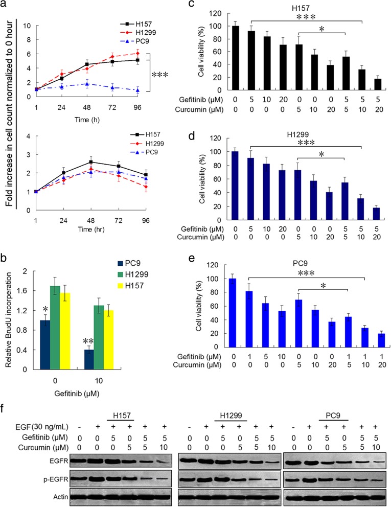
Background: Epidermal growth factor receptor (EGFR) tyrosine kinase inhibitors (TKIs) are being wildly used as target therapy in non-small-cell lung cancer (NSCLC). However, NSCLC patients with wild-type EGFR and KRAS mutation are primary resistant to EGFR-TKIs such as gefitinib. Curcumin has been known as a potential therapeutic agent for several major human cancers. In this study, we investigated the effect of curcumin on the reversal of gefitinib resistance in NSCLC cells as well as their molecular bases.

Methods: H157 (wild-type EGFR and KARS mutation) and H1299 (wild-type EGFR and HRAS mutation) cells were treated with gefitinib or curcumin alone, or the two combination, and then cell viability, EGFR activity, expressions of Sp1 and Sp1-dependent proteins and receptor tyrosine kinases, markers of autophagy and apoptosis were examined by using CCK-8, colony formation, immunoblot, quantitative PCR, immunofluoscence, and flow cytometry assays. Also xenograft experiments were conduced to test the synergism of curcumin to gefitinib.

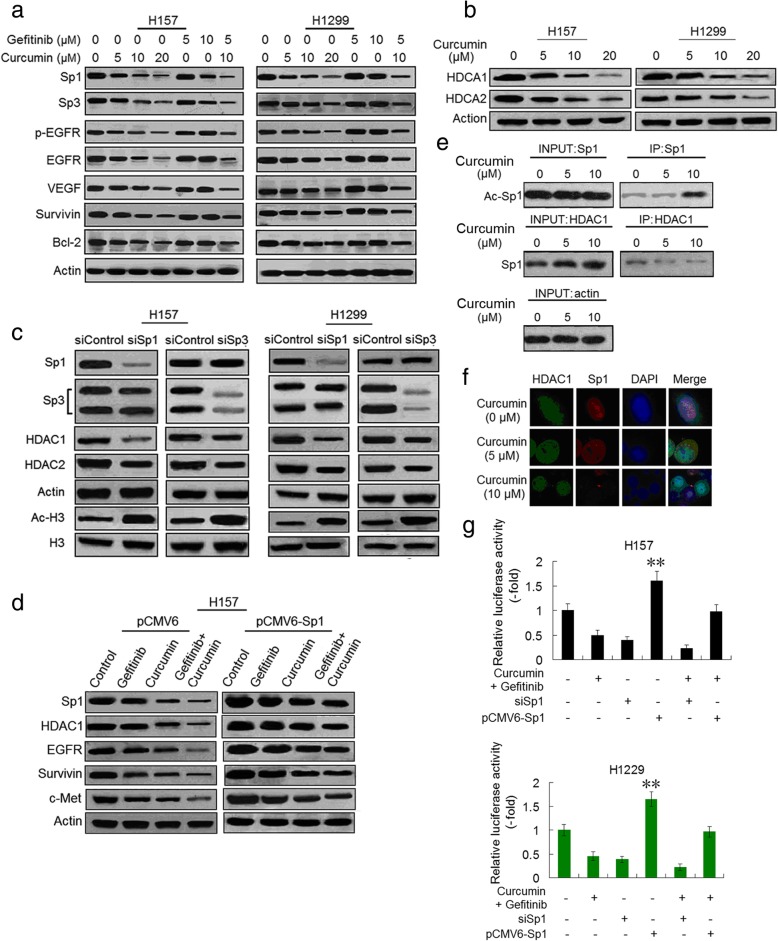
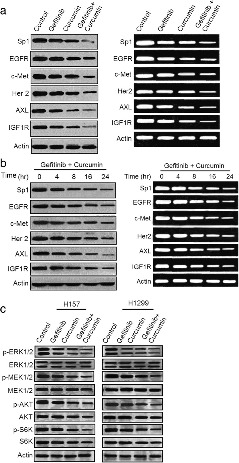
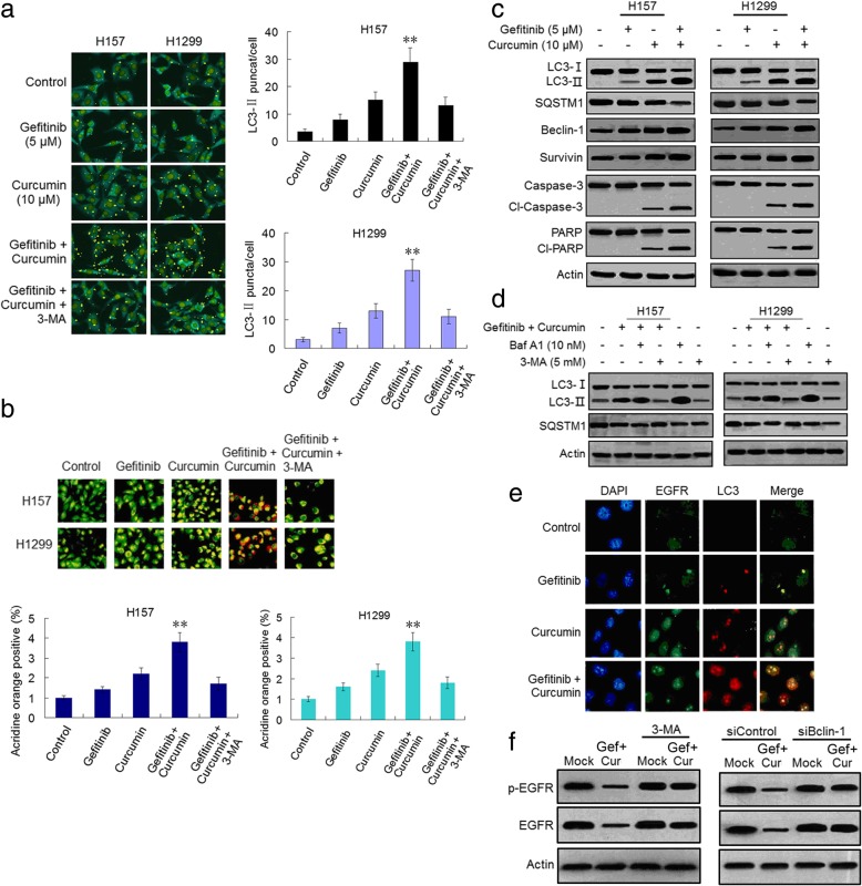
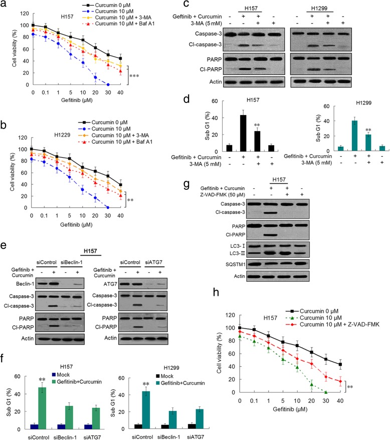
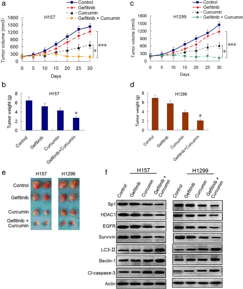
Results: Our results showed that curcumin significantly enhanced inhibitory effect of gefitinib on primary gefitinib-resistant NSCLC cell lines H157 and H1299. Combination treatment with curcumin and gefitinib markedly downregulated EGFR activity through suppressing Sp1 and blocking interaction of Sp1 and HADC1, and markedly suppressed receptor tyrosine kinases as well as ERK/MEK and AKT/S6K pathways in the resistant NSCLC cells. Meanwhile, combination treatment of curcumin and gefitinib caused dramatic autophagy induction, autophagic cell death and autophagy-mediated apoptosis, compared to curcumin or gefitinib treatment alone, as evidenced by the findings that curcumin and gefitinib combination treatment-produced synergistic growth inhibition and apoptosis activation can be reversed by pharmacological autophagy inhibitors (Baf A1 or 3-MA) or knockdown of Beclin-1 or ATG7, also can be partially returned by pan-caspase inhibitor (Z-VAD-FMK) in H157 and H1299 cells. Xenograft experiments in vivo yielded similar results.

Conclusions: These data indicate that the synergism of curcumin on gefitinib was autophagy dependent. Curcumin can be used as a sensitizer to enhance the efficacy of EGFR-TKIs and overcome the EGFR-TKI resistance in NSCLC patients with wild-type EGFR and/or KRAS mutation.

Keywords: Curcumin; Epidermal growth factor receptor (EGFR); Gefitinib; Non-small-cell lung cancer; Resistance; Tyrosine kinase inhibitor (TKI).


Conflict of interest statement

The authors declare that they have no competing interests.

Figures

Fig. 1
Fig. 1
Curcumin enhances anticancer effect of gefitinib on NSCLC cell and suppresses EGFR activity. a H157, H1299 and PC9 cell lines were growth in complete media in the presence of 5 μM gefitinib (top), or 5 μM curcumin (nether) for 24, 48, 72, 96 h. Fold increase in cell counts normalized to zero hour counts of respective cell lines are represent (***P<0.001). b The three cell lines were grown in the presence DMSO or 10 μM gefitinib in complete media. BrdU substrate was added 48 h after drug treatment and assayed after 24 h. H157 c, H1299 d and PC9 e cells were treated with gefitinib, or curcumin alone, or the two combination at indicated concentrations for 48 h. Cell viability was measured by CCK-8 assay (*P<0.05; ***P<0.001). f H157, H1299 and PC9 cell lines were pre-treated with curcumin or gefitinib alone, or the two combination at indicated concentrations for 12 h, and then EGF (30 ng/mL) was added for 1 h. Immunoblot analysis was used to determine p-EGFR and total EGFR expression. Actin was used as aloading control in immunoblots. Similar results were obtained from three independent experiments. Typical immunoblots were presented in the Figure
Fig. 2
Fig. 2
Combination of curcumin and gefitinib downregulates EGFR through inhibiting SP1 and HDAC1 binding induced-EGFR transcription activity. a H157 and H1299 cells were treated with gefitinib, or curcumin alone, or the two combination at indicated concentrations for 48 and evaluated for the protein expression as indicated by immunoblot analysis. b Curcumin-induced suppression of HDCA1 and HDCA2 proteins in H157 and H1299 cells. Protein expression was determined as outlined above in A. c Expressions of Sp1 and other indicated proteins in control and Sp1 or Sp3 knocked down cells were determined by immunoblotting in H157 and H1299 cells. d H157 cells were transfected with Sp1 plasmids (PCMV6-Sp1) and control plasmid DNA (PCMV6). After 8 h, cells were treated with 5 μM gefitinib, or 10 μM curcumin alone, or two drug combination for an additional 48 h. And then the expressions of Sp1, HDAC1, EGFR, Survivin and c-Met proteins were determined by immunoblotting. Similar results were obtained from three independent experiments. Typical immunoblots were presented in the Figure. e H157 cells were pre-incubated with curcumin and nuclear extracts were prepared. The immunoprecipitation was performed using anti-Sp1 and anti-HDAC1 antibodies. The immunoprecipitated pellets were analyzed by immunoblotting with anti-acetyl-lysine and anti-Sp1 antibodies. f H157 cells treated with DMSO or curcumin at indicated concentrations were stained for HDAC1 (green) and Sp1 (red) using immunofluorescence assay. g Effect of Sp1 on EGFR promoter activity. Cells were treated with curcumin (10 μM) plus gefitinib (5 μM), or transfected with siSp1 or plasmids containing Sp1 (PCMV6-Sp1), and luciferase activity was determined as described in Materials and Methods. (**P<0.01 compared with gefitinib plus curcumin, or siSp1, or gefitinib plus curcumin in combination with pCMV6-Sp1, respectively)
Fig. 3
Fig. 3
Combination of curcumin and gefitinib markedly depresses the receptor tyrosine kinase signaling pathways in gefiinib-resistant NSCLC cells. a H157 cells were treated with 5 μM gefitinib, or 5 μM curcumin alone, or the two combination for 48 h. Cell lysates and total mRNA were prepared and subjected to immunoblotting and RT-PCR analysis for detecting proteins and mRNA as indicated. b The combination of curcumin (5 μM) and gefitinib (5 μM) time-dependently suppressed expressions of proteins and mRNAs as indicated. c H157 and H1299 cells were treated with gefitinib (5 μM), or curcumin (5 μM) alone, or the two combination for 48 h. The whole-cell lysates were prepared and subjected to immuoblotting analysis for detecting phosphorylations of ERK1/2, MEK1/2, AKT and S6K proteins. Similar results were obtained from three independent experiments. Typical immunoblots were presented in the Figure
Fig. 4
Fig. 4
Combination of curcumin and gefitinib significantly potentiates induction of autophagy and apoptosis. a H157 and H1299 cells were treated with gefitinib, or curcumin alone, or the two combination at indicated concentration for 48 h. Then the LC3-II puncta formation was determined using immunofluorescence analysis and imaged by confocal microscopy (left). The number of LC3-II puncta/cell was quantified by Image-Pro Plus 5.1 software (right). (**P<0.01 compared with gefitinib, or curcumin alone, or the two combination plus 3-MA, respectively). b H157 and H1299 cells were stained with acridine orange after drug treatments (5 μM of gefitinib or 10 μM of curcumin alone, or the two combination for 48 h) and analyzed by confocal fluorescent microscopy. Development of AVOs (orange) was observed (left). Quantification of AVOs using FACSanalysis (right) (** P<0.01 compared with gefitinib, or curcumin alone, or the two combination plus 3-MA, respectively). c H157 and H1299 cells were treated with gefitinib, or curcumin alone, or gefitinib plus curcumin at indicated concentration for 48 h. The autophagy-related and apoptosis activation-related proteins were analyzed by immunoblotting. d H157 and H1299 cells were treated with gefitinib plus curcumin in the absence and presence of Baf A1 or 3-MA. The autophagy-related proteins were analyzed by immunoblotting. e H157 and H1299 cells were treated with gefitinib, or curcumin alone, or gefitinib plus curcumin at indicated concentration for 48 h and then were fixed with methanol, immunostained with anti-EGFR (green), anti-LC3 (red), and DAPI (blue), and analyzed by confocal microscopy to determine the intracellular locations of EGFR and LC3. f H157 cells were treated with 5 μM gefitinib (Gef) plus 10 μM curcumin (Cur) for 48 h in the presence or absence of 3-MA or Beclin-1 knokdown. The phosphorylated and total EGFR were detected by immunoblotting. Similar results were obtained from three independent experiments. Typical immunoblots were presented in the Figure
Fig. 5
Fig. 5
Curcumin reverses gefitinib resistance in NSCLC cells through induction of autophagic cell death and autophagy-mediated apoptosis. a-b H157 and H1299 cells were treated with gefitinib plus curcumin at indicated concentration for 48 h in the absence or presence of Baf A1 (10 nM) or 3-MA (5 mM), cell viability was detected by CCK-8 assay. c H157 and H1299 cells were treated with gefitinib (5 μM) plus curcumin (10 μM) for 48 h in the absence or presence of 3-MA (5 mM). Cleaved (Cl)-caspase-3 and Cl-PARP were determined by immunoblotting. d Flow cytometry was used to analyze sub-G1 population in H157 and H1299 cells after drug treatment (5 μM gefitinib plus 10 μM curcumin in combination with or without 5 mM 3-MA). e After transfection with siBeclin-1 or siATG7 or siControl, H157 cells were treated with gefitinib (5 μM) plus curcumin (10 μM) for 48 h, Cl-caspase-3 and Cl-PARP were determined by immunoblotting. Similar results were obtained from three independent experiments. Typical immunoblots were presented in the Figure. f The sub-G1 population was analyzed by flow cytometry in H157 and H1299 after transfection with siBeclin-1 or siATG7 or siControl . g H157 cells were treated with gefitinib (5 μM) plus curcumin (10 μM) in the presence and absence of Z-VAD-MFK, immunoblotting was used to detect apoptosis- and autophagy-related proteins. h H157 cells were treated with gefitinib plus curcumin at indicated concentration in the presence or absence of Z-VAD-MFK, cell viability was determined by CCK-8 assay
Fig. 6
Fig. 6
Curcumin enhances therapeutic efficacy of gefitinib in vivo through Sp1/EGFR downregulation-mediated autophagy induction. a-d H157 and H1299 cells were subcutaneously injected to the flanks of BALBL/c mice respectively. The mice were treated with vehicle (control), gefitinib, or curcumin, or the two drug combination as described in Materials and Methods. Tumor volume was measured at the time indicated after the onset of treatment. (*P<0.05 and ***P<0.001). The tumor weight was measured at the end of the experiment (*P<0.05 compared with gefitinib, or curcumin treatment alone). e Representive pictures of tumor samples from the mice bearing H157 and H1299 cell tumors receiving different treatments as indicated. f Immunoblot analysis of the expressions of Sp1, HDAC1, EGFR, survivin, LC3-II, Beclin-1 and cleaved-caspase-3 in H157 and H1299 cell tumor samples from the mice in each treatment group. All experiments were repeated three times. Similar results were obtained from three independent experiments. Typical immunoblots were presented in the Figure

Similar articles

Cited by

References

    1. Shama SV, Bell DW, Settleman J, Haber DA. Epidermal growth factor receptor mutation in lung cancer. Nat Rev Cancer. 2007;7:169–181. doi: 10.1038/nrc2088. - DOI - PubMed
    1. Pao W, Chmielechi J. Rational biologically based treatment of EGFR-mutation non-small-cell lung cancer. Nat Rev Cancer. 2010;10:760–774. doi: 10.1038/nrc2947. - DOI - PMC - PubMed
    1. Catado VD, Gibbons DL, pérez-Solar R, Quintás-Cardama A. Treatment of non-small-cell lung cancer with erlotinib or gefitinib. N Engl J Med. 2011;64:947–955. doi: 10.1056/NEJMct0807960. - DOI - PubMed
    1. Stella GM, Luisetti M, Inghilleri S, Cemmi F, Scabini R, Zorzetfo M, Pozzi E. Targeting EGFR in non-small-cell lung cancer: lessons experiences, strategies. Respir Med. 2012;106:173–183. doi: 10.1016/j.rmed.2011.10.015. - DOI - PubMed
    1. Kobayashi S, Boggon TJ, Dayaram T, Jänne DA, kocher O, Meyerson M, Jonhnson BE, Eck MJ, Tene DG, Halmos B. EGFR mutation and resistance of non-small-cell lung cancer to gefitinib. N Engl J Med. 2005;352:786–792. doi: 10.1056/NEJMoa044238. - DOI - PubMed

MeSH terms