Fat extract improves fat graft survival via proangiogenic, anti-apoptotic and pro-proliferative activities
- PMID: 31196213
- PMCID: PMC6567564
- DOI: 10.1186/s13287-019-1290-1
Fat extract improves fat graft survival via proangiogenic, anti-apoptotic and pro-proliferative activities
Abstract
Background: Our previous study proved that nanofat could enhance fat graft survival by promoting neovascularization. Fat extract (FE), a cell-free component derived from nanofat, also possesses proangiogenic activity.
Objectives: The aim of this study was to investigate whether FE could improve fat graft survival and whether FE and nanofat could work synergistically to promote fat graft survival. The underlying mechanism was also investigated.
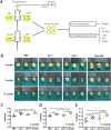
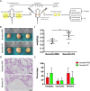
Methods: In the first animal study, human macrofat from lipoaspirate was co-transplanted into nude mice with FE or nanofat. The grafts were evaluated at 2, 4 and 12 weeks post-transplantation. In the second animal study, nude mice were transplanted with a mixture of macrofat and nanofat, followed by intra-graft injection of FE at days 1, 7, 14, 21 and 28 post-transplantation. The grafts were evaluated at 12 weeks post-transplantation. To detect the mechanism by which FE impacts graft survival, the proangiogenic, anti-apoptotic and pro-proliferative activities of FE were analysed in grafts in vivo and in cultured human vascular endothelial cells (HUVECs), adipose-derived stem cells (ADSCs) and fat tissue in vitro.
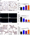
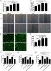
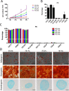
Results: In the first animal study, the weights of the fat grafts in the nanofat- and FE-treated groups were significantly higher than those of the fat grafts in the control group. In addition, higher fat integrity, more viable adipocytes, more CD31-positive blood vessels, fewer apoptotic cells and more Ki67-positive proliferating cells were observed in the nanofat- and FE-treated groups. In the second animal study, the weights of the fat grafts in the nanofat+FE group were significantly higher than those of the fat grafts in the control group. In vitro, FE showed proangiogenic effects on HUVECs, anti-apoptotic effects on fat tissue cultured under hypoxic conditions and an ability to promote ADSC proliferation and maintain their multiple differentiation capacity.
Conclusions: FE could improve fat graft survival via proangiogenic, anti-apoptotic and pro-proliferative effects on ADSCs. FE plus nanofat-assisted fat grafting is a new strategy that could potentially be used in clinical applications.
Keywords: Anti-apoptotic; Fat extract; Fat transplantation; Nanofat; Pro-proliferative; Proangiogenic.
Conflict of interest statement
The authors declare that they have no competing interests.
Figures



Similar articles
-
Co-Transplantation of Nanofat Enhances Neovascularization and Fat Graft Survival in Nude Mice.Aesthet Surg J. 2018 May 15;38(6):667-675. doi: 10.1093/asj/sjx211. Aesthet Surg J. 2018. PMID: 29161346
-
Co-transplantation of exosomes derived from hypoxia-preconditioned adipose mesenchymal stem cells promotes neovascularization and graft survival in fat grafting.Biochem Biophys Res Commun. 2018 Feb 26;497(1):305-312. doi: 10.1016/j.bbrc.2018.02.076. Epub 2018 Feb 8. Biochem Biophys Res Commun. 2018. PMID: 29428734
-
Salvianolic acid-B improves fat graft survival by promoting proliferation and adipogenesis.Stem Cell Res Ther. 2021 Sep 17;12(1):507. doi: 10.1186/s13287-021-02575-4. Stem Cell Res Ther. 2021. PMID: 34535194 Free PMC article.
-
Adipose tissue and the vascularization of biomaterials: Stem cells, microvascular fragments and nanofat-a review.Cytotherapy. 2020 Aug;22(8):400-411. doi: 10.1016/j.jcyt.2020.03.433. Epub 2020 Jun 2. Cytotherapy. 2020. PMID: 32507607 Review.
-
Fat Grafting for Facial Rejuvenation with Nanofat Grafts.Clin Plast Surg. 2020 Jan;47(1):53-62. doi: 10.1016/j.cps.2019.08.006. Epub 2019 Oct 28. Clin Plast Surg. 2020. PMID: 31739897 Review.
Cited by
-
Human adipose-derived stem cells enriched with VEGF-modified mRNA promote angiogenesis and long-term graft survival in a fat graft transplantation model.Stem Cell Res Ther. 2020 Nov 19;11(1):490. doi: 10.1186/s13287-020-02008-8. Stem Cell Res Ther. 2020. PMID: 33213517 Free PMC article.
-
Investigation of the Effect of Mobile and Immobile Regions on Fat Graft Viability: An Experimental Study in a New Model.Aesthetic Plast Surg. 2024 Dec;48(23):5155-5161. doi: 10.1007/s00266-024-04267-9. Epub 2024 Aug 8. Aesthetic Plast Surg. 2024. PMID: 39117873 Free PMC article.
-
Small extracellular vesicles from human adipose-derived mesenchymal stromal cells: a potential promoter of fat graft survival.Stem Cell Res Ther. 2021 May 3;12(1):263. doi: 10.1186/s13287-021-02319-4. Stem Cell Res Ther. 2021. PMID: 33941279 Free PMC article.
-
Autologous Cell-Free Fat Extract: A Novel Approach for Infraorbital Rejuvenation-A Pilot Study.J Cosmet Dermatol. 2025 Feb;24(2):e16682. doi: 10.1111/jocd.16682. Epub 2024 Dec 8. J Cosmet Dermatol. 2025. PMID: 39645645 Free PMC article.
-
Enhanced adipose-derived stem cells with IGF-1-modified mRNA promote wound healing following corneal injury.Mol Ther. 2023 Aug 2;31(8):2454-2471. doi: 10.1016/j.ymthe.2023.05.002. Epub 2023 May 10. Mol Ther. 2023. PMID: 37165618 Free PMC article.
References
-
- Coleman SR. Structural fat grafting: more than a permanent filler. Plast Reconstr Surg. 2006;118(3):108s–120s. - PubMed
-
- Zhu M, Cohen SR, Hicok KC, Shanahan RK, Strem BM, Yu JC, et al. Comparison of three different fat graft preparation methods: gravity separation, centrifugation, and simultaneous washing with filtration in a closed system. Plast Reconstr Surg. 2013;131(4):873–880. - PubMed
-
- Peer LA, Walker JC., Jr The behavior of autogenous human tissue grafts: a comparative study. Plast Reconstr Surg (1946) 1951;7(1):6–23. - PubMed
-
- Coleman SR. Structural fat grafts: the ideal filler? Clin Plast Surg. 2001;28(1):111–119. - PubMed
-
- Eto H, Kato H, Suga H, Aoi N, Doi K, Kuno S, et al. The fate of adipocytes after nonvascularized fat grafting: evidence of early death and replacement of adipocytes. Plast Reconstr Surg. 2012;129(5):1081–1092. - PubMed
Publication types
MeSH terms
LinkOut - more resources
Full Text Sources

