. 2019:247:375-436.
doi: 10.1016/bs.pbr.2019.03.012. Epub 2019 Apr 17.

Dispositional negativity, cognition, and anxiety disorders: An integrative translational neuroscience framework

Affiliations

Dispositional negativity, cognition, and anxiety disorders: An integrative translational neuroscience framework

Juyoen Hur et al. Prog Brain Res. 2019.

Abstract

When extreme, anxiety can become debilitating. Anxiety disorders, which often first emerge early in development, are common and challenging to treat, yet the underlying mechanisms have only recently begun to come into focus. Here, we review new insights into the nature and biological bases of dispositional negativity, a fundamental dimension of childhood temperament and adult personality and a prominent risk factor for the development of pediatric and adult anxiety disorders. Converging lines of epidemiological, neurobiological, and mechanistic evidence suggest that dispositional negativity increases the likelihood of psychopathology via specific neurocognitive mechanisms, including attentional biases to threat and deficits in executive control. Collectively, these observations provide an integrative translational framework for understanding the development and maintenance of anxiety disorders in adults and youth and set the stage for developing improved intervention strategies.

Keywords: Affective neuroscience; Amygdala; Attentional biases; Developmental psychopathology; Emotion; Fear and anxiety; Individual differences; Neuroimaging.


Figures

Figure 1.
Figure 1.. Simplified schematic of amygdala circuitry relevant to dispositional negativity, attentional biases, and hyper-vigilance to threat.
The amygdala is a heterogeneous collection of nuclei buried beneath the temporal lobe. It receives inputs from sensory (yellow), contextual (blue), and regulatory (green) systems and, as shown by the translucent white arrow at the center of the figure, information generally flows from the more ventral basal regions of the amygdala shown at the lower left toward the central (Ce) nucleus of the amygdala (magenta) and the neighboring bed nucleus of the stria terminalis (BST) at the upper right. The Ce and BST are, in turn, poised to orchestrate or trigger specific physiological, behavioral, and cognitive components of negative affect via their projections to downstream effector regions (orange). Prioritized processing of threat-related and other kinds of cues can occur through two mechanisms: directly, via projections from the basolateral (BL) nucleus to relevant areas of sensory cortex (e.g., fusiform face area) and indirectly, via projections from the Ce and BST to neuromodulatory systems in the basal forebrain and brainstem that, in turn, can modulate sensory cortex. Abbreviations—Basolateral (BL), Basomedial (BM), Central (Ce), Lateral (La), and Medial (Me) nuclei of the amygdala; Bed nucleus of the stria terminalis (BST). BM is often termed the ‘accessory basal’ (AB) nucleus. The term ‘basolateral amygdala’ (BLA) is often used to refer to the basal and lateral nuclei. Figure adapted with permission from (Tillman et al., 2018).
Figure 2.
Figure 2.. Elevated dispositional negativity is associated with increased activity in the dorsal amygdala in the region of the Ce. Adults.
Meta-analysis of six published imaging studies reveals consistently elevated activation bilaterally in the dorsal amygdala among adults with a more negative disposition (Calder, Ewbank, & Passamonti, 2011). Significant relations with dispositional negativity (trait) are shown in blue; significant relations with momentary negative affect (state) are depicted in red; and the overlap is shown in purple. Adults with an extreme childhood history. Meta-analysis of seven published imaging studies reveals consistently elevated activation in the dorsal amygdala (black ring) in adults with a childhood history of elevated dispositional negativity (Fox & Kalin, 2014a). Six of eight amygdala peaks overlapped (yellow) in the dorsal amygdala; four of the peaks extended into the region shown in red. Youth. Using arterial spin labeled (ASL) functional MRI acquired in the absence of an explicit task (‘at rest’) from 878 youth (M = 16.5 years, range = 12–23 years), Kaczkurkin and colleagues (2016) demonstrated that individuals with a more negative disposition show elevated perfusion in the dorsal amygdala (black ring). Panel depicts the results of a voxelwise regression analysis. Young monkeys. Using high-resolution 18-fluorodeoxyglucose-positron emission tomography (FDG-PET) acquired from 592 young rhesus monkeys, Fox and colleagues (2015) showed that threat-related metabolic activity in the dorsal amygdala (black ring) is increased among individuals with a more negative disposition. Abbreviations—L: left hemisphere, R: right hemisphere. Panel depicts the results of a voxelwise regression analysis. Portions of this figure were adapted with permission from (Calder et al., 2011; Fox & Kalin, 2014a; Fox et al., 2015a; Kaczkurkin et al., 2016a).
Figure 3.
Figure 3.. Elevated amygdala activity is a shared substrate for different phenotypic presentations of dispositional negativity.
Shackman and colleagues (2013) used a well-established young nonhuman primate model of childhood dispositional negativity and high-resolution FDG-PET to demonstrate that individuals with divergent phenotypic presentations of their extreme disposition show increased activity in the Ce (orange rings). Divergent phenotypic presentations: To illustrate this, phenotypic profiles are plotted for groups (N = 80/group) selected to be extreme on a particular dimension of the phenotype (Top tercile: solid lines; Bottom tercile: broken lines). The panels on the left illustrate how this procedure sorts individuals into groups with divergent presentations of dispositional negativity. Convergent neural activity: To illustrate the consistency of Ce activity across divergent phenotypic presentations, mean neural activity for the extreme groups (± SEM) is shown on the right. Individuals with high levels of cortisol, freezing, or vocal reductions (and intermediate levels of the other two responses) were characterized by greater metabolic activity in the Ce. Figure adapted with permission from (Shackman et al., 2013).
Figure 4.
Figure 4.. Elevated dispositional negativity is associated with alterations in Ce functional connectivity.
A. Ce-BST connectivity. Fox and colleagues (2018) used fMRI to demonstrate that functional connectivity between the Ce (red rings) and BST (black rings) is associated with elevated dispositional negativity in a sample of 378 young monkeys drawn from an extended 8-generation pedigree (N = 1,928). They also showed that Ce-BST functional connectivity is genetically correlated with individual differences in dispositional negativity, indicating an overlapping pattern of intergenerational transmission. Inset depicts the corresponding plane of the rhesus brain atlas. B. Amygdala-Hippocampal connectivity. Kirkby and colleagues (2018) used a combination of intracranial electrophysiological recordings, experience sampling, and machine learning techniques to identify an amygdala-hippocampal functional network (i.e. temporal variability of coherence in the β band; 13–30 Hz) that reliably predicted momentary fluctuations in negative mood among treatment-resistant, adult epilepsy patients with elevated levels of dispositional negativity. Figure depicts the spatially normalized centroid locations of amygdala (magenta) and hippocampal (orange) recording electrodes. C. Ce-dlPFC connectivity. Birn and colleagues (2014) demonstrated that young monkeys with elevated levels of dispositional negativity (top) and children with anxiety disorders (bottom) show a similar pattern of reduced functional connectivity between the Ce (red rings) and dorsolateral PFC (dlPFC; black arrows). Pediatric imaging data were collected while patients were quietly resting. Nonhuman primate data were collected under anesthesia, eliminating potential individual differences in scanner-elicited apprehension or neuroendocrine activation (cf. Shackman et al., 2016c). Abbreviations—L: left hemisphere, R: right hemisphere. Portions of this figure were adapted with permission from (Birn et al., 2014; Fox et al., 2018c; Kirkby et al., 2018).
Figure 5.
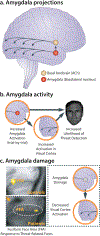
Figure 5.. The amygdala plays a key role in enhancing attention to threat-relevant information.
a. Amygdala projections. Anatomical tracing studies in monkeys and mechanistic studies in rodents indicate that the amygdala can enhance vigilance and prioritize the processing of threat-relevant information directly, via monosynaptic projections from the basolateral nucleus (BL; see Figure 1) to sensory cortex, and indirectly, via projections from the basal nuclei and central nucleus (Ce) to ascending neuromodulatory systems in the basal forebrain and brain stem. In turn, these transmitter systems can enhance the signal-to-noise ratio of neuronal processing in cortical sensory regions. In this simplified illustration, select projections from the basal forebrain cholinergic (ACh) system to the visual cortex are depicted. b. Amygdala activity. Using fMRI, Lim and colleagues demonstrated that amygdala activation predicts trial-by-trial fluctuations in threat detection (Lim et al., 2009). Mediation analyses revealed that relations between amygdala activation and detection performance were explained by increased activation in the visual cortex, consistent with work in animals. c. Amygdala damage. In a seminal study, Vuilleumier and colleagues (2004) showed that individuals with amygdala damage do not show increased activation to threat-related facial expressions in the fusiform face area (FFA) of the visual cortex, indicating that the amygdala causally contributes to the enhanced processing of threat-related stimuli in humans. This observation has since been replicated using more selective chemical lesions in monkeys (Hadj-Bouziane et al., 2012). Abbreviations—ACh: acetylcholine; FFA: fusiform face area. Portions of this figure were adapted with permission from (Tang, Holzel, & Posner, 2015; Vuilleumier et al., 2004).
Figure 6.
Figure 6.. The amygdala plays a key role in orienting overt attention to potentially threat-diagnostic information in the environment. a. Attentional exploration of faces.
Eye tracking data reveal a strong bias for scanning the eye and brow region, particularly for fearful faces (Scheller et al., 2012). This bias is evident in both the density of fixations over time (top panel: warmer colors indicate higher density) and the likelihood of reflexive saccades toward the facial feature presented in the visual periphery (bottom panel). b. Amygdala activation and attentional orienting. Individuals with increased activation in the right amygdala (indicated by the red ring) are more likely to orient their gaze to the eye and brow region of fearful faces (Gamer & Buchel, 2009). C. Amygdala damage impairs reflexive orienting. Patient MW has selective damage to the right amygdala (red ring) and shows a profound reduction in reflexive saccades to the eye region of the face (Gamer et al., 2013). Abbreviations—L: left hemisphere, R: right hemisphere. Portions of this figure were adapted with permission from (Shackman et al., 2016a).
Figure 7.
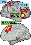
Figure 7.. Executive control networks.
The frontoparietal (blue) and cingulo-opercular (green)networks are sensitive to a broad spectrum of executive function and cognitive control tasks. Abbreviations—AI: anterior insula; dlPFC: dorsolateral prefrontal cortex; FO: frontal operculum; IPS: intraparietal sulcus; MCC: midcingulate cortex; SMA: supplementary motor area. This figure were adapted with permission from (Li et al., 2017)

Similar articles

Cited by

References

    1. Abdellaoui A, Sanchez-Roige S, Sealock J, Treur JL, Dennis J, Fontanillas P, … Boomsma DI. (2018). Phenome-wide investigation of health outcomes associated with genetic predisposition to loneliness. bioRxiv. - PMC - PubMed
    1. Abend R, de Voogd L, Salemink E, Wiers RW, Perez-Edgar K, Fitzgerald A, … Bar-Haim Y (2018). Association between attention bias to threat and anxiety symptoms in children and adolescents. Depression and Anxiety, 35, 229–238. - PMC - PubMed
    1. Abercrombie HC, Schaefer SM, Larson CL, Oakes TR, Lindgren KA, Holden JE, … Davidson RJ (1998). Metabolic rate in the right amygdala predicts negative affect in depressed patients. Neuroreport, 9, 3301–3307. - PubMed
    1. Adams MJ, Howard DM, Luciano M, Clarke T-K, Davies GM, Hill WD, … McIntosh AM. (2019). Stratifying depression by neuroticism: revisiting a diagnostic tradition using GWAS data. bioRxiv. - PMC - PubMed
    1. Admon R, Lubin G, Stern O, Rosenberg K, Sela L, Ben-Ami H, & Hendler T (2009). Human vulnerability to stress depends on amygdala’s predisposition and hippocampal plasticity. Proceedings of the National Academy of Sciences of the United States of America, 106, 14120–14125. - PMC - PubMed

Publication types