Review
. 2019:83:353-396.
doi: 10.1016/bs.ctm.2019.01.001. Epub 2019 Feb 18.

Ion channels and transporters in diabetic kidney disease

Affiliations
Review

Ion channels and transporters in diabetic kidney disease

Denisha Spires et al. Curr Top Membr. 2019.

Abstract

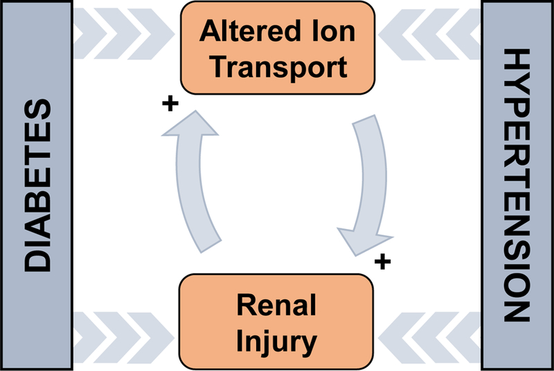
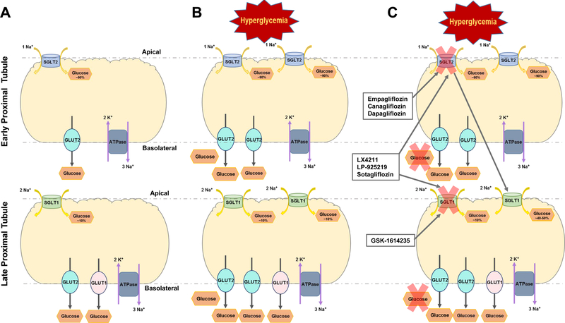
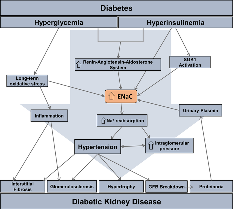
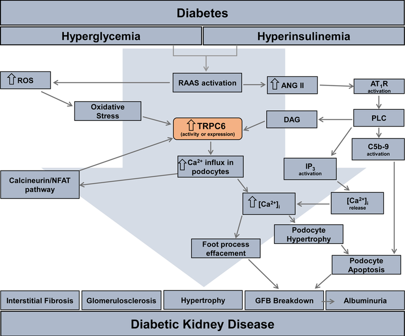
Type 1 and 2 diabetes mellitus are major medical epidemics affecting millions of patients worldwide. Diabetes mellitus is the leading cause of diabetic kidney disease (DKD), which is the most common cause of end-stage renal disease (ESRD). DKD is associated with significant changes in renal hemodynamics and electrolyte transport. Alterations in renal ion transport triggered by pathophysiological conditions in diabetes can exacerbate hypertension, accelerate renal injury, and are integral to the development of DKD. Renal ion transporters and electrolyte homeostasis play a fundamental role in functional changes and injury to the kidney during DKD. With the large number of ion transporters involved in DKD, understanding the roles of individual transporters as well as the complex cascades through which they interact is essential in the development of effective treatments for patients suffering from this disease. This chapter aims to gather current knowledge of the major renal ion transporters with altered expression and activity under diabetic conditions, and provide a comprehensive overview of their interactions and collective functions in DKD.

Keywords: Diabetic kidney disease; Diabetic nephropathy; ENaC; K(ATP) channel; NHE; SGLT2; TRPC6; TRPM6.


Figures

Figure 1.
Figure 1.
The positive feedback relationship between diabetes, hypertension, altered ion transport, and renal injury. This represents a simplified explanation for a very complex relationship that defines diabetic kidney disease (DKD).
Figure 2.
Figure 2.
Schematic for pathogenesis of diabetic kidney disease (DKD). Hyperglycemia and hyperinsulinemia induced by diabetes leads to the activation and expression of the different channels and transporters located along the various nephron segments. These changes are exercised either directly or indirectly upon the channel. The multitude of these channel alterations result in interstitial fibrosis, glomerulosclerosis, hypertrophy, breakdown of the glomerular filtration barrier (GFB) and albuminuria. The culmination of the damage to various portions of the nephron is the development of DKD. Abbreviations: calcium activated potassium channel 3.1 (KCa3.1); glucose transporters (GLUT1 and 2); sodium glucose cotransporter (SGLT1 and 2); large conductance Ca2+-activated K+ channel (BKCa); epithelial Na+ channel (ENaC); transient receptor potential canonical (TRPC) channel.
Figure 3.
Figure 3.
Tubular view of glucose transport within the proximal tubule of the kidneys. A. Healthy proximal tubule segments and normal function of sodium glucose cotransporters (SGLT1 and SGLT2) and their glucose transporter (GLUT1 and GLUT2) counterparts. B. The altered function of SGLT and GLUT transporters under DKD conditions. C. Examples of the inhibitors specific for SGLT2, SGLT1, and dual inhibitors for both transporters and their general effects under DKD conditions.
Figure 4.
Figure 4.
Schematic for epithelial Na+ channel (ENaC) induced tubular renal injury in DKD. Hyperglycemia and hyperinsulinemia induced via diabetes cause over-activation of the renin-angiotensin-aldosterone system (RAAS), long-term oxidative stress, and serum and glucocorticoid-regulated kinase (SGK) 1 activation that all directly cause the increase in the ENaC activation and/or expression. This increase in ENaC leads to various factors shown in the above pathway, such as hypertension. The culmination of these factors result in the development of the major characteristics of DKD.
Figure 5.
Figure 5.
Schematic for TRPC6-induced glomerular renal injury in the progression of DKD. Activation of the renin-angiotensin-aldosterone system (RAAS) cause an increase in angiotensin II (ANG II) that acts through the angiotensin II receptor type 1 (AT1R). This receptor activates phospholipase C (PLC) on the podocyte cell membrane. PLC activates 3 additional targets/pathways in the podocyte: diacylglycerol (DAG) that cause calcium influx through the transient receptor potential canonical 6 (TRPC6) channel which in turn increases intracellular calcium [Ca2+]i; inositol triphosphate (IP3) activates calcium release from the intracellular stores; the membrane attack complex of complement C (C5b-9), a transmembrane channel involved in some immune responses, induces podocyte apoptosis. Increases in RAAS also leads to an increase in reactive oxygen species (ROS) that begets oxidative stress activating the calcineurin/NFAT pathway that increases the transcription of Trpc6. The culmination of the increase in [Ca2+]I from over-activation of TRPC6 leads to podocyte hypertrophy/apoptosis and foot process effacement ending in breakdown of the glomerular filtration barrier (GFB). The ultimate result of this pathway is the development of the DKD characteristic albuminuria.

Similar articles

Cited by

References

    1. Abdul-Ghani MA, Norton L, & DeFronzo RA (2015). Renal sodium-glucose cotransporter inhibition in the management of type 2 diabetes mellitus. Am J Physiol Renal Physiol, 309(11), F889–900. 10.1152/ajprenal.00267.2015 - DOI - PMC - PubMed
    1. Abramowitz J, & Birnbaumer L (2009). Physiology and pathophysiology of canonical transient receptor potential channels. FASEB J, 23(2), 297–328. 10.1096/fj.08-119495 - DOI - PMC - PubMed
    1. Adebiyi A, Soni H, John TA, & Yang F (2014). Lipid rafts are required for signal transduction by angiotensin II receptor type 1 in neonatal glomerular mesangial cells. Exp Cell Res, 324(1), 92–104. 10.1016/j.yexcr.2014.03.011 - DOI - PubMed
    1. Anders H-J, Huber TB, Isermann B, & Schiffer M (2018). CKD in diabetes: diabetic kidney disease versus nondiabetic kidney disease. Nature Reviews Nephrology, 14(6), 361–377. 10.1038/s41581-018-0001-y - DOI - PubMed
    1. Andersen H, Friis UG, Hansen PB, Svenningsen P, Henriksen JE, & Jensen BL (2015). Diabetic nephropathy is associated with increased urine excretion of proteases plasmin, prostasin and urokinase and activation of amiloride-sensitive current in collecting duct cells. Nephrol Dial Transplant, 30(5), 781–789. 10.1093/ndt/gfu402 - DOI - PubMed

Publication types