

Assessing Postconcussive Reaction Time Using Transport-Based Morphometry of Diffusion Tensor Images
- PMID: 31196860
- PMCID: PMC7048545
- DOI: 10.3174/ajnr.A6087
Assessing Postconcussive Reaction Time Using Transport-Based Morphometry of Diffusion Tensor Images
Abstract
Background and purpose: Cognitive deficits are among the most commonly reported post-concussive symptoms, yet the underlying microstructural injury is poorly understood. Our aim was to discover white matter injury underlying reaction time in mild traumatic brain injury DTI by applying transport-based morphometry.
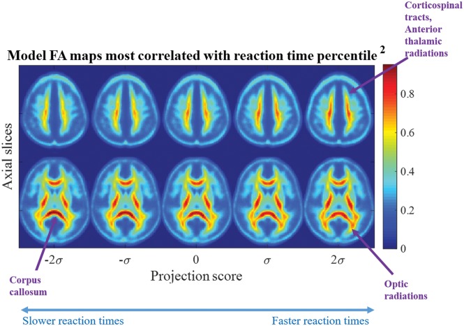
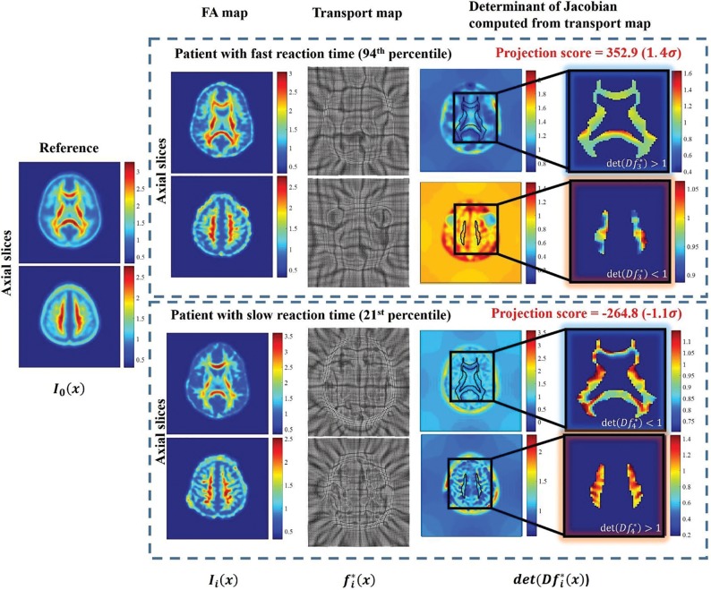
Materials and methods: In this retrospective study, we performed DTI on 64 postconcussive patients (10-28 years of age; 69% male, 31% female) between January 2006 and March 2013. We measured the reaction time percentile by using Immediate Post-Concussion Assessment and Cognitive Testing. Using the 3D transport-based morphometry technique we developed, we mined fractional anisotropy maps to extract the common microstructural injury associated with reaction time percentile in an automated manner. Permutation testing established statistical significance of the extracted injuries. We visualized the physical substrate responsible for reaction time through inverse transport-based morphometry transformation.
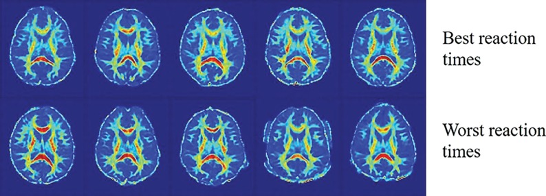
Results: The direction in the transport space most correlated with reaction time was significant after correcting for covariates of age, sex, and time from injury (Pearson r = 0.44, P < .01). Inverting the computed direction using transport-based morphometry illustrates physical shifts in fractional anisotropy in the corpus callosum (increase) and within the optic radiations, corticospinal tracts, and anterior thalamic radiations (decrease) with declining reaction time. The observed shifts are consistent with biologic pathways underlying the visual-spatial interpretation and response-selection aspects of reaction time.
Conclusions: Transport-based morphometry discovers complex white matter injury underlying postconcussive reaction time in an automated manner. The potential influences of edema and axonal loss are visualized in the visual-spatial interpretation and response-selection pathways. Transport-based morphometry can bridge the gap between brain microstructure and function in diseases in which the structural basis is unknown.
© 2019 by American Journal of Neuroradiology.
Figures
Similar articles
-
Microstructural neuroimaging of white matter tracts in persistent post-concussion syndrome: A prospective controlled cohort study.Neuroimage Clin. 2019;23:101842. doi: 10.1016/j.nicl.2019.101842. Epub 2019 May 6. Neuroimage Clin. 2019. PMID: 31108457 Free PMC article.
-
Differences in Callosal and Forniceal Diffusion between Patients with and without Postconcussive Migraine.AJNR Am J Neuroradiol. 2017 Apr;38(4):691-695. doi: 10.3174/ajnr.A5073. Epub 2017 Jan 26. AJNR Am J Neuroradiol. 2017. PMID: 28126745 Free PMC article.
-
Longitudinal Changes in Diffusion Tensor Imaging Following Mild Traumatic Brain Injury and Correlation With Outcome.Front Neural Circuits. 2019 May 7;13:28. doi: 10.3389/fncir.2019.00028. eCollection 2019. Front Neural Circuits. 2019. PMID: 31133818 Free PMC article.
-
Diffusion tensor imaging (DTI) findings in adult civilian, military, and sport-related mild traumatic brain injury (mTBI): a systematic critical review.Brain Imaging Behav. 2018 Apr;12(2):585-612. doi: 10.1007/s11682-017-9708-9. Brain Imaging Behav. 2018. PMID: 28337734
-
The role of diffusion tensor imaging and fractional anisotropy in the evaluation of patients with idiopathic normal pressure hydrocephalus: a literature review.Neurosurg Focus. 2016 Sep;41(3):E12. doi: 10.3171/2016.6.FOCUS16192. Neurosurg Focus. 2016. PMID: 27581308 Review.
Cited by
-
Machine intelligence in healthcare-perspectives on trustworthiness, explainability, usability, and transparency.NPJ Digit Med. 2020 Mar 26;3:47. doi: 10.1038/s41746-020-0254-2. eCollection 2020. NPJ Digit Med. 2020. PMID: 32258429 Free PMC article.
-
Enabling early detection of osteoarthritis from presymptomatic cartilage texture maps via transport-based learning.Proc Natl Acad Sci U S A. 2020 Oct 6;117(40):24709-24719. doi: 10.1073/pnas.1917405117. Epub 2020 Sep 21. Proc Natl Acad Sci U S A. 2020. PMID: 32958644 Free PMC article.
-
Discovering the gene-brain-behavior link in autism via generative machine learning.Sci Adv. 2024 Jun 14;10(24):eadl5307. doi: 10.1126/sciadv.adl5307. Epub 2024 Jun 12. Sci Adv. 2024. PMID: 38865470 Free PMC article.
References
-
- Himanen L, Portin R, Isoniemi H, et al. . Cognitive functions in relation to MRI findings 30 years after traumatic brain injury. Brain Inj 2005;19:93–100 - PubMed
-
- Hellawell DJ, Taylor RT, Pentland B. Cognitive and psychosocial outcome following moderate or severe traumatic brain injury. Brain Inj 1999;13:489–504 - PubMed
Publication types
MeSH terms
Grants and funding
LinkOut - more resources
Full Text Sources
Miscellaneous