

Review
doi: 10.3174/ajnr.A6089.
Epub 2019 Jun 13.
A Practical Review of Functional MRI Anatomy of the Language and Motor Systems
Affiliations
- PMID: 31196862
- PMCID: PMC6754743
- DOI: 10.3174/ajnr.A6089
Item in Clipboard
Review
A Practical Review of Functional MRI Anatomy of the Language and Motor Systems
AJNR Am J Neuroradiol.
2019 Jul.
Erratum in
-
Erratum.AJNR Am J Neuroradiol. 2020 Aug;41(8):E72. doi: 10.3174/ajnr.A6660. Epub 2020 Jul 2. AJNR Am J Neuroradiol. 2020. PMID: 32616584 Free PMC article. No abstract available.
Abstract
Functional MR imaging is being performed with increasing frequency in the typical neuroradiology practice; however, many readers of these studies have only a limited knowledge of the functional anatomy of the brain. This text will delineate the locations, anatomic boundaries, and functions of the cortical regions of the brain most commonly encountered in clinical practice-specifically, the regions involved in movement and language.
© 2019 by American Journal of Neuroradiology.
Figures
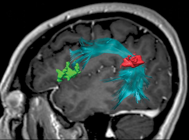
Sagittal 3D-FLAIR image with a superimposed diffusion tensor image of the arcuate fasciculus between the inferior parietal lobule (pars opercularis and pars triangularis) (green) and the angular gyrus (red). The frontotemporal segment of the arcuate fasciculus connects the Broca area at the inferior frontal gyrus with the Wernicke area more posteriorly, traditionally at the posterior superior temporal gyrus. However, this connection is variable, and in this case, it is at the angular gyrus/posterior superior temporal area.

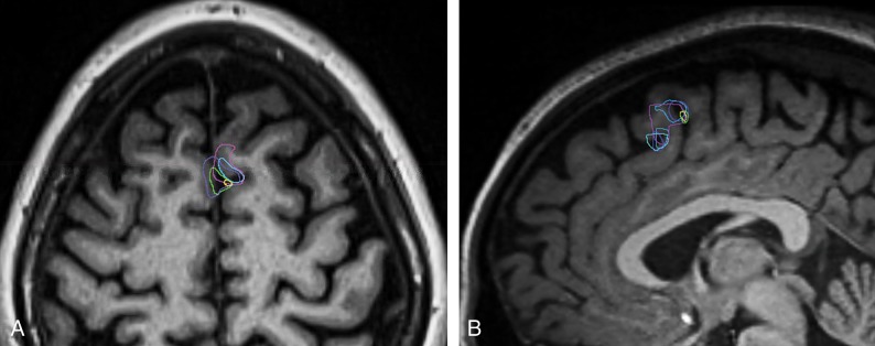
A, Axial MPRAGE image through the sensorimotor cortex with functional areas labeled in the right cerebral hemisphere. B, Coronal contrast-enhanced MPRAGE image with superimposed diffusion tensor imaging view of the corticospinal tract. DLPC, dorsolateral prefrontal cortex; SMA, supplementary motor area.
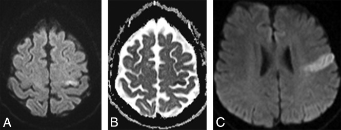
DWI (A) and ADC map (B) demonstrate a left hand/arm motor infarct in a patient with sudden onset of right-arm weakness. C, DWI demonstrates a left facial motor infarct in a patient with facial weakness.
Functional MR imaging blood oxygen level–dependent activation of the SMA superimposed on axial (A) and sagittal (B) MPRAGE images. Dark blue designates activation during antonym generation; turquoise, during picture naming; purple, during rhyming; light blue, during silent word generation; yellow, during finger movement; and green, during lip movement.
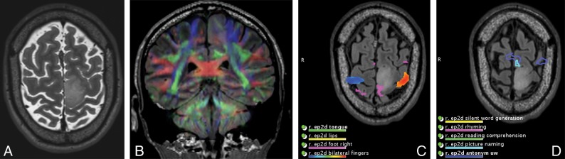
A patient with a left precentral gyrus/paracentral lobule and an SMA mass who initially presented with seizure. A, Axial T2-weighted MR imaging shows a left SMA mass. The patient underwent subtotal resection complicated by transient postoperative right hemiparesis (SMA syndrome). Pathology showed a diffuse infiltrating astrocytoma, isocitrate dehydrogenase-mutant, World Health Organization grade II. DTI superimposed on coronal FLAIR (B) and axial functional MR imaging show right foot (C) and language (D) SMA activation at the margins of the mass.
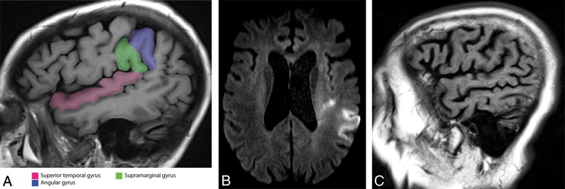
A, Sagittal MPRAGE image through the inferior parietal lobule demonstrates the supramarginal gyrus and angular gyrus. The superior temporal gyrus also is highlighted. Axial DWI (B) and sagittal T1-weighted (C) images of a left IPL infarct. The patient presented with the inability to understand and could not produce any comprehensible speech. The IPL is involved in semantic and phonologic processing. Patients with an infarct in this region have severe word- and sentence-comprehension deficits due to a transcortical sensory aphasia. Subdivisions of the arcuate fasciculus connect the IPL to the posterior temporal area as well as the IPL to the Broca area.
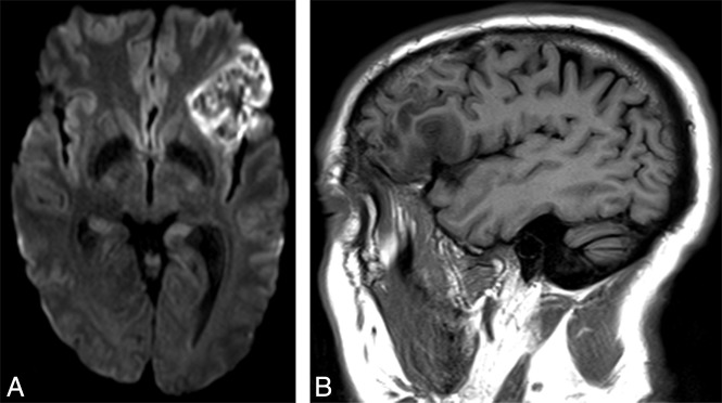
A left Broca infarct with classic expressive aphasia. A, Axial diffusion-weighted image demonstrates restricted diffusion at the classic Broca area. B, Sagittal T1-weighted image demonstrates hypointensity due to edema from the infarct.
Similar articles
-
Brain Functional Imaging Anatomy.Neuroimaging Clin N Am. 2022 Aug;32(3):491-505. doi: 10.1016/j.nic.2022.04.001. Neuroimaging Clin N Am. 2022. PMID: 35843658 Review.
-
Cortical and subcortical mapping of language areas: correlation of functional MRI and tractography in a 3T scanner with intraoperative cortical and subcortical stimulation in patients with brain tumors located in eloquent areas.Radiologia. 2013 Nov-Dec;55(6):505-13. doi: 10.1016/j.rx.2012.01.004. Epub 2012 Apr 21. Radiologia. 2013. PMID: 22521686 English, Spanish.
-
The role of functional magnetic resonance imaging in brain surgery.Neurosurg Focus. 2010 Feb;28(2):E4. doi: 10.3171/2009.12.FOCUS09251. Neurosurg Focus. 2010. PMID: 20121439 Review.
-
Impairment of preoperative language mapping by lesion location: a functional magnetic resonance imaging, navigated transcranial magnetic stimulation, and direct cortical stimulation study.J Neurosurg. 2015 Aug;123(2):314-24. doi: 10.3171/2014.10.JNS141582. Epub 2015 Apr 17. J Neurosurg. 2015. PMID: 25884257
-
Neuroanatomical Considerations in Preoperative Functional Brain Mapping.Top Magn Reson Imaging. 2019 Aug;28(4):213-224. doi: 10.1097/RMR.0000000000000213. Top Magn Reson Imaging. 2019. PMID: 31385901 Review.
Cited by
-
The utility of diffusion tractography for speech preservation in laser ablation of the dominant insula: illustrative case.J Neurosurg Case Lessons. 2021 May 10;1(19):CASE21113. doi: 10.3171/CASE21113. eCollection 2021 May 10. J Neurosurg Case Lessons. 2021. PMID: 35854831 Free PMC article.
-
Identifying the neural correlates of anticipatory postural control: A novel fMRI paradigm.Hum Brain Mapp. 2023 Jul;44(10):4088-4100. doi: 10.1002/hbm.26332. Epub 2023 May 10. Hum Brain Mapp. 2023. PMID: 37162423 Free PMC article.
-
Resting-state fMRI functional connectivity of the left temporal parietal junction is associated with visual temporal order threshold.Sci Rep. 2022 Sep 24;12(1):15933. doi: 10.1038/s41598-022-20309-1. Sci Rep. 2022. PMID: 36153359 Free PMC article.
-
Tumor-associated alterations in white matter connectivity have prognostic significance in MGMT-unmethylated glioblastoma.J Neurooncol. 2022 Jul;158(3):331-339. doi: 10.1007/s11060-022-04018-3. Epub 2022 May 7. J Neurooncol. 2022. PMID: 35525907
-
Iron accumulation/overload and Alzheimer's disease risk factors in the precuneus region: A comprehensive narrative review.Aging Med (Milton). 2024 Oct 22;7(5):649-667. doi: 10.1002/agm2.12363. eCollection 2024 Oct. Aging Med (Milton). 2024. PMID: 39507230 Free PMC article. Review.
References
-
- Ungerleider LG, Mishkin M. Two cortical visual systems. In: Ingle D, Goodale MA, Mansfield RJ, eds. Analysis of Visual Behavior. Cambridge: MIT Press; 1982:549–86
Publication types
MeSH terms
Grants and funding
LinkOut - more resources
Full Text Sources
Medical