. 2019 Jun 13;2(3):e201800267.
doi: 10.26508/lsa.201800267. Print 2019 Jun.

CRISPR-induced double-strand breaks trigger recombination between homologous chromosome arms

Affiliations

CRISPR-induced double-strand breaks trigger recombination between homologous chromosome arms

Erich Brunner et al. Life Sci Alliance. .

Abstract

CRISPR-Cas9-based genome editing has transformed the life sciences, enabling virtually unlimited genetic manipulation of genomes: The RNA-guided Cas9 endonuclease cuts DNA at a specific target sequence and the resulting double-strand breaks are mended by one of the intrinsic cellular repair pathways. Imprecise double-strand repair will introduce random mutations such as indels or point mutations, whereas precise editing will restore or specifically edit the locus as mandated by an endogenous or exogenously provided template. Recent studies indicate that CRISPR-induced DNA cuts may also result in the exchange of genetic information between homologous chromosome arms. However, conclusive data of such recombination events in higher eukaryotes are lacking. Here, we show that in Drosophila, the detected Cas9-mediated editing events frequently resulted in germline-transmitted exchange of chromosome arms-often without indels. These findings demonstrate the feasibility of using the system for generating recombinants and also highlight an unforeseen risk of using CRISPR-Cas9 for therapeutic intervention.


Conflict of interest statement

The authors declare that they have no conflict of interest.

Figures

Figure 1.
Figure 1.. CIGAR design and activation.
(A) CIGAR consists of four elements: 1. Ubiquitin-p63E promoter (brown box), 2. “shifter” sequence, 3. linker sequence (orange), 4. reporter cDNA (ORF) lacking a translational start site followed by the tubulin 3′UTR (grey open arrow). The shifter sequence contains optimized translation initiation codons covering all three frames (light blue boxes) and a guide RNA target region (purple box) followed by a protospacer adjacent motif (PAM) (black box). In the inactive CIGAR, translation from each ATG (grey arrows) is terminated 5′ of the ORF by a STOP codon (red asterisks) preventing the translation of the downstream ORF. (B) Activation of two CIGAR variants harboring either an eGFP or a monomeric Cherry (mCherry). Activation is achieved by Cas9-induced DNA cleavage within stopT (pink), the most upstream STOP codon which is in-frame with the downstream ORF. The resulting double-strand break (DSB; the putative site of the DSB, 3 bp upstream of the PAM site is indicated with a pink, open arrowhead) is mended by NHEJ-mediated repair concomitantly eliminating stopT (indel; open white box) and shifting one of the ATGs in-frame with the ORF. Flies that inherited a translationally activated CIGAR appear uniformly green or red. The eye-specific red fluorescence marks the attP target site into which the CIGAR constructs have been inserted. (C) Comparison of the 20-nt sgRNA target sequences of CIGAReGFP and CIGARmCherry, respectively. The 9-bp substitutions in the two target sites are indicated. (D) For specific and simultaneous targeting of both reporters, a tRNA-spaced tandem array (U6:3-sgRNACIGAR(1,2) harboring sgRNA-1 (targeting CIGAReGFP) and sgRNA-2 (targeting CIGARmCherry) is used.
Figure 2.
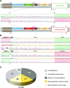
Figure 2.. Detailed CIGAReGFP and CIGARmCherry reporter design and illustration of recombination events on the sequence level.
(A) Design and sequence details of un-CRISPRed CIGAReGFP (top) and CIGARmCherry reporters (bottom). The sequences of the sgRNAs and the ORFs are shaded in green and red, respectively. Note that except for the sgRNAs and the ORFs, the sequences of the reporters are identical. The targeted STOP codon (stopT; pink) differ in sequence. The CRISPR target sites are delineated in the sequence context (pink, open arrows). Analysis of the shifter sequence is performed using primer pairs specific for the Ubi promoter and the 5′ end of the respective ORF (purple arrows). (B) The shifter region of flies harboring a single copy of one of the CIGAR reporters on the X chromosome (attP 5D) was analyzed by single fly PCR and Sanger sequencing. Shown are recombination events from a CIGAReGFP/CIGARmCherry co-targeting experiment visualized on the sequence level. As in (A), the sequences of the sgRNAs and the ORFs are shaded in green and red, respectively. Recombinants exhibit a rearranged sequential arrangement (green-red or red-green) of sgRNA and reporter cDNA. Note that recombination events may or may not be accompanied by indels at the target site. (C) Co-targeting experiments using CIGAR reporters on the fourth chromosome (attP 102F). 172 animals were analyzed by single fly PCR and Sanger sequencing. The yellow sections represent the number of recombinants with or without indel.
Figure S1.
Figure S1.. CIGAR constructs inserted @ ZH attP 5D (X chromosome) or ZH attP 102F (fourth chromosome).
(A) Sequence comparison of the target sequences of CIGAReGFP and CIGARmCherry. (B) Image illustrating the position of the two reporters in the fly genome.
Figure S2.
Figure S2.. Analysis of the shifter region of F1 animals from a CIGAReGFP and CIGARmCherry coactivation experiments.
The shifter region of flies harboring a single copy of one of the CIGAR reporters on the X chromosome (ZH attP 5D) was analyzed by single fly PCR and Sanger sequencing. Shown are the results for a total of 104 animals. See also Table S1 for complementary information.
Figure 3.
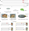
Figure 3.. Cas9-induced recombination between two phenotypic markers on the fourth chromosome.
(A) Each of the two markers (w+ [mini-white] and svspa-pol) is located on a different homologous chromosome. The two markers are separated by about 100 kb. The svspa-pol chromosome is marked with y+ due to a duplication of X-chromosomal material to the short, left arm of chromosome four. The Cas9 cut site is represented by a red dashed line and red open arrow heads. The repair of CRISPR-induced DSB may lead to targeted recombination events (TR) between the two markers.(B) Embryos with the genotype yw; CIGARmCherry,102F,w+/Dp(1;4)1021,y+, svspa-pol were injected with recombinant Cas9 RNPs containing in vitro–translated sgRNA-3. (C) Cas9 RNP injected G0 animals are backcrossed to animals with the genotype yw; Dp(1;4)1021,y+, svspa-pol/Dp(1;4)1021,y+, svspa-pol to be able to visually score putative recombinants. The phenotype of the animals is shown. (D) A total of 8,604 animals were screened and 253 putative recombinants were recovered. (E) Unrecombined animals (UR) appeared phenotypically as y+; w+ (UR-A) or y+; svspa-pol (UR-B). Putative recombinants (boxed) presented either as y+; w+; svspa-pol (TR-A) or y+, sv+ animals (TR-B).
Figure S3.
Figure S3.. CRISPR-induced recombination between two phenotypic markers on chromosome 4.
The CRISPR target site, the sequence, and position of sgRNA-3 located in the 3′ UTR of the toy gene, as well as the position of the two phenotypic markers in the genome (w+ and svspa-pol) are shown. The image is based on screenshots from the Ensembl database (https://www.ensembl.org).
Figure 4.
Figure 4.. CRISPR-induced DSBs may lead to loss of chromosomal structures.
(A) Of the putative 257 recombinants (see Fig 3D), 57 were characterized in more detail. 21 were true recombinants, whereas 36 animals had a loss of chromosomal structures distal to the CRISPR target site which manifested in PEV, that is, variable expression of the mini-white gene. (B) Fly exhibiting PEV is shown. The genotype of the fly and the loss of chromosomal structures distal to the cut site are shown below the image. Important to note is that only the loss of distal chromosome structures of the w+ chromosome can be scored from the TR screen (described in Fig 3). The corresponding loss of svspa-pol on the Dp(1;4)1021,y+ chromosome is phenotypically identical to UR-B (Fig 3E) and will not be recovered from the screen. (C) Control experiment to assess if CRISPR–Cas9–induced DSBs in general enhances the frequency of recombination away from the Cas9 cut site (dashed line and red open arrowheads). (D) Cas9 RNP–injected animals (same as in Fig 3B) are backcrossed to animals with the genotype yw to be able to visually score NSR events between w+ and y+. (E) No NSR between y+ and w+ marker (NSR-A and NSR-B) were recovered amongst 13,220 animals screened (see text and the Materials and Methods section for more details).

Similar articles

Cited by

References

    1. Jinek M, Chylinski K, Fonfara I, Hauer M, Doudna JA, Charpentier E (2012) A programmable dual-RNA-guided DNA endonuclease in adaptive bacterial immunity. Science 337: 816–821. 10.1126/science.1225829 - DOI - PMC - PubMed
    1. Doudna JA, Charpentier E (2014) Genome editing. The new frontier of genome engineering with CRISPR-Cas9. Science 346: 1258096 10.1126/science.1258096 - DOI - PubMed
    1. Hsu PD, Lander ES, Zhang F (2014) Development and applications of CRISPR-Cas9 for genome engineering. Cell 157: 1262–1278. 10.1016/j.cell.2014.05.010 - DOI - PMC - PubMed
    1. Bosley KS, Botchan M, Bredenoord AL, Carroll D, Charo RA, Charpentier E, Cohen R, Corn J, Doudna J, Feng G, et al. (2015) CRISPR germline engineering: The community speaks. Nat Biotechnol 33: 478–486. 10.1038/nbt.3227 - DOI - PubMed
    1. Sternberg SH, Doudna JA (2015) Expanding the biologist’s toolkit with CRISPR-Cas9. Mol Cell 58: 568–574. 10.1016/j.molcel.2015.02.032 - DOI - PubMed

Publication types

Substances

LinkOut - more resources