. 2019 Jun 13;2(3):e201900382.
doi: 10.26508/lsa.201900382. Print 2019 Jun.

Evidence for vesicle-mediated antigen export by the human pathogen Babesia microti

Affiliations

Evidence for vesicle-mediated antigen export by the human pathogen Babesia microti

Jose Thekkiniath et al. Life Sci Alliance. .

Abstract

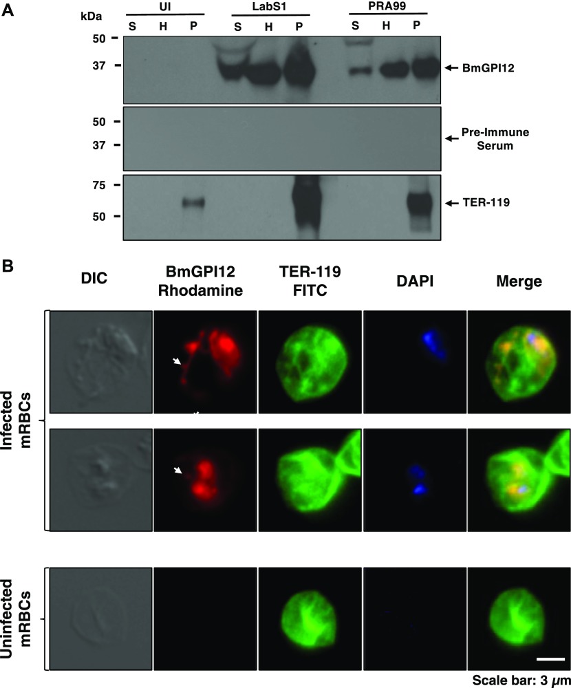
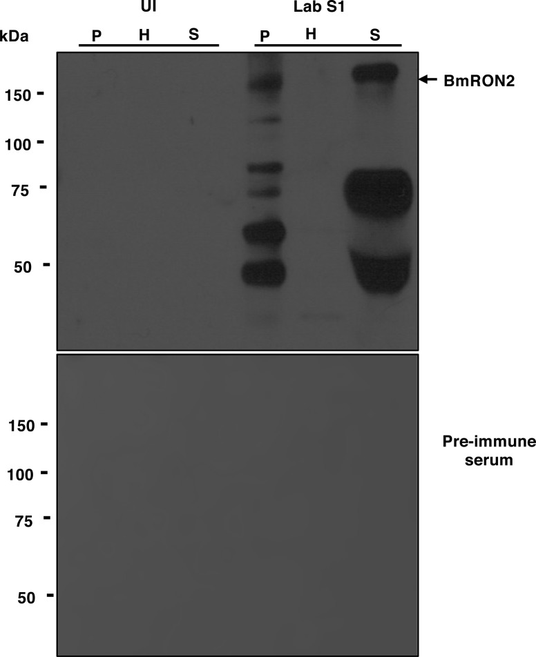
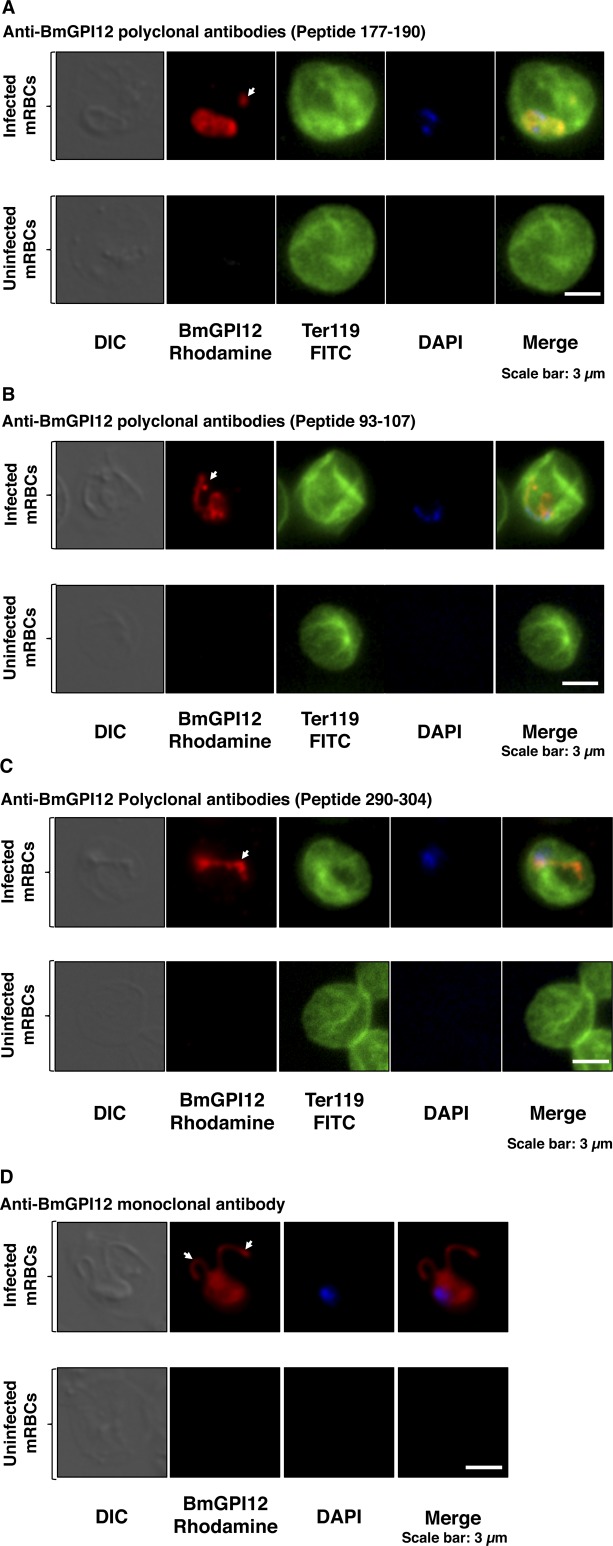
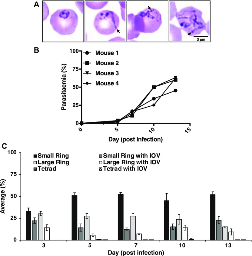
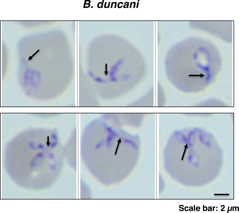
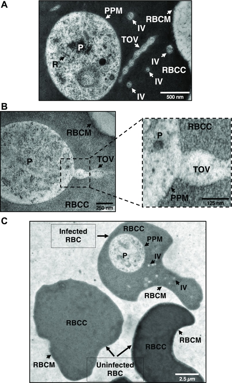
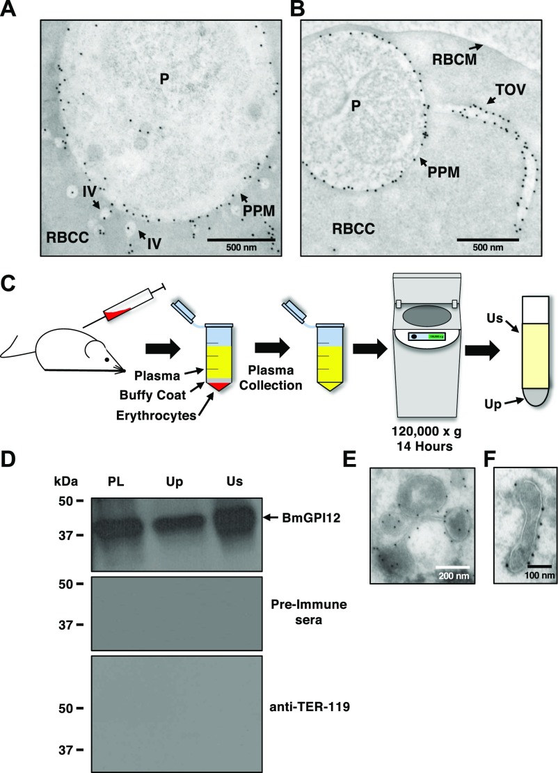
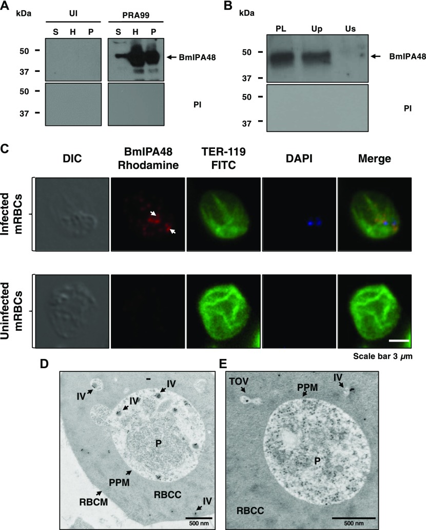
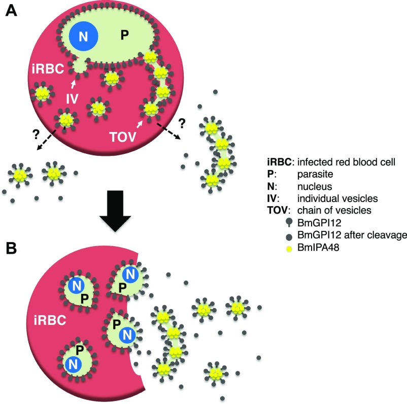
The apicomplexan parasite Babesia microti is the primary agent of human babesiosis, a malaria-like illness and potentially fatal tick-borne disease. Unlike its close relatives, the agents of human malaria, B. microti develops within human and mouse red blood cells in the absence of a parasitophorous vacuole, and its secreted antigens lack trafficking motifs found in malarial secreted antigens. Here, we show that after invasion of erythrocytes, B. microti undergoes a major morphogenic change during which it produces an interlacement of vesicles (IOV); the IOV system extends from the plasma membrane of the parasite into the cytoplasm of the host erythrocyte. We developed antibodies against two immunodominant antigens of the parasite and used them in cell fractionation studies and fluorescence and immunoelectron microscopy analyses to monitor the mode of secretion of B. microti antigens. These analyses demonstrate that the IOV system serves as a major export mechanism for important antigens of B. microti and represents a novel mechanism for delivery of parasite effectors into the host by this apicomplexan parasite.


Conflict of interest statement

The authors declare that they have no conflict of interest.

Figures

Figure 1.
Figure 1.. BmGPI12 is secreted into the erythrocyte cytoplasm and subsequently into the extracellular environment of the B. microti–infected erythrocyte.
(A) Immunoblotting analysis using preimmune (PI) and anti-BmGPI12 immune rabbit sera on fractions of uninfected erythrocytes (UI) or erythrocytes infected with either PRA99 or LabS1 strain of B. microti. In uninfected erythrocytes, the P fraction consists primarily of erythrocyte membranes. In B. microti–infected erythrocytes, the P fraction includes both erythrocyte membranes and protein extracts from isolated parasites. The erythrocyte membrane protein TER-119 (52 kD) was detected only in the P fractions from uninfected and B. microti–infected red blood cells using anti–TER-119 monoclonal antibody. (B) Immunofluorescence assay on mouse erythrocytes infected with the PRA99 strain of B. microti. BmGPI12 was labeled with anti-BmGPI12 polyclonal antibodies and could be observed within the parasite cytoplasm, the PPM as well as in the erythrocyte cytoplasm and within IV and TOVs (indicated by arrowheads). Anti–TER-119 monoclonal antibody was used to label the plasma membrane of the infected mouse erythrocytes. The DAPI staining was applied to verify the presence of parasites within the erythrocytes by labeling parasitic nuclear DNA. Staining of control uninfected red blood cells using the same antibodies is shown (lower panel). Scale bar: 3 μm. H, hemolysate; mRBC, mouse red blood cells; P, membrane fractions; S, mouse plasma.
Figure S1.
Figure S1.. Distribution of the apical end protein BmRON2 in B. microti–infected cells.
(A) Immunoblotting analysis using preimmune (PI) and anti-BmRON2 polyclonal antibodies on fractions of uninfected erythrocytes (UI) or erythrocytes infected with B. microti strain LabS1. Consistent with previous studies (Ord et al, 2016), BmRON2 (163 kD) undergoes proteolytic degradation (asterisks indicated degradation products). The 163-kD band is found in the P and S fractions but not in the H fraction consistent with the presence of BmRON2 on the surface of daughter parasites and release upon rupture of the infected erythrocyte. No signal was detected using preimmune sera. H, hemolysate; P, membrane fractions collected after saponin treatment of erythrocytes; S, mouse plasma.
Figure S2.
Figure S2.. BmGPI12 localization in B. microti–infected erythrocytes.
Immunofluorescence analysis on uninfected or B. microti (PRA99)-infected mouse erythrocytes. (A–D) BmGPI12 was labeled with three polyclonal anti-BmGPI12 antibodies (A, B, and C) and one monoclonal antibody (D) and could be observed within the parasite cytoplasm, the PPM and within IV and TOVs (indicated by arrowheads). Anti–TER-119 monoclonal antibody was used to label the plasma membrane of mouse erythrocytes, and DAPI was used to stain the parasite nucleus. Scale bars: 3 μm (A, B).
Figure 2.
Figure 2.. B. microti develops an IOV system in the cytoplasm of the infected erythrocytes.
(A) Representative images of Giemsa-stained blood smears from B. microti (LabS1)–infected erythrocytes. Long membranous structures within the erythrocyte cytoplasm are indicated by arrowheads. Scale bar: 3 μm. (B, C) Analysis of blood from four B. microti–infected SCID mice over a 13-d period after infection with the LabS1 strain. (B) Parasitemia levels in individual mice. For each sample, a total of 5,000 erythrocytes were analyzed. (C) Proportion of each morphological form detected in the blood smears at days 3, 5, 7, 10, and 13 postinfection. A total of 20 images were analyzed per smear at a given day (Mean ± SEM).
Figure S3.
Figure S3.. IOVs are present in erythrocytes parasitized with different Babesia species.
Representative images of Giemsa-stained thin blood smears of B. duncani–infected human erythrocytes. Arrowheads indicate components of the IOV protruding from the parasites. Scale bar: 3.5 μm.
Figure 3.
Figure 3.. B. microti develops an IOV system in the cytoplasm of the infected erythrocytes.
(A) EPON-embedded LabS1-infected erythrocytes revealed the presence of the IOV system in the erythrocyte cytoplasm. The IOV system contains the same electron-dense structures as the parasite, indicating that these structures are of parasitic origin. (A, B) Various structures of parasites and erythrocytes are shown by arrows (A and B). (C) Comparison of ultrathin sections of EPON-embedded infected and uninfected erythrocytes (C). Scale bars: 500 nm (A), 250 nm and 125 nm (B), 2.5 μm (C). P, parasite; R, ribosomes; RBCC, red blood cell cytoplasm; RBCM, red blood cell membrane.
Figure S4.
Figure S4.. Electron microscopy evidence for IOVs emerging from the PPM.
(A, B) Ultrathin EPON sections of LabS1 B. microti–infected mouse erythrocytes showing IVs (A) as well as TOVs (B) emerging from the PPM. Scale bars: 250 nm (A and B). P, parasite; PPM, parasite plasma membrane; RBC, red blood cell cytoplasm; RBCM, red blood cell membrane; TOVs, tube of vesicles.
Figure 4.
Figure 4.. BmGPI12 is localized to the PPM and associated with vesicles and tubules.
(A, B) Immunoelectron microscopic analysis of B. microti LabS1–infected mouse erythrocytes. Ultrathin sections of high-pressure frozen and Durcupan resin–embedded infected erythrocytes were immunolabeled with anti-BmGPI12 polyclonal antibodies (amino acid 1–302). Scale bars: 500 nm (A, B). (C) Schematic diagram showing the steps in the UC of plasma samples collected from B. microti–infected mice. (D) Immunoblot analyses using preimmune (PI) serum, and anti-BmGPI12 or anti–TER-119 antibodies on either intact plasma (PL) collected from mice infected with B. microti LabS1 strain or on two fractions (supernatant: Us and pellet: Up) of plasma after UC at 120,000g. (E, F) Immunoelectron microscopic analysis of the plasma membrane fraction (Up) from mice infected with B. microti LabS1 using anti-BmGPI12 antibodies coupled to 10-nm gold particles. Scale bars: 200 nm (E), 100 nm (F). P, parasite; RBCC, red blood cell cytoplasm; RBCM, red blood cell membrane.
Figure 5.
Figure 5.. Vesicle-mediated secretion of BmIPA48 antigen by B. microti.
(A) Distribution of BmIPA48 in the plasma (S), hemolysate (H), and membrane (P) fractions isolated from blood of uninfected or B. microti (PRA99)–infected erythrocytes was determined by Western blotting using polyclonal antibodies against BmIPA48 (48 kD). Preimmune (PI) sera were used as control. (B) Immunoblot analysis using preimmune and anti-BmIPA48 antibodies on either intact plasma (PL) collected from mice infected with B. microti PRA99 strain or on two fractions (supernatant: Us and pellet: Up) of plasma after UC at 120,000g. (C) Immunofluorescence assay using anti-BmIPA48 in PRA99-infected erythrocytes. BmIPA48 was labeled with polyclonal antibodies and could be detected within the parasite and in discrete IVs within the cytoplasm of the infected erythrocyte. Anti–TER-119 monoclonal antibody was used to label the plasma membrane of mouse erythrocytes and DAPI was used to stain the parasite nucleus. Staining of control uninfected red blood cells is shown. Scale bar: 3 μm. (D, E) Representative images of immunoelectron micrographs of B. microti LabS1–infected mouse erythrocytes. Ultrathin sections of high-pressure frozen and Durcupan resin–embedded infected erythrocytes were immunolabeled with anti-BmIPA48 antibodies coupled to 10-nm gold particles. Scale bars: 500 nm (D and E). mRBC, mouse red blood cells; P, parasite; RBCC, red blood cell cytoplasm; RBCM, red blood cell membrane.
Figure 6.
Figure 6.. Model of vesicular-mediated export of antigens by B. microti during development and after rupture.
(A) Dashed lines indicate yet-to-be-identified vesicle export pathways during B. microti intraerythrocytic development (rings) before cell division. (B) Model of export of TOVs and IVs after parasite division and rupture of the infected erythrocyte. The tetrad (The Maltese cross) stage is a transient developmental stage rarely detected in infected animals.

Similar articles

Cited by

References

    1. Abraham A, Brasov I, Thekkiniath J, Kilian N, Lawres L, Gao R, DeBus K, He L, Yu X, Zhu G, et al. (2018) Establishment of a continuous in vitro culture of Babesia duncani in human erythrocytes reveals unusually high tolerance to recommended therapies. J Biol Chem 293: 19974–19981. 10.1074/jbc.ac118.005771 - DOI - PMC - PubMed
    1. Baranyai T, Herczeg K, Onodi Z, Voszka I, Modos K, Marton N, Nagy G, Mager I, Wood MJ, El Andaloussi S, et al. (2015) Isolation of exosomes from blood plasma: Qualitative and quantitative comparison of ultracentrifugation and size exclusion chromatography methods. PLoS One 10: e0145686 10.1371/journal.pone.0145686 - DOI - PMC - PubMed
    1. Ben Mamoun C, Allred DR (2018) Babesiosis In eLS. Chichester: John Wiley & Sons, Ltd.
    1. Boddey JA, O’Neill MT, Lopaticki S, Carvalho TG, Hodder AN, Nebl T, Wawra S, van West P, Ebrahimzadeh Z, Richard D, et al. (2016) Export of malaria proteins requires co-translational processing of the PEXEL motif independent of phosphatidylinositol-3-phosphate binding. Nat Commun 7: 10470 10.1038/ncomms10470 - DOI - PMC - PubMed
    1. Chen TK, Ben Mamoun C, Krause PJ (2015) Human babesiosis In Clinical Infectious Disease. Schlossberg D. (ed) Cambridge: Cambridge University Press; pp 1295–1301.

Publication types

Substances

LinkOut - more resources