. 2019 Jul 2;476(13):1911-1926.
doi: 10.1042/BCJ20190240.

Nitration-induced ubiquitination and degradation control quality of ERK1

Affiliations

Nitration-induced ubiquitination and degradation control quality of ERK1

Yuanya Zhang et al. Biochem J. .

Abstract

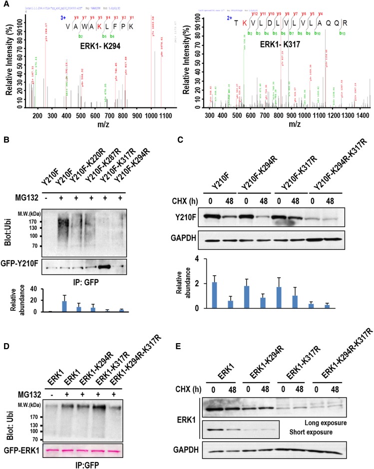
The mitogen-activated protein kinase ERK1/2 (ERKs, extracellular-regulated protein kinases) plays important roles in a wide spectrum of cellular processes and have been implicated in many disease states. The spatiotemporal regulation of ERK activity has been extensively studied. However, scarce information has been available regarding the quality control of the kinases to scavenge malfunctioning ERKs. Using site-specific mutagenesis and mass spectrometry, we found that the disruption of the conserved H-bond between Y210 and E237 of ERK1 through point mutation at or naturally occurring nitration on Y210 initiates a quality control program dependent on chaperon systems and CHIP (C-terminal of Hsp70-interacting protein)-mediated ubiquitination and degradation. The H-bond is also important for the quality control of ERK2, but through a distinct mechanism. These findings clearly demonstrate how malfunctioning ERKs are eliminated when cells are in certain stress conditions or unhealthy states, and could represent a general mechanism for scavenging malfunctioning kinases in stress conditions.

Keywords: ERK; mass spectrometry; phosphorylation; quality control; tyrosine nitration; ubiquitination.


Conflict of interest statement

The authors declare that there are no competing interests associated with the manuscript.

Figures

Figure 1.
Figure 1.. Y210F mutation abrogates ERK1 nuclear localization.
(A) HEK293T cells expressing GFP-ERK1 (ERK1) or GFP-ERK1-Y210F (Y210F) were serum-starved overnight and then treated with the nuclear export inhibitor LMB (40 mM) or vehicle (DMSO) for 3 h as indicated. The cells were then stimulated with 10% FBS or unstimulated (Starve) for 10 min as indicated. The cells were then fixed, stained with DAPI, and imaged with fluorescence microscopy (upper panel). Bar = 10 µm. The ERK phosphorylation level (TEY motif phosphorylation) in each treatment was detected with Western blotting, and total ectopically expressed ERK1 was also probed as the loading control (lower panel). Shown is the representative result of three biological replicates. (B) HEK293T cells expressing phosphomimetic mutant of the ERK1 nuclear translocation signal (NTS) (ERK1-EPE), the unphosphorylable mutant of NTS (ERK1-APA), and the triple-mutant Y210F-EPE were serum-starved and then stimulated with 10% FBS for 10 min or unstimulated (Starve). The cells were then fixed, stained with DAPI, and imaged with fluorescence microscopy. Bar = 25 µm.
Figure 2.
Figure 2.. The H-bond between Y210 and E237 is critical for ERK1 phosphorylation and nuclear localization.
(A) A part of ERK1 crystal structure (PDB accession: 2ZOQ) showing an H-bond between Y210 and E237 (panel a, dashed line). Simulations of the structures for Y210F and E237A mutants show that the H-bond between Y210 and E237 in wild-type ERK1 is disrupted in the mutants (panels b,c). (B) HEK293T cells expressing GFP, ERK1, Y210F, or E237A were serum-starved and stimulated with 10% FBS for 10 min or unstimulated (Starve), WCL were immunoblotted for phosphor-ERK and total ERK, respectively. Shown is the representative result of three biological replicates. (C) Serum-starved cells described in (B) were stimulated with 10% FBS or unstimulated (Starve). The cells were then fixed, stained with DAPI, and imaged with fluorescence microscopy. (D) The mass spectrum shows the identification of Y210 nitration. HEK293 cells expressing GFP-ERK1 were serum-starved overnight and then stimulated with 10% FBS for 10 min, GFP-ERK1 was then immunoprecipitated from the harvested cell lysates and analyzed wtih LC–MS/MS. The residue in red in the peptide sequence indicates the tyrosine nitration site (upper panel). The spectrum was annotated with the software pFind, and the score and mass accuracy were also shown (bottom panel). (E) In vitro tyrosine nitration assay of ERK1. GST-ERK1 was pulled down from WCL and then incubated with freshly collected WCL of 293T cells in the presence or absence of 500 mM peroxynitrite. The fusion protein was then separated with SDS–PAGE and probed for nitrotyrosine, ubiquitination, and GST using respective specific antibodies. The total level of GST-ERK1 was also probed as the loading control using anti-GST antibody. This experiment was performed in triplicates and only the representative result was shown. (F) Endogenous ERK1 was IPed from serum-starved HEK293T cells treated with 50 mM peroxynitrite for 4 h. ERK1 ubiquitination was detected by Western blotting and the results were quantified from triplicated experiments (mean ± s.d.).
Figure 3.
Figure 3.. ERK1 Y210F mutation promotes the recruitment of Hsp90.
(A) Ectopically expressed GFP-ERK1 (ERK1) or GFP-ERK1 Y210 (Y210) in 293T cells were IPed from the WCL with anti-GFP antibody and silver-stained after separation with SDS–PAGE. The protein bands indicated by the arrows were excised from the gel and analyzed with MS. (B) GFP fusion of ERK1 or Y210F in serum-starved or MG132-treated (5 µM) HEK293 cells was IPed with anti-GFP antibody and probed for Hsp90 by Western blotting. Total levels of Hsp90 in the WCL were also probed by Western blotting as the loading control. The bars indicate the quantified Hsp90 level from three IP replicates (mean ± s.d.). The bands in red were stained by Ponceau in this and all the other figures. (C) 293T cells expressing the GFP fusion of ERK1 or Y210F were transfected with siRNA targeting for Hsp90 (siHsp90) or non-targeting siRNA (siCtrl). The cells were fixed 72 h after the transfection, then stained with DAPI, and imaged with fluorescence microscopy. (D) The knockdown efficiency of Hsp90 was detected by Western blotting using anti-Hsp90 antibody. β-Actin was also probed as the loading control. Shown is the representative result of three biological replicates.
Figure 4.
Figure 4.. Y210F mutation promotes ERK1 ubiquitination and degradation.
293T cells expressing GFP-ERK1 (ERK1) or GFP-Y210F (Y210F) were treated with CHX for the indicated times. (A) The expression level of the fusion proteins was probed by Western blotting using anti-GFP antibody and quantified from triplicated experiments (mean ± s.d.). The up-shifted bands are probably the ubiquitinated ERK1 or Y210F. (B) HEK293T cells expressing GFP, GFP-ERK1 (ERK1), GFP-Y210F (Y210F), or GFP-E237A (E237A) were treated with MG132 or vehicle (DMSO) for 4 h and then lysed. GFP and the other GFP fusion proteins were IPed from the WCL and probed for ubiquitination by Western blotting using anti-ubi antibody. Quantification of ubiquitination was performed using results from triplicated experiments (mean ± s.d.).
Figure 5.
Figure 5.. CHIP is the major E3 ubiquitin ligase mediating Y210F ubiquitination and degradation.
(A) GFP-ERK1 (ERK1) or GFP-Y210F (Y210F) was IPed from 293T cells. The co-IPed proteins were identified by MS, and the spectra count of each protein co-IPed with GFP-ERK1 was used as the standard for normalization. The bars indicate the fold changes of the spectra counts of the identified proteins over the standards. (B) 293T cells expressing GFP-Y210F were transfected with siRNA targeting for MEKK1 (siMEKK1) or CHIP (siCHIP) or non-targeting siRNA (siCtrl) as indicated. The cells were imaged with fluorescence microscopy, and the numbers of cells with fluorescent aggregates were counted with Image J. The bars indicate the numbers of cells with fluorescence aggregates per field (mean ± s.d.). The knockdown efficiency of MEKK1 and CHIP was detected by RT-PCR. (C) The same transfected cells in (B) were treated with CHX for the indicated times and then lysed. The WCL was probed for Y210F by Western blotting using anti-GFP antibody. GAPDH was probed as the loading control (upper panel). The bars show the quantitative results from three independent experiments (mean ± s.d.) and were aligned with the corresponding lane in the upper panel (lower panel). (D) The same transfected cells in (B) were treated with MG132 or untreated (Starve) for 4 h and then harvested. GFP-Y210F was IPed from the WCL and probed for ubiquitination by Western blotting using anti-ubi antibody (upper panel). The bars in the lower panel show the quantified results of Y210F ubiquitination from three independent experiments (mean ± s.d.) and were aligned to the corresponding lanes in the upper panel. (E and G) The results from the same experiments described in (C) and (D), respectively, except that the cells used were expressing GFP-ERK1 instead of GFP-Y210F. (F) HEK293T cells were transfected with siRNA targeting for CHIP (siCHIP) or non-targeting siRNA (siCtrl) as indicated. The cells were treated with CHX for 24 h, 48 h, or untreated (0 h). The total level of endogenous ERK1 was detected by Western blotting using anti-ERK1 antibody. GAPDH was probed as the loading control. Quantification was performed using results from triplicated experiments (mean ± s.d.).
Figure 6.
Figure 6.. K294 and K317 ubiquitination mediates ERK1 degradation.
(A) The mass spectra show the identification of the two peptides from ERK1 ubiquitinated at K294 and K317, respectively. The residues in red in the peptide sequences indicate ubiquitination sites. (B) 293T cells expressing site-specific mutants generated from Y210F were treated with MG132 for 4 h or untreated as indicated. The cells were then lysed and the GFP fusion proteins were IPed using anti-GFP antibody before probed for ubiquitination by Western blotting using anti-ubi antibody. (C) 293T cells expressing GFP fusion of different site-specific mutants generated from Y210F were treated with CHX for 48 h or untreated (0 h). The total level of the fusion proteins was detected by Western blotting using anti-GFP antibody. GAPDH was probed as the loading control. (D and E) 293T cells expressing site-specific mutants generated from ERK1 as indicated were treated and assayed as described in (B) and (C), respectively. Quantifications for Western blot results, when applicable, were performed from three independent experiments (mean ± s.d.).
Figure 7.
Figure 7.. Y193F mutation promotes ERK2 ubiquitination and degradation but via a distinct mechanism from that of ERK1.
(A) Western blotting detection of the total and the phosphorylation levels of GFP fused ERK2 or Y193F in transfected 293T cells treated with 10% FBS or untreated as indicated. The cells expressing GFP was used as the control. (B) Western blotting detection of the expression levels of GFP-ERK2 and GFP-Y193F in transfected 293T cells treated with CHX for the indicated times. GAPDH was also probed for the loading control. (C) 293T cells expressing GFP, GFP-ERK2, and GFP-Y193F were treated with MG132 or vehicle (DMSO). The ectopically expressed proteins were IPed with anti-GFP antibody and probed for ubiquitination by Western blotting using anti-Ubi antibody. (D) GFP fusions of indicated proteins or their point mutants were IPed from the corresponding transfected 293T cells, and co-IPed Hsp90 was probed by Western blotting. Total Hsp90 in the transfected cells was also probed as the loading control. The results were quantified from triplicated experiments (mean ± s.d.). (E) Fluorescence images show the subcellular localization of the GFP fusions of ERK2 and its point mutants in transfected 293T cells. The cells were either treated with 10% FBS or starved as indicated. (F) GFP-ERK2 (ERK2) or GFP-Y210F (Y193F) was IPed from 293T cells. The co-IPed proteins were identified by MS, and the spectra count of each protein co-IPed with GFP-ERK2 was used as the standard for normalization. The bars indicate the fold changes in the spectra counts of the identified proteins over the standards.
Figure 8.
Figure 8.. The diagramed model shows Y210 modification-induced ERK1 ubiquitination and degradation.
Once Y210 is modified, the H-bond between Y210 and E237 is disrupted resulting in aberrant ERK1 folding and subsequent recruitment of Hsp90. Binding of Hsp90 can facilitate either the refolding and degradation of the misfolded ERK1, depending on whether the niche where ERK1 resides favor refolding or degradation. If the PTM conjugated on Y210 can be removed and the H-bond can be reformed, the refolding attempt could result in renatured proteins with full function. However, if the disrupted H-bond cannot be re-connected due to the failure of removing the PTM on Y210, Hsp90 will facilitate the recruitment of the other proteins such as the Hsp70–CHIP complex, and the latter can ubiquitinate and target ERK1 for degradation through the 26S proteasome system. The binding of Hsp90 also inhibits ERK1 nuclear entry, a secondary effect.

Similar articles

Cited by

References

    1. Plotnikov A., Zehorai E., Procaccia S. and Seger R. (2011) The MAPK cascades: signaling components, nuclear roles and mechanisms of nuclear translocation. Biochim. Biophys. Acta 1813, 1619–1633 10.1016/j.bbamcr.2010.12.012 - DOI - PubMed
    1. Yoon S. and Seger R. (2006) The extracellular signal-regulated kinase: multiple substrates regulate diverse cellular functions. Growth Factors 24, 21–44 10.1080/02699050500284218 - DOI - PubMed
    1. Fukuda M., Gotoh Y. and Nishida E. (1997) Interaction of MAP kinase with MAP kinase kinase: its possible role in the control of nucleocytoplasmic transport of MAP kinase. EMBO J. 16, 1901–1908 10.1093/emboj/16.8.1901 - DOI - PMC - PubMed
    1. Marais R., Wynne J. and Treisman R. (1993) The SRF accessory protein Elk-1 contains a growth factor-regulated transcriptional activation domain. Cell 73, 381–393 10.1016/0092-8674(93)90237-K - DOI - PubMed
    1. Khokhlatchev A.V., Canagarajah B., Wilsbacher J., Robinson M., Atkinson M., Goldsmith E. et al. (1998) Phosphorylation of the MAP kinase ERK2 promotes its homodimerization and nuclear translocation. Cell 93, 605–615 10.1016/S0092-8674(00)81189-7 - DOI - PubMed

Publication types

MeSH terms

Substances