

Detection of NTRK Fusions: Merits and Limitations of Current Diagnostic Platforms
- PMID: 31196931
- PMCID: PMC6606326
- DOI: 10.1158/0008-5472.CAN-19-0372
Detection of NTRK Fusions: Merits and Limitations of Current Diagnostic Platforms
Abstract
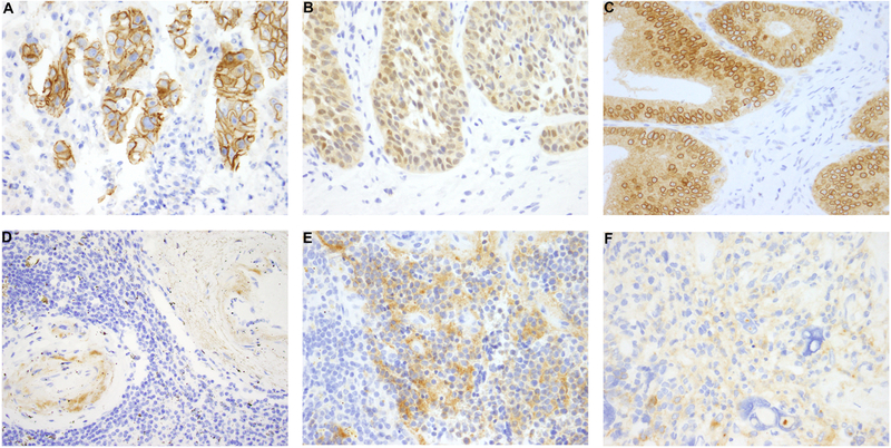
Oncogenic fusions involving NTRK1, NTRK2, and NTRK3 with various partners are diagnostic of infantile fibrosarcoma and secretory carcinoma yet also occur in lower frequencies across many types of malignancies. Recently, targeted small molecular inhibitor therapy has been shown to induce a durable response in a high percentage of patients with NTRK fusion-positive cancers, which has made the detection of NTRK fusions critical. Several techniques for NTRK fusion diagnosis exist, including pan-Trk IHC, FISH, reverse transcription PCR, DNA-based next-generation sequencing (NGS), and RNA-based NGS. Each of these assays has unique features, advantages, and limitations, and familiarity with these assays is critical to appropriately screen for NTRK fusions. Here, we review the details of each existing methodology.
©2019 American Association for Cancer Research.
Figures
Similar articles
-
Pan-Trk Immunohistochemistry Is an Efficient and Reliable Screen for the Detection of NTRK Fusions.Am J Surg Pathol. 2017 Nov;41(11):1547-1551. doi: 10.1097/PAS.0000000000000911. Am J Surg Pathol. 2017. PMID: 28719467 Free PMC article.
-
The oncogenic roles of NTRK fusions and methods of molecular diagnosis.Cancer Genet. 2021 Nov;258-259:110-119. doi: 10.1016/j.cancergen.2021.10.005. Epub 2021 Oct 18. Cancer Genet. 2021. PMID: 34710798 Review.
-
Pan-tumor screening for NTRK gene fusions using pan-TRK immunohistochemistry and RNA NGS fusion panel testing.Cancer Genet. 2022 Apr;262-263:47-52. doi: 10.1016/j.cancergen.2021.12.010. Epub 2022 Jan 2. Cancer Genet. 2022. PMID: 35007853
-
ESMO recommendations on the standard methods to detect NTRK fusions in daily practice and clinical research.Ann Oncol. 2019 Sep 1;30(9):1417-1427. doi: 10.1093/annonc/mdz204. Ann Oncol. 2019. PMID: 31268127
-
Detection of Tumor NTRK Gene Fusions to Identify Patients Who May Benefit from Tyrosine Kinase (TRK) Inhibitor Therapy.J Mol Diagn. 2019 Jul;21(4):553-571. doi: 10.1016/j.jmoldx.2019.03.008. Epub 2019 May 7. J Mol Diagn. 2019. PMID: 31075511 Free PMC article. Review.
Cited by
-
Immunohistochemistry as a screening tool for NTRK gene fusions: results of a first Belgian ring trial.Virchows Arch. 2021 Feb;478(2):283-291. doi: 10.1007/s00428-020-02921-6. Epub 2020 Sep 11. Virchows Arch. 2021. PMID: 32915263 Free PMC article. Clinical Trial.
-
NTRK Fusions in Central Nervous System Tumors: A Rare, but Worthy Target.Int J Mol Sci. 2020 Jan 23;21(3):753. doi: 10.3390/ijms21030753. Int J Mol Sci. 2020. PMID: 31979374 Free PMC article. Review.
-
Infantile fibrosarcoma of the perineum with dorsal metastasis in a neonate: a case report original.BMC Pediatr. 2023 Jun 29;23(1):327. doi: 10.1186/s12887-023-04129-4. BMC Pediatr. 2023. PMID: 37386422 Free PMC article.
-
Characterization of an ETV6-NTRK3 rearrangement with unusual, but highly significant FISH signal pattern in a secretory carcinoma of the salivary gland: a case report.Diagn Pathol. 2021 Aug 9;16(1):73. doi: 10.1186/s13000-021-01133-z. Diagn Pathol. 2021. PMID: 34372873 Free PMC article.
-
Pan-TRK positive uterine sarcoma in immunohistochemistry without neurotrophic tyrosine receptor kinase gene fusions: A case report.World J Clin Cases. 2025 Jan 16;13(2):96876. doi: 10.12998/wjcc.v13.i2.96876. World J Clin Cases. 2025. PMID: 39823101 Free PMC article. Review.
References
-
- Kheder ES and Hong DS, Emerging Targeted Therapy for Tumors with NTRK Fusion Proteins. Clin Cancer Res, 2018. 24(23): p. 5807–5814. - PubMed
Publication types
MeSH terms
Substances
Grants and funding
LinkOut - more resources
Full Text Sources
