. 2019 Jul 2;116(27):13508-13516.
doi: 10.1073/pnas.1903165116. Epub 2019 Jun 13.

Treg-mediated prolonged survival of skin allografts without immunosuppression

Affiliations

Treg-mediated prolonged survival of skin allografts without immunosuppression

Nina Pilat et al. Proc Natl Acad Sci U S A. .

Erratum in

Abstract

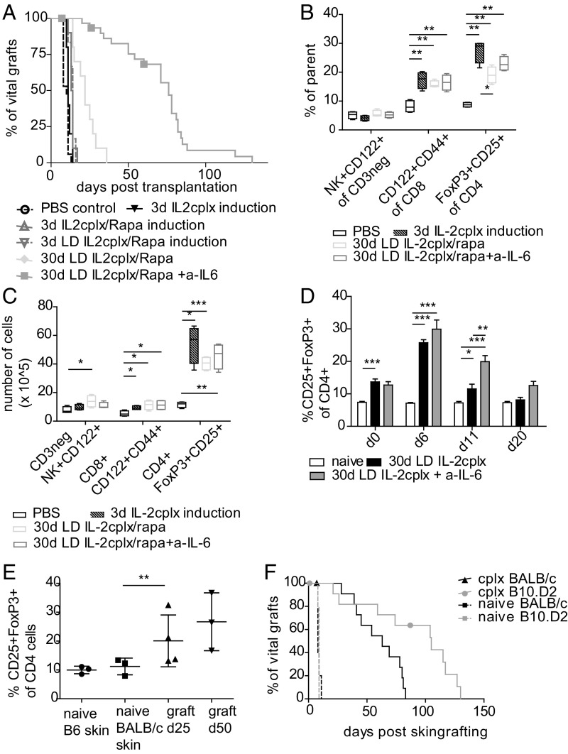
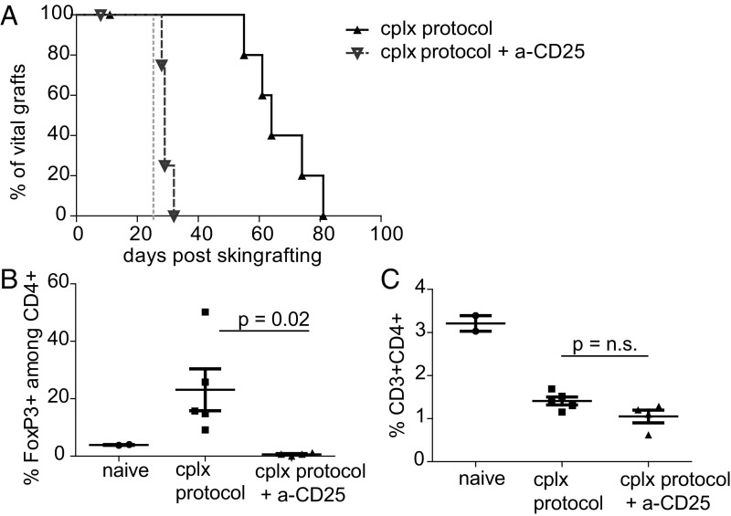
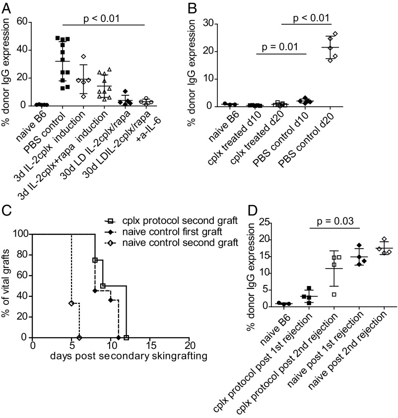
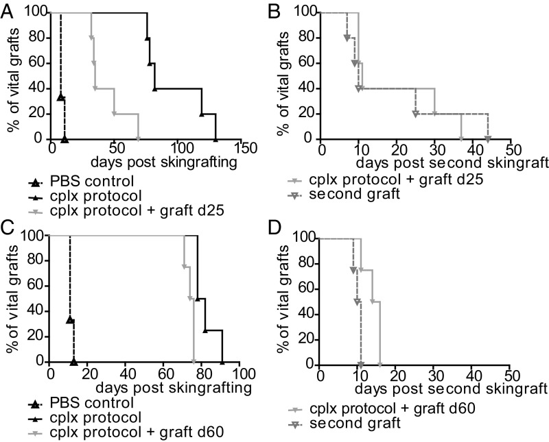
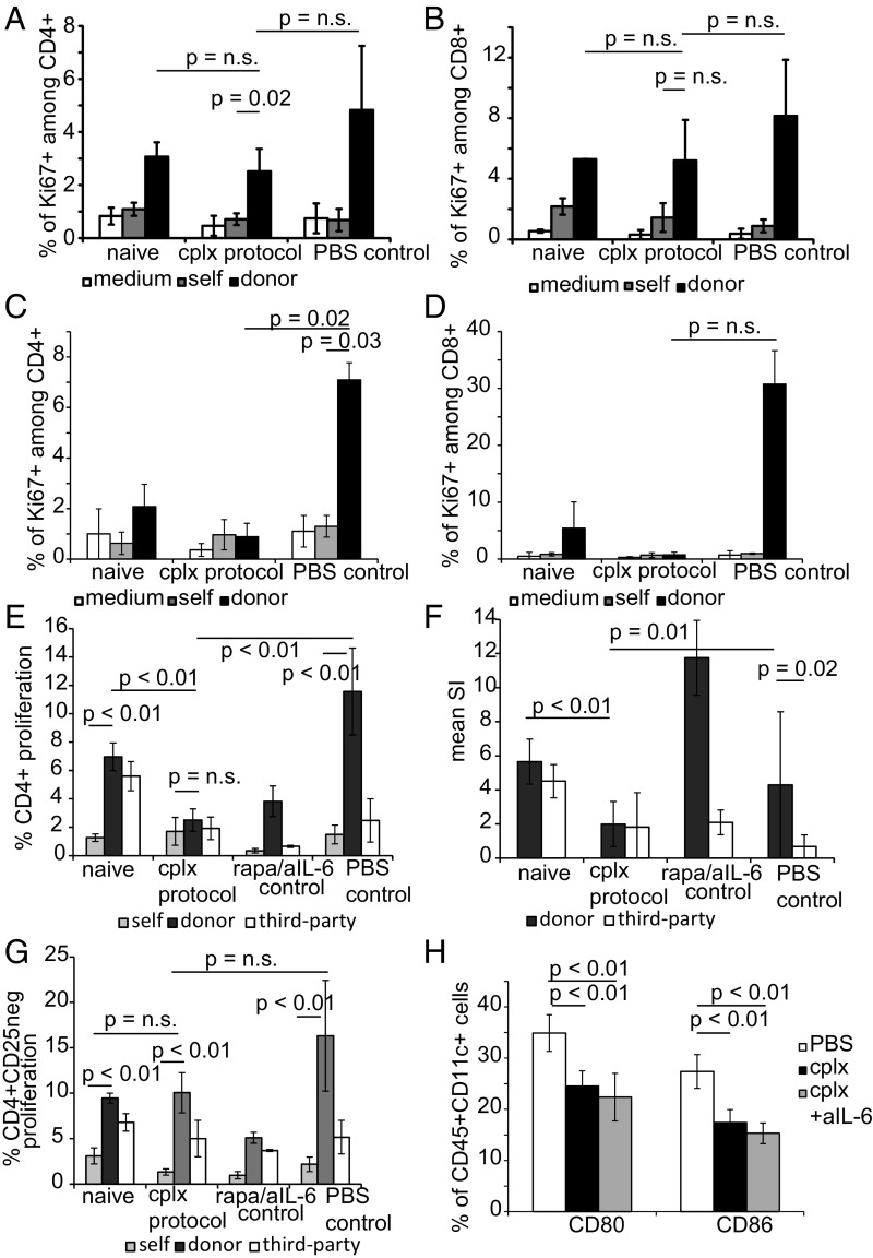
Injection of Interleukin-2 (IL-2) complexed with a particular anti-IL-2 monoclonal antibody (mab) JES6-1 has been shown to selectively expand CD4+Foxp3+ T regulatory T cells (Tregs) in vivo. Although the potency of this approach with regard to transplantation has already been proven in an islet transplantation model, skin graft survival could not be prolonged. Since the latter is relevant to human allograft survival, we sought to improve the efficiency of IL-2 complex (cplx) treatment for skin allograft survival in a stringent murine skin graft model. Here, we show that combining low doses of IL-2 cplxs with rapamycin and blockade of the inflammatory cytokine IL-6 leads to long-term (>75 d) survival of major histocompatibility complex-different skin allografts without the need for immunosuppression. Allograft survival was critically dependent on CD25+FoxP3+ Tregs and was not accompanied by impaired responsiveness toward donor alloantigens in vitro after IL-2 cplx treatment was stopped. Furthermore, second donor-type skin grafts were rejected and provoked rejection of the primary graft, suggesting that operational tolerance is not systemic but restricted to the graft. These findings plus the lack of donor-specific antibody formation imply that prolonged graft survival was largely a reflection of immunological ignorance. The results may represent a potentially clinically translatable strategy for the development of protocols for tolerance induction.

Keywords: interleukin-2; regulatory T cells; tolerance; transplantation.


Conflict of interest statement

The authors declare no conflict of interest.

Figures

Fig. 1.
Fig. 1.
Combined treatment with low dose IL-2 cplxs, rapamycin, and anti–IL-6 potentiates Treg function and prolongs skin allograft survival. Groups of mice were treated with IL-2 cplx-based tolerance regimens and grafted with fully mismatched BALB/c skin. Groups of mice received PBS (control; n = 17 [5]; MST = 10 d), IL-2 cplxs only (cplx; n = 5 [1]; MST = 11 d), IL-2 cplx combined with rapamycin (cplx/rapa; n = 10; MST = 13.5 d; low-dose (LD) cplx/rapa; n = 6 [1]; MST = 14 d) or prolonged treatment with cplx and rapamycin with (30 d LD cplx/rapa + a–IL-6 n = 31 [5]; MST = 76 d) and without anti–IL-6 (30 d LD cplx/rapa n = 10; MST = 22). (A) Survival curves of cumulative data of several independent experiments are shown (indicated in brackets), log-rank test. (B and C) Changes of leukocyte subsets were analyzed in the spleen on day 6 (d6) (*P < 0.05, **P < 0.005, ***P < 0.0005 2-tailed t test with unequal variances) and (D) in peripheral blood over time at indicated timepoints (two-way ANOVA *P < 0.05, **P < 0.01, ***P < 0.001). Mean percentages (B and D) and total cell numbers (C) are shown. Error bars indicate SD. (E) Mice were treated with IL-2 cplx-based tolerance protocol were grafted with 2 BALB/c grafts on contralateral sides of the back. Macroscopically intact grafts were harvested for analysis on d25 and d50. Graft-infiltrating T cells subsets were analyzed for percentage of CD25+FoxP3+ (Tregs; gated on CD45+CD4+). Mean percentages are shown, error bars indicate SD, groups were compared using a 2-tailed t test with unequal variances (**P < 0.01). (F) Mice were grafted with BALB/c (full mismatch) or B10.D2 (MHC mismatch, with minimal minor H antigen difference) skin, and groups were treated with IL-2 cplx-based tolerance regime. Naïve recipients uniformly rejected allografts within 10 d (BALB/c graft MST = 8 d; B10.D2 graft MST = 9 d; n = 5 per group), whereas graft survival was prolonged after IL-2 cplx-based treatment in all groups (BALB/c graft MST = 61.5 d; B10.D2 graft MST = 105 d, P = 0.0075 vs. BALB/c; n = 12 per group). Data were pooled from 2 independent experiments.
Fig. 2.
Fig. 2.
Depletion of Tregs during IL-2 cplx treatment leads to rapid graft rejection. (A) Mice were treated with IL-2 cplx-based tolerance protocol (cplx protocol) and grafted with BALB/c skin (n = 6; MST = 64 d); some mice were treated with anti-CD25 mab (day 25, indicated with dashed gray line) to deplete Tregs (cplx protocol + a-CD25: n = 5; MST = 29 d, P = 0.0029). (B and C) Peripheral blood was analyzed on day 30 to measure frequency of (B) FoxP3+ Tregs among CD4+ subset and (C) total CD4 T cells among leukocytes (naïve n = 2, cplx protocol n = 5, cplx protocol + a-CD25 n = 4; 2-tailed t test with unequal variances).
Fig. 3.
Fig. 3.
Treatment with IL-2 cplxs prevents alloantibody production humoral response and impairs memory cell differentiation. (A) Sera of mice were collected ∼2 wk post skin-graft rejection and analyzed for the presence of donor graft-specific IgG (naïve B6 n = 5; PBS control n = 11; 3 d cplx = 5; 3 d cplx/rapa n = 10; 30 d LD cplx/rapa n = 5; 30 d LD cplx/rapa + a–IL-6 n = 4). (B) Sera of naïve, cplx-treated or PBS-treated control mice were analyzed for donor-specific IgG early after skin grafting (day 10, day 20; n = 3–5 per group). (C) Rejection of a second BALB/c graft applied without additional treatment ∼2 wk after rejection of the first graft (naïve first: n = 11, MST = 8 d; naïve second n = 4, MST = 5 d, P < 0.0001 vs. naïve first; cplx second: n = 4, MST = 10.5 d, P = 0.0058 vs. naïve second; P = 0.1383 vs. naïve first; log-rank test). (D) Sera of mice were analyzed for the presence of anti-donor IgG after the first and second rejection (n = 2–4 per group). Groups were compared with a 2-tailed t test with unequal variances.
Fig. 4.
Fig. 4.
Challenge with a second donor-type graft breaks tolerance. Groups of mice received BALB/c skin grafts under tolerogeneic IL-2 cplx protocol. Operationally tolerant mice (graft macroscopically intact) were grafted with a second graft of BALB/c strain (A and B) at day 25 (d25) (PBS control n = 3, cplx protocol n = 5, cplx protocol + graft d25 n = 5; P = 0.0018 ± second graft) or (C and D) at d60 (PBS control n = 3, cplx protocol n = 4, second graft: cplx protocol + graft d60 n = 4; P = 0.0091 ± second graft) after primary skin grafting. Overall survival of the primary grafts with or without additional antigen challenge on (A) d25 or (C) d60 and survival of the grafts after placing a second donor-type graft transplanted on (B) d25 and (D) d60 (second graft d25 MST = 10 d; second graft d60 MST = 10.5 d; P = n.s. vs. PBS control) are shown. Log-rank test was used for comparison of survival curves.
Fig. 5.
Fig. 5.
IL-2 cplx-based tolerance protocol does not induce systemic (donor) hyporesponsiveness. (AD) Responsiveness to graft alloantigens was evaluated by in vitro T cell proliferation assays. LN cells were prepared from groups of B6 recipients of BALB/c skin grafts at day 50 (A and B) or day 20 (C and D) after skin transplantation; LN cells from normal B6 mice (naïve) and grafted mice not given IL-2 cplx (PBS control) were used as controls. The 4 × 10⁵ unseparated LN cells were cultured alone (medium) or with 4 × 10⁵ irradiated spleen cells from B6 (self) or BALB/c (donor) mice for 4 d and then assessed for Ki67 expression by flow cytometry. Mean expression of Ki67 expression in CD4 (A and C) and CD8 (B and D) T cells in triplicate cultures is shown (naïve = 2, cplx protocol = 4, PBS control = 3; 2-tailed t test with unequal variances for P values). (EG) Responsiveness to graft versus third-party alloantigens. LN cells were prepared from B6 recipients of BALB/c skin grafts at day 14 after grafting and starting treatment with IL-2 cplx (cplx protocol) or a mixture of rapamycin and anti–IL-6 mab (rapa/IL-6 control), with untreated grafted mice (PBS control) as a control. LN cells were labeled with VPD and 4 × 10⁵ cells were cultured with 4 × 10⁵ irradiated spleen cells from B6 (self), BALB/c (donor), or C3H (third party) mice for 4d and then assessed for VPD dilution by flow cytometry. Responder cells were either unseparated (E and F) or depleted of CD25+ cells to remove Tregs (G) before culture. (E and G) Mean percentage of CD4 cells that expressed a low density of VPD (indicating proliferation) is shown for triplicate cultures. (F) The mean (SI ratio of responses to BALB/c and C3H relative to responses to B6 stimulators) of unseparated LN cells is shown. Results are pooled from 2 separate experiments and involve cells taken from multiple mice (naïve n = 4, cplx protocol n = 6, rapa/anti–IL-6 n = 2, PBS control n = 5). (H) B7 expression on host DCs. Expression of B7 molecules CD80 and CD86 was measured at day 14 in CD11+ splenocytes of PBS-treated (n = 4), cplx-treated (n = 7), and cplx/anti–IL-6 treated (n = 7) mice (data pooled from 2 independent experiments). Data are shown as mean percentage, error bars depict SD, P values were calculated using a 2-tailed t test with unequal variances.

References

    1. Vignali D. A., Collison L. W., Workman C. J., How regulatory T cells work. Nat. Rev. Immunol. 8, 523–532 (2008). - PMC - PubMed
    1. Tang Q., Bluestone J. A., Regulatory T-cell therapy in transplantation: Moving to the clinic. Cold Spring Harb. Perspect. Med. 3, a015552 (2013). - PMC - PubMed
    1. Joffre O., et al. , Prevention of acute and chronic allograft rejection with CD4+CD25+Foxp3+ regulatory T lymphocytes. Nat. Med. 14, 88–92 (2008). - PMC - PubMed
    1. Tsang J. Y., et al. , Conferring indirect allospecificity on CD4+CD25+ Tregs by TCR gene transfer favors transplantation tolerance in mice. J. Clin. Invest. 118, 3619–3628 (2008). - PMC - PubMed
    1. Nadig S. N., et al. , In vivo prevention of transplant arteriosclerosis by ex vivo-expanded human regulatory T cells. Nat. Med. 16, 809–813 (2010). - PMC - PubMed

Publication types