. 2019 Jul 2;116(27):13621-13630.
doi: 10.1073/pnas.1811825116. Epub 2019 Jun 13.

Region-specific and activity-dependent regulation of SVZ neurogenesis and recovery after stroke

Affiliations

Region-specific and activity-dependent regulation of SVZ neurogenesis and recovery after stroke

Huixuan Liang et al. Proc Natl Acad Sci U S A. .

Abstract

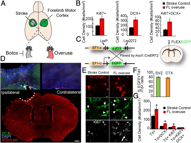
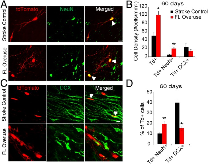
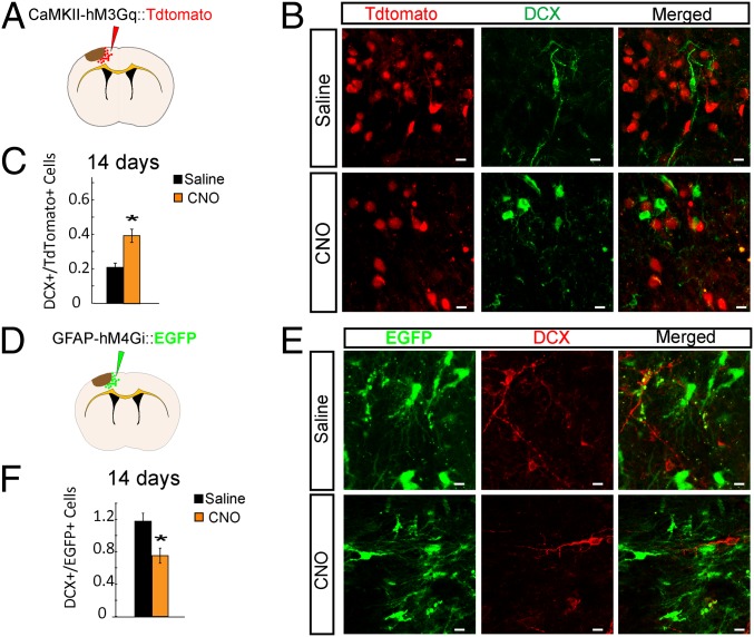
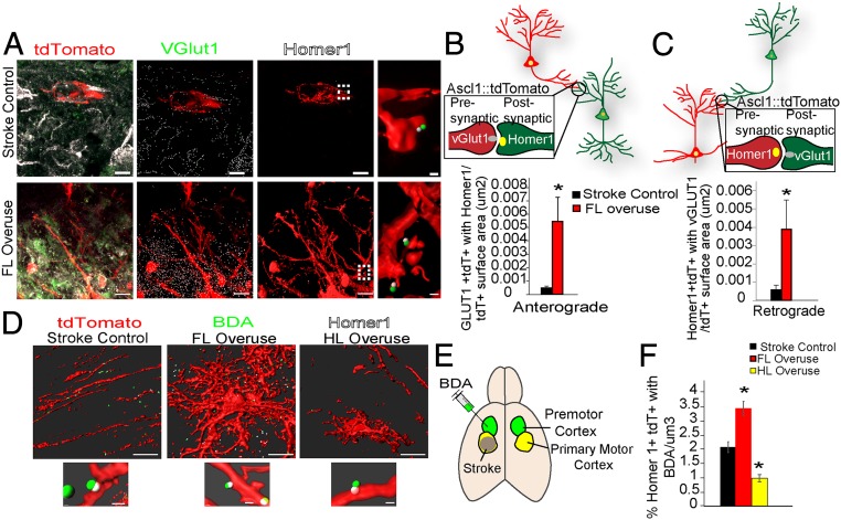
Stroke is the leading cause of adult disability. Neurogenesis after stroke is associated with repair; however, the mechanisms regulating poststroke neurogenesis and its functional effect remain unclear. Here, we investigate multiple mechanistic routes of induced neurogenesis in the poststroke brain, using both a forelimb overuse manipulation that models a clinical neurorehabilitation paradigm, as well as local manipulation of cellular activity in the peri-infarct cortex. Increased activity in the forelimb peri-infarct cortex via either modulation drives increased subventricular zone (SVZ) progenitor proliferation, migration, and neuronal maturation in peri-infarct cortex. This effect is sensitive to competition from neighboring brain regions. By using orthogonal tract tracing and rabies virus approaches in transgenic SVZ-lineage-tracing mice, SVZ-derived neurons synaptically integrate into the peri-infarct cortex; these effects are enhanced with forelimb overuse. Synaptic transmission from these newborn SVZ-derived neurons is critical for spontaneous recovery after stroke, as tetanus neurotoxin silencing specifically of the SVZ-derived neurons disrupts the formation of these synaptic connections and hinders functional recovery after stroke. SVZ-derived neurogenesis after stroke is activity-dependent, region-specific, and sensitive to modulation, and the synaptic connections formed by these newborn cells are functionally critical for poststroke recovery.

Keywords: astrocyte; motor; neurorehabilitation; plasticity; synapse.


Conflict of interest statement

The authors declare no conflict of interest.

Figures

Fig. 1.
Fig. 1.
Forelimb overuse increases neurogenesis in the peri-infarct cortex after stroke. (A) Schematic demonstration of stroke induction and forelimb overuse. Focal ischemic stroke in the forelimb motor cortex was induced by photothrombosis. To induce overuse of the paretic forelimb, Botox was injected to paralyze the nonparetic forelimb 24 h after stroke. (B) Density of Ki67+, DCX+, and Ki67+DCX+ cells. *P < 0.05. (C) Schematic demonstration of the EF1-α–FLEX–EGFP plasmid and lateral ventricle injection of the lentivirus expressing EF1-α–FLEX–EGFP. Lentivirus EF1-α–FLEX–EGFP was injected into the lateral ventricle of Ascl1–CreERT2::tdTomato animals 7 d before stroke. Tamoxifen was administered daily to the mice from days 2 to 7 after stroke. (D) Low-magnification confocal micrograph of the whole-brain coronal section with tdTomato, DCX, and DAPI 14 d after stroke. (Scale bar: 500 µm.) (E) Confocal micrographs of tdTomato+, DCX+, and Ki67+ cells in the peri-infarct cortex of the Ascl1–CreERT2::tdTomato mice 14 d after stroke. Filled arrowheads, tdTomato+DCX+ cells; open arrowheads, tdTomato+Ki67+ cells. (Scale bar: 5 µm.) (F) Percentage of EGFP+ tdTomato+ cells among total tdTomato+ cells in the SVZ and in the peri-infarct cortex 14 d after stroke. (G) Density of tdTomato+, tdTomato+DCX+, tdTomato+Ki67+, and tdTomato+Ki67+DCX+ cells in the peri-infarct region 14 d after stroke. *P < 0.05.
Fig. 2.
Fig. 2.
Forelimb overuse increases long-term survival and neuronal differentiation of SVZ-derived progeny into the peri-infarct cortex 60 d after stroke. (A) Confocal micrographs of tdTomato+ and NeuN+ cells in the peri-infarct cortex of the Ascl1–CreERT2::tdTomato mice 60 d after stroke. Arrowheads, tdTomato+NeuN+ cells. (Scale bar: 5 µm.) (B) Quantification of density of tdTomato+, tdTomato+NeuN+, and tdTomato+DCX+ cells in the peri-infarct region. (C) Confocal micrographs of tdTomato+ and DCX+ cells in the peri-infarct cortex of the Ascl1–CreERT2::tdTomato mice 60 d after stroke. Arrowheads: tdTomato+DCX+ cells. (Scale bar: 5 µm.) (D) Percentages of tdTomato cells that coexpress NeuN and DCX in the peri-infarct region. *P < 0.05.
Fig. 3.
Fig. 3.
Modulation of neuronal and glial activity in the peri-infarct affects poststroke neurogenesis. (A) Schematic of lentivirus approach with CaMKII–hM3Gq tdTomato injection. Lentivirus expressing the CaMKII–hM3Gq tdTomato was injected into the cortex 14 d before stroke. Saline or CNO was administered twice daily from day 3 until euthanizing at day 14 after stroke. (B) Confocal micrographs of CaMKII–hM3Gq tdTomato+ and DCX+ cells in the peri-infarct cortex, 14 d after stroke in C57BL/6 mice. (Scale bars: 5 µm.) (C) Ratio of numbers of DCX+ cells to numbers of tdTomato+ cells in the peri-infarct cortex. (D) Schematic of lentivirus approach with GFAP–hM4Gi–EGFP injection, with the same methods as in A. (E) Confocal micrographs of GFAP–hM4Gi–EGFP+ and DCX+ cells in the peri-infarct cortex, 14 d after stroke in the C57BL/6 animals. (Scale bars: 5 µm.) (F) Numbers of DCX+ cells to numbers of EGFP+ cells in the peri-infarct cortex. *P < 0.05.
Fig. 4.
Fig. 4.
Hindlimb overuse decreases neurogenesis in the peri-infarct forelimb motor cortex and partially offsets the neurogenic effect induced by forelimb overuse. (AD) C57BL/6 stroke animals were treated with no Botox (stroke control), FL overuse, or HL overuse before euthanizing at 14 d after stroke. (A) Confocal micrographs of Ki67+ and DCX+ cells in the peri-infarct forelimb motor cortex. (Scale bar: 10 µm.) (B) Density of DCX+, Ki67+, and DCX+Ki67+ cells in the peri-infarct forelimb motor cortex. For all group comparisons, ANOVA: P < 0.05. (C) Confocal micrographs of Ki67+ and DCX+ cells in the hindlimb motor cortex. (Scale bar: 10 µm.) (D) Density of DCX+, Ki67+, and Ki67+DCX+ cells in the hindlimb motor cortex. For all group comparisons, ANOVA: P < 0.05. (E) Schematic of lentivirus CaMKII–hM3Gq tdTomato injection into hindlimb motor cortex. HL DREADD control group received stroke, HL CAMKII-Gq injection, and saline injection; HL DREADD activation group received stroke, HL CAMKII-Gq injection, and CNO injection; and HL DREADD activation+FL overuse group received stroke, HL CAMKII-Gq injection, CNO injection, and FL overuse. (F) Density of DCX+, Ki67+, and DCX+Ki67+ cells in the peri-infarct forelimb motor cortex. For all group comparisons, ANOVA: P < 0.05. (G) No Botox (stroke control), FL overuse, or HL overuse before euthanizing at 60 d after stroke in Ascl1–CreERT2::tdTomato mice. Confocal micrographs of tdTomato+ and NeuN+ in the peri-infarct forelimb motor cortex are shown. Filled arrowheads: tdTomato+ and NeuN+ mature neurons differentiated from SVZ progenitors. (Scale bar: 10 µm.) (H) Quantification for the percentage of percent tdTomato+ NeuN+ among all tdTomato cells in the peri-infarct region. For all group comparisons, ANOVA: P < 0.05. *P < 0.05.
Fig. 5.
Fig. 5.
Forelimb overuse increases synaptogenesis and neural connectivity from the SVZ-derived progenitors. (AC) Ascl1–CreERT2(+);tdTomato animals induced with stroke and tamoxifen were treated with no Botox (control) or FL overuse before euthanizing at 60 d after stroke. (A) Confocal micrographs of tdTomato+ (red), VGlut1+ (green) presynaptic proteins, and Homer1+ (white) postsynaptic proteins in the peri-infarct cortex of Ascl1–CreERT2::tdTomato mice. Images are segmented (Imaris, Bitplane). (Scale bars: 10 μm.) The rightmost images show a higher magnification of colocation of VGLUT1 with the postsynaptic Homer1, from the corresponding regions highlighted with the dashed lines. (Scale bars: 2 μm.) (B) Anterograde synapses identified by the colocalization of tdTomato+VGLUT1+ with Homer1. Quantification is number of anterograde synaptic formations per tdTomato cell surface (μm2). (C) Retrograde synapses identified by colocalization of tdTomato+Homer1+ with VGLUT1. Quantifications is the number of retrograde synaptic formation per tdTomato cell surface (μm2). (DF) BDA was injected into the premotor cortex of Ascl1–CreERT2(+);tdTomato stroke animals 7 d before euthanizing at the poststroke day-60 time point. These animals received either no Botox (stroke control), FL overuse, or HL overuse. (D) Segmented images (Imaris) of colocation of Homer1 within the tdTomato cells with BDA axons. (D, Lower) Higher-magnification images. [Scale bars: 10 μm (Upper) and 2 μm (Lower).] (E) Schematic illustration of stroke induction and BDA injection. (F) Number of colocalizations between tdTomato+Homer1+ and BDA+ per μm3. For all group comparisons, ANOVA: P < 0.05. *P < 0.05.
Fig. 6.
Fig. 6.
Forelimb overuse increases the monosynaptic neural connectivity of SVZ-derived neurons, and synaptic connectivity is required for functional recovery. (A) AAV Synapsin-Cre and AAV FLEXBTG were co-injected into the peri-infarct cortex 7 d before stroke. Animals were allowed to survive 2 months after stroke and the rabies BFP was injected into the same region 4 d before sacrifice. Arrowheads show GFP+BFP+ starter cells. (Scale bar: 5 µm.) (B) Confocal micrographs of tdTomato+, GFP+ and BFP+ SVZ-derived neurons forming monosynaptic connections in the peri-infarct. Arrowheads show GFP+BFP+ starter cells. (Scale bar: 5 µm.) (C) Quantification of synaptic connection with the cortical strength index from either cortical neurons or SVZ-derived neurons. *P < 0.05. n.s., not significant. (D) EYFP cell density in the peri-infarct regions 2 mo after stroke. (E) Quantification for the number of VAMP2 located within EYFP+ cells per EYFP cell surface. (F) Quantification for percentage of foot faults from the total number of steps, based on the grid-walk task. (G) Quantification of (right paw duration − left paw duration)/total duration, based on the pasta-handling task. *P < 0.05.

References

    1. Bi B., et al. , Cortical glial fibrillary acidic protein-positive cells generate neurons after perinatal hypoxic injury. J. Neurosci. 31, 9205–9221 (2011a). - PMC - PubMed
    1. Buffo A., et al. , Origin and progeny of reactive gliosis: A source of multipotent cells in the injured brain. Proc. Natl. Acad. Sci. U.S.A. 105, 3581–3586 (2008). - PMC - PubMed
    1. Ohira K., et al. , Ischemia-induced neurogenesis of neocortical layer 1 progenitor cells. Nat. Neurosci. 13, 173–179 (2010). - PubMed
    1. Donega V., Raineteau O., Postnatal neural stem cells: Probing their competence for cortical repair. Neuroscientist 23, 605–615 (2017). - PubMed
    1. Arvidsson A., Collin T., Kirik D., Kokaia Z., Lindvall O., Neuronal replacement from endogenous precursors in the adult brain after stroke. Nat. Med. 8, 963–970 (2002). - PubMed