. 2019 Jul 1;218(7):2185-2197.
doi: 10.1083/jcb.201803041. Epub 2019 Jun 13.

SFI1 promotes centriole duplication by recruiting USP9X to stabilize the microcephaly protein STIL

Affiliations

SFI1 promotes centriole duplication by recruiting USP9X to stabilize the microcephaly protein STIL

Andrew Kodani et al. J Cell Biol. .

Abstract

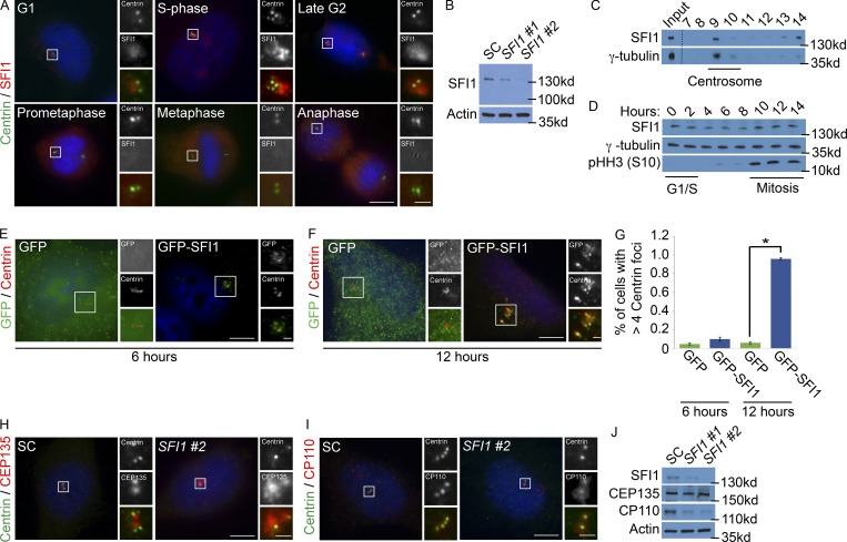
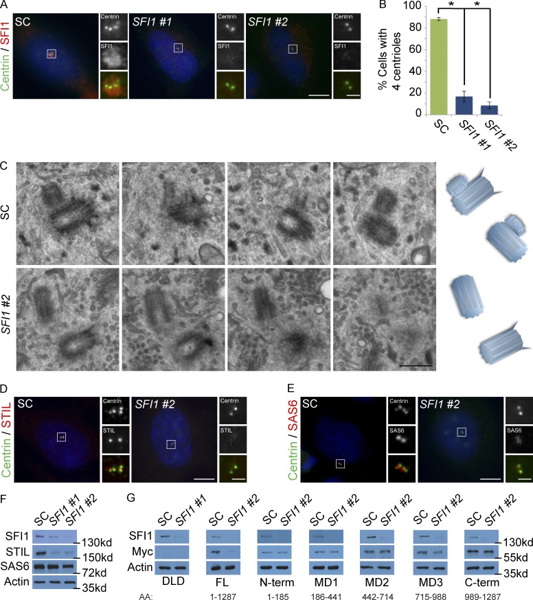
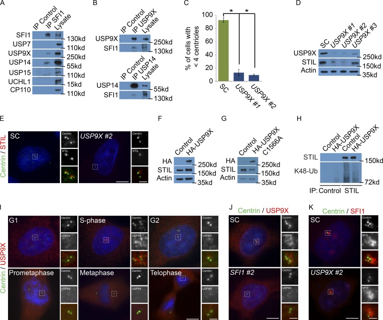
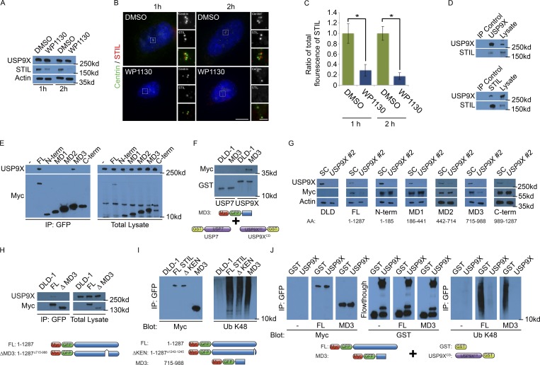
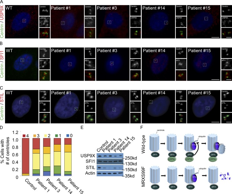
In mammals, centrioles participate in brain development, and human mutations affecting centriole duplication cause microcephaly. Here, we identify a role for the mammalian homologue of yeast SFI1, involved in the duplication of the yeast spindle pole body, as a critical regulator of centriole duplication in mammalian cells. Mammalian SFI1 interacts with USP9X, a deubiquitylase associated with human syndromic mental retardation. SFI1 localizes USP9X to the centrosome during S phase to deubiquitylate STIL, a critical regulator of centriole duplication. USP9X-mediated deubiquitylation protects STIL from degradation. Consistent with a role for USP9X in stabilizing STIL, cells from patients with USP9X loss-of-function mutations have reduced STIL levels. Together, these results demonstrate that SFI1 is a centrosomal protein that localizes USP9X to the centrosome to stabilize STIL and promote centriole duplication. We propose that the USP9X protection of STIL to facilitate centriole duplication underlies roles of both proteins in human neurodevelopment.


Figures

Figure 1.
Figure 1.
SFI1 localizes to the centrosome. (A) Asynchronously growing HeLa cells costained for Centrin (green), SFI1 (red), and DNA (blue). The number of Centrin foci and DNA condensation were used to determine the cell cycle stage. (B) Total cell lysates of HeLa cells treated with one of two siRNAs targeting SFI1 (#1 or #2) or an SC were immunoblotted for SFI1. Actin served as a loading control. (C) Sucrose gradient fractions of HeLa cell lysates were immunoblotted for the centrosomal protein γ-tubulin and SFI1. Dashed line represents spliced blot. (D) Total cell lysate from synchronized HeLa cells collected every 2 h after release of a double thymidine block were immunoblotted for SFI1, γ-tubulin, and phospho-Histone H3 (serine 10), which served as a marker for mitosis. (E and F) Immunofluorescence of HeLa cells expressing GFP or GFP-SFI1 costained for GFP (green) and Centrin (red) 6 or 12 h after transfection. (G) Quantification of cells with greater than four Centrin foci 6 or 12 h after transfection with GFP or GFP-SFI1. For all quantifications, ≥100 cells were counted per experiment (n = 3); *P < 0.005 (paired t test). Error bars represent ± SD. (H) S phase SC and SFI1 #2 siRNA-transfected cells were stained for CEP135 (red) and Centrin (green). (I) S phase HeLa cells transfected with SC or SFI #2 siRNA costained for Centrin (green) and the distal centriole component CP110 (red). Scale bars represent 5 µm for all images and 1 µm for inset images. (J) Total cell lysates of SC and SFI1-depleted cells were immunoblotted for SFI1, CEP135, and CP110. Actin served as a loading control.
Figure 2.
Figure 2.
Depletion of SFI1 destabilizes STIL. (A) S phase HeLa cells transfected with SFI1 siRNA #1, siRNA #2, or SC were costained with SFI1 (red) and Centrin (green). (B) Quantification of S phase SC and SFI1-depleted HeLa cells with four Centrin foci. 100 cells or more were counted per experiment (n = 3). *P < 0.005 (paired t test) for SC versus SFI1 siRNA transfected cells. Error bars represent ± SD. (C) Serial ultrathin section electron micrographs of centrioles in SC and SFI1 #2 siRNA-treated HeLa cells in S phase. Scale bar represents 400 nm. (D) Immunofluorescence of SC and SFI1 #2 siRNA-transfected HeLa cells in S phase costained for Centrin (green) and STIL (red). (E) S phase HeLa cells transfected with SC or SFI1#2 siRNA costained for Centrin (green) and SAS6 (red). Scale bar represents 5 µm for all images and 1 µm for inset images. (F) Total cell lysate of SC and SFI1-depleted HeLa cells immunoblotted for SFI1, STIL, and SAS6. Actin served as a loading control. (G) Total cell lysate of DLD-1 cells transfected with SC or SFI1 siRNA, induced to express full-length (FL) GFP-Myc-tagged STIL and fragments, and analyzed by immunoblot using antibodies to SFI1 and Myc. Actin served as a loading control.
Figure 3.
Figure 3.
SFI1 localizes USP9X to the centrosome to stabilize STIL. (A) We immunoprecipitated (IP) endogenous SFI1 from HeLa total cell lysates and immunoblotted precipitant for SFI1, USP7, USP9X, USP14, USP15, and UCHL1. CP110 served as a negative control. (B) HeLa total cell lysate was subjected to immunoprecipitation of USP9X, USP14, and c-Myc, which served as a negative control throughout. Precipitating proteins were immunoblotted for USP9X, SFI1, and USP14. (C) Percentage of S phase cells with four centrioles in SC, USP9X #1, and USP9X #2 siRNA-treated HeLa cells. For all quantifications, ≥100 cells were counted per experiment (n = 3); *P < 0.005 (paired t test). Error bars represent ± SD. (D) Total cell lysates from HeLa cells transfected with SC or one of three nonoverlapping siRNAs to USP9X were immunoblotted for USP9X and STIL. Actin served as a loading control. (E) SC or USP9X #2 siRNA-transfected HeLa cells were stained for Centrin (green) and STIL (red). (F and G) Total cell lysates of HeLa cells transfected with control or HA-tagged USP9X or USP9X C1566A were analyzed with antibodies to HA and STIL. Actin served as a loading control. (H) STIL was immunoprecipitated from HeLa cells expressing HA-USP9X 6 h after transfection. Precipitating proteins were immunoblotted for STIL and K48-linked ubiquitin (K48-Ub). (I) Asynchronously growing HeLa cells costained for Centrin (green), USP9X (red), and DNA (blue). (J) S phase HeLa cells transfected with SC or USP9X #2 siRNA costained for Centrin (green) and USP9X (red). (K) HeLa cells transfected with SC or SFI1 #2 siRNA were costained for Centrin (green) and USP9X (red). Scale bars represent 5 µm for all images and 1 µm for inset images.
Figure 4.
Figure 4.
USP9X deubiquitylates STIL. (A) Total cell lysates from HeLa cells treated with DMSO or 5 µM WP1130 for 1 or 2 h were immunoblotted for USP9X and STIL. Actin served as a loading control. (B) S phase HeLa cells treated with DMSO or WP130 for 1 or 2 h were stained for Centrin (green) and STIL (red). Scale bars represent 5 µm for all images and 1 µm for inset images. (C) Quantification of the centrosomal fluorescence intensity of STIL in DMSO- and WP1130-treated HeLa cells expressed as the ratio ± SD to the fluorescence intensities of control cells. For all quantifications, 10 cells were analyzed per experiment (n = 3). *P < 0.005 (t test). (D) Reciprocal coimmunoprecipitation (IP) of endogenous USP9X and STIL from HeLa total cell lysates. Efficient precipitation and coprecipitation were detected using antibodies for USP9X and STIL. c-Myc served as a negative control. (E) Control (parental) DLD-1 or stable DLD-1 cells expressing full-length GFP-Myc–tagged STIL or the indicated STIL fragments were immunoprecipitated using antibody to GFP. STIL or its fragments were detected by immunoblotting for Myc, respectively. Asterisk denotes nonspecific band. Coimmunoprecipitating endogenous USP9X was detected by immunoblotting for USP9X. (F) Total cell lysate from doxycycline-induced DLD-1 cells or cells expressing GFP-Myc–tagged MD3 domain of STIL were incubated with recombinant GST-USP7 or GST fused to the catalytic domain (aa 1,531–1,972) of USP9X (GST-USP9XCD). Proteins precipitating with GST-USP7 or GST-USP9XCD were analyzed by immunoblot using antibodies to Myc and GST. (G) Total cell lysates of DLD-1 cells transfected with SC or USP9X siRNA, induced to express GFP-Myc–tagged STIL fragments, and immunoblotted for SFI1 and Myc. Actin served as a loading control. (H) Lysates from doxycycline-induced control DLD-1 cells or cells expressing GFP-Myc–tagged full-length STIL or STIL lacking the MD3 domain (STILΔMD3) were immunoprecipitated using antibody to GFP. Precipitating proteins were immunoblotted for USP9X and Myc. (I) From total cell lysates of doxycycline-induced control (parental) DLD-1 or DLD-1 cells expressing GFP-Myc–tagged STIL constructs, we immunoprecipitated full-length (FL) STIL, ΔKEN STIL, or the MD3 domain of STIL with an antibody to GFP. Precipitating proteins were detected with antibodies to Myc and K48-linked ubiquitin (Ub). (J) A negative control, full-length (FL), or the MD3 fragment of STIL were immunoprecipitated with immobilized GFP-trap beads, incubated with recombinant GST or the catalytic domain of USP9X, and immunoblotted for Myc, GST, and ubiquitylated (Ub) K48.
Figure 5.
Figure 5.
Loss-of-function USP9X mutations destabilize STIL. (A–C) S phase fibroblasts from control individual or USP9X mutant MRXS99F patients were stained for Centrin (green) and USP9X, SFI1, or STIL (red). Scale bars represent 5 µm for all images and 1 µm for inset images. (D) Percentage of S phase fibroblasts with one, two, three, or four centrioles in control and MRXS99F patient fibroblasts. For all quantifications, ≥100 cells were counted per experiment (n = 3); *P < 0.005 (paired t test). Error bars represent ± SD. (E) Immunoblot assessment of USP9X, SFI1, and STIL levels in control and MRXS99F patient fibroblasts. Actin served as a loading control. (F) SFI1 recruits USP9X to the centrosome during S phase and removes the K48-ubiquitin chain on STIL. USP9X fails to localize to the centrosome during S phase in MRXS99F cells, leading to STIL degradation and attenuated centriole duplication.

Similar articles

Cited by

References

    1. Al-Hakim A.K., Bashkurov M., Gingras A.C., Durocher D., and Pelletier L.. 2012. Interaction proteomics identify NEURL4 and the HECT E3 ligase HERC2 as novel modulators of centrosome architecture. Mol. Cell. Proteomics. 11:M111.014233. - PMC - PubMed
    1. Arquint C., and Nigg E.A.. 2014. STIL microcephaly mutations interfere with APC/C-mediated degradation and cause centriole amplification. Curr. Biol. 24:351–360. 10.1016/j.cub.2013.12.016 - DOI - PubMed
    1. Arquint C., Cubizolles F., Morand A., Schmidt A., and Nigg E.A.. 2018. The SKP1-Cullin-F-box E3 ligase βTrCP and CDK2 cooperate to control STIL abundance and centriole number. Open Biol. 8:170253 10.1098/rsob.170253 - DOI - PMC - PubMed
    1. Balestra F.R., Strnad P., Flückiger I., and Gönczy P.. 2013. Discovering regulators of centriole biogenesis through siRNA-based functional genomics in human cells. Dev. Cell. 25:555–571. 10.1016/j.devcel.2013.05.016 - DOI - PubMed
    1. Bayless B.A., Giddings T.H. Jr., Winey M., and Pearson C.G.. 2012. Bld10/Cep135 stabilizes basal bodies to resist cilia-generated forces. Mol. Biol. Cell. 23:4820–4832. 10.1091/mbc.e12-08-0577 - DOI - PMC - PubMed

Publication types