. 2020 Apr;105(4):914-924.
doi: 10.3324/haematol.2018.215160. Epub 2019 Jun 13.

Ephrin/Eph receptor interaction facilitates macrophage recognition of differentiating human erythroblasts

Affiliations

Ephrin/Eph receptor interaction facilitates macrophage recognition of differentiating human erythroblasts

Lea A Hampton-O'Neil et al. Haematologica. 2020 Apr.

Abstract

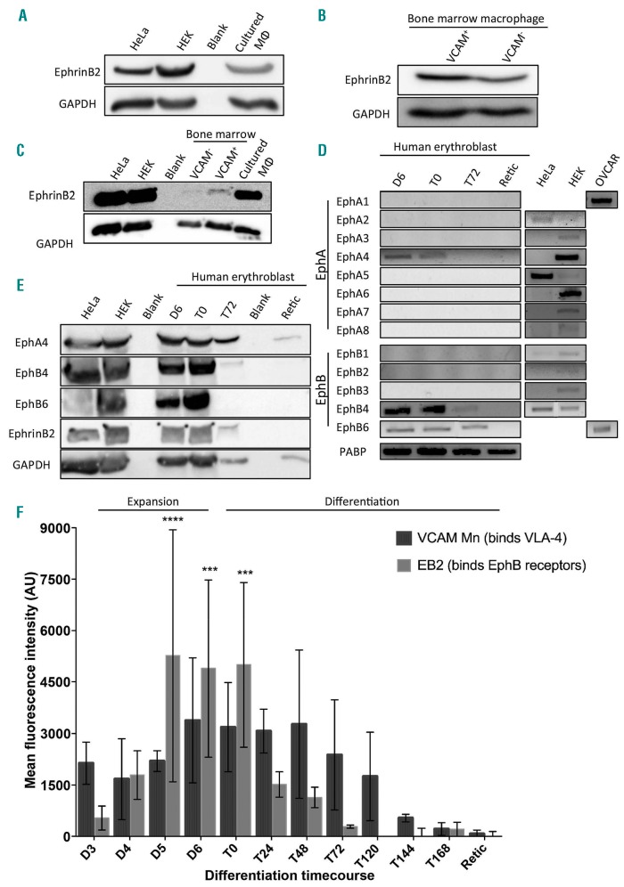
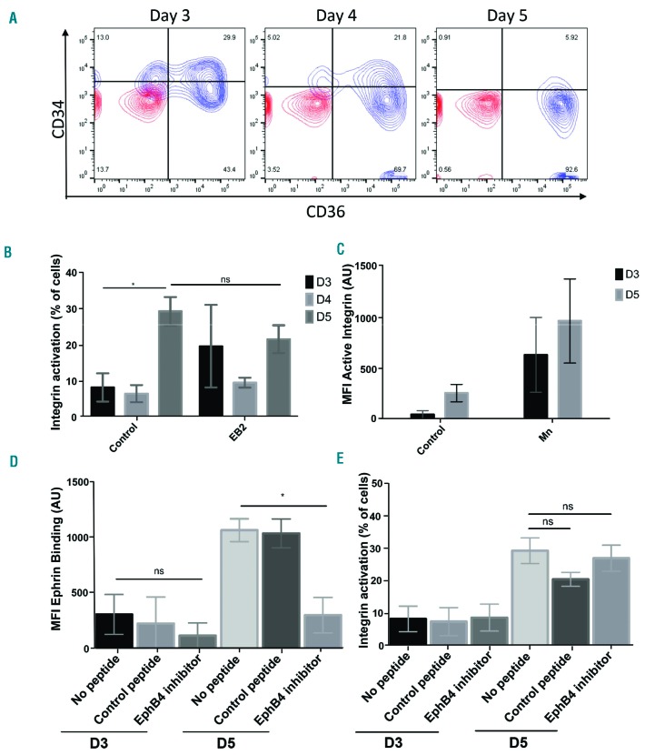
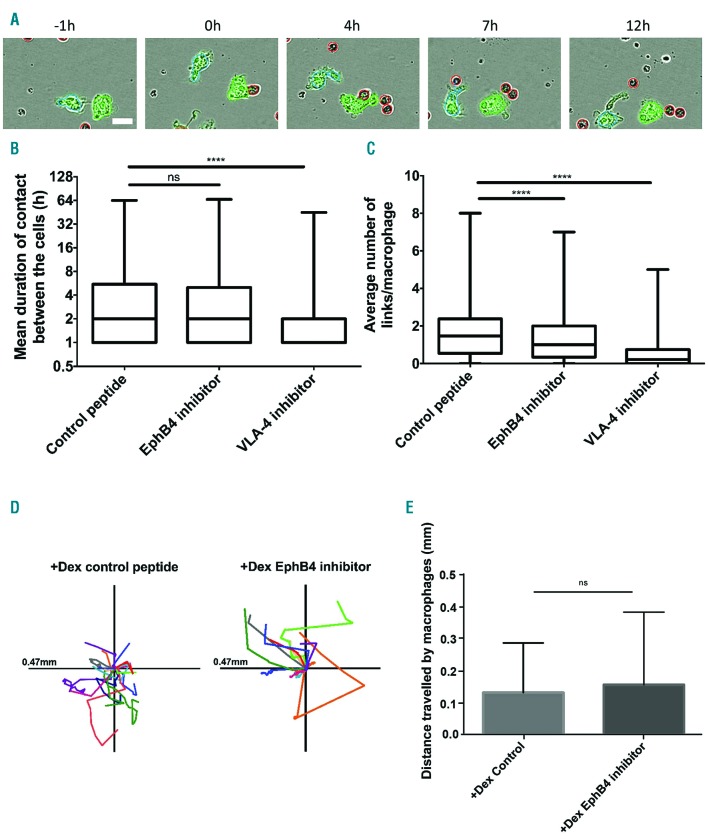
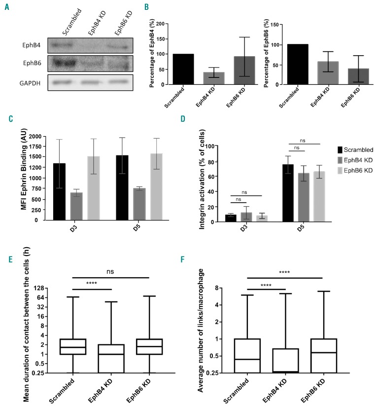
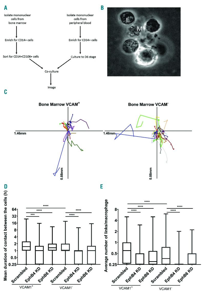
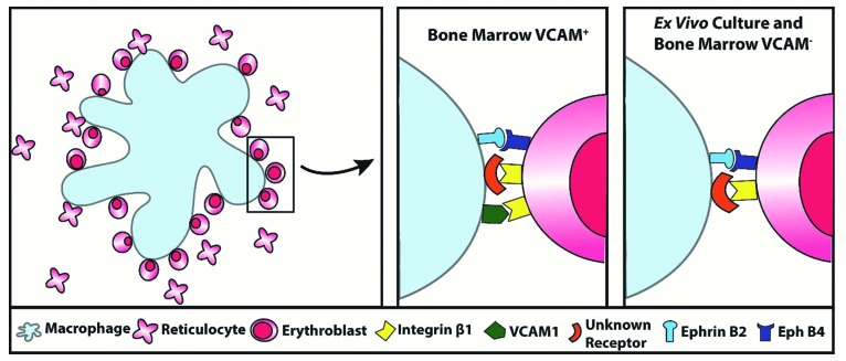
Erythropoiesis is one of the most efficient cellular processes in the human body producing approximately 2.5 million red blood cells every second. This process occurs in a bone marrow niche comprised of a central resident macrophage surrounded by differentiating erythroblasts, termed an erythroblastic island. It is not known what initially attracts the macrophage to erythroblasts to form these islands. The ephrin/Eph receptor family are known to regulate heterophilic cell-cell adhesion. We find that human VCAM1+ and VCAM1- bone marrow macrophages and in vitro cultured macrophages are ephrin-B2 positive, whereas differentiating human erythroblasts express EPHB4, EPHB6 and EPHA4. Furthermore, we detect a rise in integrin activation on erythroblasts at the stage at which the cells bind which is independent of EPH receptor presence. Using a live cell imaging assay, we show that specific inhibitory peptides or shRNA depletion of EPHB4 cause a significant reduction in the ability of macrophages to interact with erythroblasts but do not affect integrin activation. This study demonstrates for the first time that EPHB4 expression is required on erythroblasts to facilitate the initial recognition and subsequent interaction with macrophages, alongside the presence of active integrins.


Figures

Figure 1.
Figure 1.
EPH receptor expression profile is specific to the cell type and its differentiation. (A) Lysates from cultured macrophages, HeLa and HEK cells were western blotted for ephrin-B2 and GAPDH. This is a representative blot (n=3). (B) Lysates from sorted VCAM+ and VCAM- bone marrow (BM) CD14+ cells were blotted for ephrin-B2 and GAPDH. This is a representative blot (n=2). (C) Lysates from HEK, HeLa, sorted BM and cultured macrophages with dexamethasone (+Dex) were probed for ephrinB2 and GAPDH. This is a representative blot (n=2). (D) Representative DNA gels showing the polymerase chain reaction products from an erythroblast differentiation course using primers against EPHA1-8 and EPHB1-6 (n=3). For each sample, a negative control was also performed to confirm the absence of genomic DNA contamination. HEK293, OVCAR 3 and HeLa cells were used as positive controls. (E) Lysates from HeLa, HEK cells and an erythroblast differentiation course were blotted for EPHB4, EPHB6, EPHA4, ephrin-B2 and GAPDH. This is a representative blot (n=3). (A, B, C and E) All lanes were loaded with 1×10⁶ cells. (F) Graph showing the mean fluorescent intensity (MFI) obtained by flow cytometry of the ephrin-B2-Fc and VCAM-Fc constructs binding to erythroblasts throughout terminal differentiation. EB2 is ephrin-B2-Fc, and VCAM Mn is VCAM-Fc with manganese activation. The ephrin constructs were pre-clustered. All points are means for ephrin-Fc constructs (n=5) and VCAM (n=3). The error bars represent the standard error of the mean. Binding to the constructs was statistically compared to the IgG control using two-way ANOVA: ***P<0.001; ****P<0.0001. The control for VCAM binding without manganese is in Online Supplementary Figure S3. D: day; T: time course; AU: arbitrary units.
Figure 2.
Figure 2.
Activated Integrin β1 on proerythroblasts is not affected by EPHB4 inhibition. (A) Representative contour plot of the erythroblasts at different stages of expansion on days (D) 3, 4 and 5 of culture (n=5). IgG is in red, and antibodies are in blue. (B) Graph showing the percentage of cells with active integrin obtained by flow cytometry of these cells with the active form of integrin β1 (clone HUTS-21) through D3, D4 and D5 of erythroblast expansion (n=4). Mixed effects analysis was run on the samples. (C) Graph showing the mean fluorescence intensity (MFI) of active integrin β1 through D3 and D5 of expanding erythroblasts and after stimulation with manganese (Mn+; n=4). (D) Graph showing the MFI value obtained by flow cytometry of the ephrin-B2 construct binding on D3 and D5 of expansion of the erythroblasts with 15-minute treatments of either no peptide, a control peptide (DYP), or an EphB4 inhibitor (TNYL) (n=3). (E) Graph showing the percentage of cells obtained by flow cytometry expressing the active form of integrin β1 (clone HUTS-21) on D3 and D5 of erythroblast expansion with 15-minute treatments of either no peptide, a control peptide (DYP), or an EphB4 inhibitor (TNYL) (n=2). The error bars represent the standard error of the mean. Comparison between the samples was conducted using one-way ANOVA: ns: not significant; *P≤0.05. AU: arbitrary units.
Figure 3.
Figure 3.
EPHB4 is required for contact inhibition of locomotion in erythroblastic island formation. (A) Example of analysis performed on an Incucyte experiment with the control peptide, DYP. Dexamethasone (+Dex) macrophages (labeled with Cell Tracker green) were grown from the peripheral blood mononuclear cells (PBMC) selected by adherence. The erythroblasts were added at a ratio of 10:1. The excess erythroblasts were gently removed by washing with media after 16 hours (h) incubation. The cells were imaged every hour. Macrophages are individually identified by the program due to their green color. The program then recognizes at each frame how many erythroblasts (red) are in contact (link) with the identified macrophage. Scale bar is 20 mm. (B) Min to max boxplot showing the mean duration of links between macrophages and erythroblasts. Kruskal-Wallis test was performed on 2,038 macrophages for +Dex EPHB4 inhibitor, 2,444 for +Dex Control peptide, and 1,841 for +Dex VLA-4 inhibitor (n=3). The y-axis is a log2 scale. (C) Min to max boxplot showing the average number of links between macrophages and erythroblasts. Kruskal-Wallis test was performed on 2,038 macrophages for +Dex EPHB4 inhibitor, 2,444 for +Dex Control peptide, and 1,841 for +Dex VLA-4 inhibitor (n=3). (D) Scaled cell-displacement vector diagrams of macrophage movement for 18 randomly selected macrophages from control peptide and 13 from the EPHB4 inhibitor condition. (E) Mean plot of total path length for 82 randomly selected macrophages from control peptide and 46 from EPHB4 inhibitor conditions. Two-tailed t-test was run on these samples. ns: not significant (P≥0.05); **** P≤0.0001.
Figure 4.
Figure 4.
Loss of EPHB4, not EPHB6, in erythroblasts impacts macrophage-erythroblast interactions. (A) Lysates from day (D)4 expanding erythroblasts, treated with either scrambled control, EPHB4 or EPHB6 shRNA, and were blotted for EPHB4, EPHB6 and GAPDH. This is a representative plot (n=5). (B) Quantification of the blots in (A) and four other repeats (n=5), normalized to GAPDH. Comparison between the samples was made with a two-way ANOVA. (C) Graph showing the mean fluorescence intensity (MFI) obtained by flow cytometry of the ephrin-B2 construct binding through three days of erythroblast expansion. Erythroblasts were treated with either scrambled, EphB4 or EphB6 shRNA prior to the binding construct experiment (n=4). (D) Graph showing the percentage of cells obtained by flow cytometry expressing the active form of integrin β1 (clone HUTS-21) through three days of expansion of shRNA-treated erythroblasts (n=4). Comparison between the samples was made with a two-way ANOVA. (E) Min to max boxplot showing the mean duration of links between macrophages and erythroblasts on D3 and D5 of erythroblast culture. Erythroblasts were transduced with either scrambled, EphB4 or EphB6 shRNA before co-culturing with macrophages and assessment of link formation. Kruskal-Wallis test was performed on 5023 macrophages for scrambled, 6060 for EPHB4 KD, and 3467 for EPHB6 KD (n=3). The y-axis is a log2 scale. (F) Min to max boxplot showing the average number of links between macrophages and erythroblasts. Kruskal-Wallis test was performed on 5,023 macrophages for Scrambled, 6060 for EPHB4 KD, 3467 for EPHB6 KD (n=3). The y-axis is a log2 scale. The error bars represent the standard error of the mean: ns: not significant (P≥0.05); *P≤0.05; ****P≤0.0001. AU: arbitrary unit.
Figure 5.
Figure 5.
Bone marrow (BM) macrophage-erythroblast interaction is sensitive to EPHB4 and EPHB6 depletion. (A) Schematic representation of workflow for island reconstitution. (B) Widefield image of a cluster in a VCAM+ sample. M: central macrophage; E: erythroblasts. (C) Scaled cell-displacement vector diagrams of macrophage movement for 11 randomly selected VCAM+ macrophages and 20 VCAM. (D) Min to max boxplot showing the mean hours (h) duration of links between BM macrophages and erythroblasts. Kruskal-Wallis test was performed on 1,123 macrophages for scrambled VCAM+ cells, 615 for EPHB4 KD VCAM+ cells, 647 for EPHB6 KD VCAM+ cells, 779 for scrambled VCAM cells, 164 for EPHB4 KD VCAM and 432 for EPHB6 KD VCAM from two separate experiments from the same donor. The y-axis is a log2 scale. (E) Min to max boxplot showing the average number of links between BM macrophages and erythroblasts. Kruskal-Wallis test was performed on 1,332 macrophages for scrambled VCAM+ cells, 875 for EPHB4 KD VCAM+ cells, 854 for EPHB6 KD VCAM+ cells, 963 for scrambled VCAM cells, 164 for EPHB4 KD VCAM and 714 for EPHB6 KD VCAM from two separate experiments from the same donor. The y-axis is a log2 scale. ***P≤0.001; ****P≤0.0001. h: hours.
Figure 6.
Figure 6.
Schematic representation of the role of receptors in erythroblastic island development. Summary diagram of the receptors involved in macrophage-erythroblast binding in erythroblastic island development in the VCAM+ cells of the bone marrow compared to ex vivo culture and VCAM cells.

Similar articles

Cited by

References

    1. Socolovsky M. Exploring the erythroblastic island. Nat Med. 2013;19(4):399–401. - PubMed
    1. Hanspal M, Hanspal JS. The association of erythroblasts with macrophages promotes erythroid proliferation and maturation: a 30-kD heparin-binding protein is involved in this contact. Blood. 1994;84(10):3494–3504. - PubMed
    1. Rhodes MM, Kopsombut P, Bondurant MC, Price JO, Koury MJ. Adherence to macrophages in erythroblastic islands enhances erythroblast proliferation and increases erythrocyte production by a dif ferent mechanism than erythropoietin. Blood. 2007;111(3):1700–1708. - PMC - PubMed
    1. Toda S, Segawa K, Nagata S. MerTK-mediated engulfment of pyrenocytes by central macrophages in erythroblastic islands. Blood. 2014;123(25):3963–3971. - PubMed
    1. Soni S, Bala S, Gwynn B, Sahr KE, Peters LL, Hanspal M. Absence of Erythroblast Macrophage Protein (Emp) Leads to Failure of Erythroblast Nuclear Extrusion. J Biol Chem. 2006;281(29):20181–20189. - PubMed

Publication types