. 2019 Jun 13;10(6):467.
doi: 10.1038/s41419-019-1690-2.

Vimentin activation in early apoptotic cancer cells errands survival pathways during DNA damage inducer CPT treatment in colon carcinoma model

Affiliations

Vimentin activation in early apoptotic cancer cells errands survival pathways during DNA damage inducer CPT treatment in colon carcinoma model

Souneek Chakraborty et al. Cell Death Dis. .

Abstract

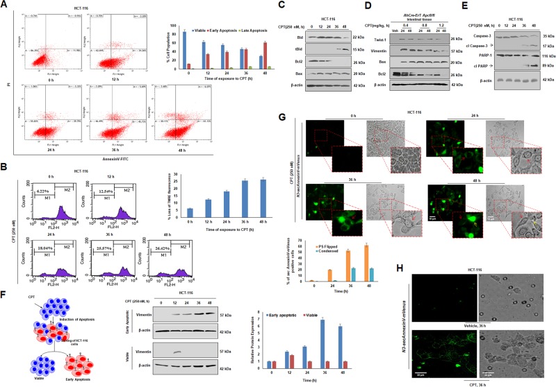
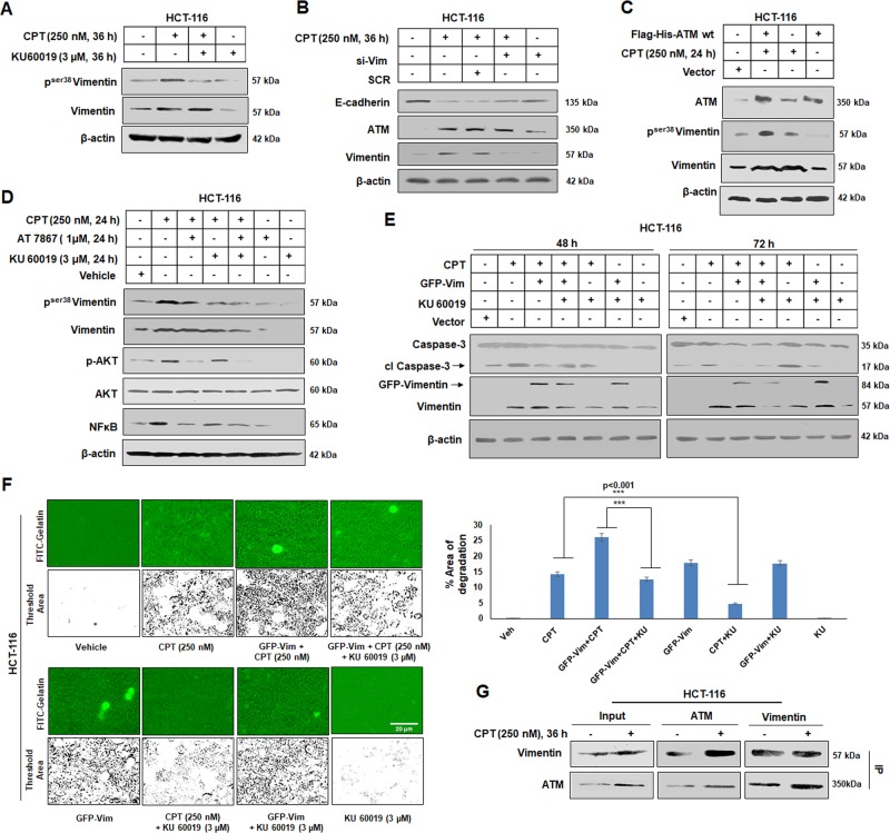
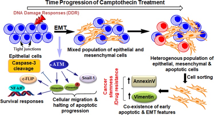
Epithelial to mesenchymal transitions (EMT) is a preparatory process for cancer cells to attain motility and further metastasis to distant sites. Majority of DNA damaging drugs have shown to develop EMT as one of the major mechanisms to attain drug resistance. Here we sought to understand the resistance/survival instincts of cancer cells during initial phase of drug treatment. We provide a tangible evidence of stimulation of EMT factors in Apc knockout colorectal carcinoma model. Our results implied that CPT-treated Apc knockout cohorts depicted increased pro-invasive and pro-survival factors (Vimentin/pser38Vimentin & NFκB). Moreover, by cell sorting experiment, we have observed the expression of Vimentin in early apoptotic cells (AnnexinV positive) from 36 to 48 h of CPT treatment. We also observed the expression of chimeric Sec-AnnexinV-mvenus protein in migrated cells on transwell membrane recapitulating signatures of early apoptosis. Notably, induction of Vimentin-mediated signaling (by CPT) delayed apoptosis progression in cells conferring survival responses by modulating the promoter activity of NFκB. Furthermore, our results unveiled a novel link between Vimentin and ATM signaling, orchestrated via binding interaction between Vimentin and ATM kinase. Finally, we observed a significant alteration of crypt-villus morphology upon combination of DIM (EMT inhibitor) with CPT nullified the background EMT signals thus improving the efficacy of the DNA damaging agent. Thus, our findings revealed a resistance strategy of cancer cells within a very initial period of drug treatment by activating EMT program, which hinders the cancer cells to achieve later phases of apoptosis thus increasing the chances of early migration.


Conflict of interest statement

The authors declare that they have no conflict of interest.

Figures

Fig. 1
Fig. 1. Activation of EMT and apoptosis in Camptothecin-mediated DNA damage response.
a HCT-116 and A549 cells were treated with 250 nM of CPT for 0, 12, 24, 36, and 48 h and checked for the expression of Vimentin, pser38Vimentin, Snail-1, ATM, β-catenin, and E-cadherin through western blot analysis. β-actin was used as loading control. b Immunocytochemistry was performed in HCT-116 cells treated with vehicle and CPT (250 nM) for 36 h for checking the expression of Vimentin, E-cadherin (green fluorescence). Nuclear staining was done with DAPI containing mounting media. Magnification of the images = ×63, c Analysis of the morphological features of HCT-116 cells through microscopic observation after exposure of cells to CPT for increasing time points (magnification = ×20). d Cells were treated with CPT (250 nM) for 24, 36, and 48 h along with vehicle and tested for their ability to degrade gelatin matrix and invadopodia formation through FITC-gelatin degradation assay. Blue stains indicate nuclear staining through DAPI mounting media. Images were taken at ×20 magnification. Bar graph showing the threshold area of degradation quantified through Image j analysis (n = 3, error bars ± s.d.). e, f Induced AhCre-ErT Apcfl/fl mice were treated with CPT (0.4, 0.8, and 12.2 mg/kg) for 24 and 48 h and the dissected intestinal tissues were sectioned and subjected to immunohistochemistry, to analyze the expression of Vimentin and pser38Vimentin. Images were taken at ×20 magnification
Fig. 2
Fig. 2. Co-existence of EMT and early-apoptosis in same population of Camptothecin treated cells.
a Cells were treated with CPT for 0, 12, 24, 36, and 48 h, tagged with AnnexinV-FITC, propidium iodide and analyzed through flow cytometry for onset of apoptosis. Bar graph showing quantification of cells in various phases of apoptosis (n = 3, error bars ± s.d.). b HCT-116 cells treated with CPT (250 nM) for 0, 12, 24, 36, and 48 h, and subjected to FACS analysis for identification of disruption of mitochondrial membrane potential by TMRE staining. Bar graph depicting loss of TMRE fluorescence indicating loss of mitochondrial membrane potential. c Western blot analysis was performed to study the expression pattern of Bid, Bcl2, Bax by exposing HCT-116 cells to 250 nM CPT for 0, 12, 24, 36, and 48 h. β-actin was used as loading control. d Western blot analysis of Apc floxed intestinal tissue treated with CPT (0.4, 0.8, and 1.2 mg/kg) for 24 and 48 h was performed to analyze the expression of Twist-1, Vimentin, Bax, and Bcl2. β-actin was used as loading control. e HCT-116 treated with 250 nM CPT for 0, 12, 24, 36, and 48 h was subjected to immunoblot analysis to check the expression of Caspase-3 and PARP-1. f HCT-116 cells were treated with CPT (250 nM) for 0, 12, 24, 36, and 48 h, stained with AnnexinV-FITC and PI and sorted using MoFlo cell sorter for the early apoptotic and viable cells (schematic). The sorted cells were then analyzed for the expression of Vimentin by immunoblot analysis; β-actin was used as loading control. Bar graph represents the densiometric analysis of expression of Vimentin in early apoptotic and viable population of sorted cells; (n = 3, error bars ± s.d.). g HCT-116 cells were transfected with N3-secAnnexinV-mVenus construct and then treated with 250 nM of CPT for 24, 36, and 48 h. Images were taken at ×20 magnification. Red arrows indicate cells with fluorescence bordering the cells and yellow arrows indicate cells having sufficient fluorescence throughout the cells. Bar graph depicting number of PS (Phosphatidyl serine) flipped cells vs condensed cells showing bright fluorescence. Cell counting was performed in Image j software (n = 3, error bars ± s.d). h HCT-116 cells transfected with N3-secAnnexinV-mVenus construct were subjected to transwell migration assay and treatment was given with 250 nM of CPT for 36 h. The migrating cells were processed further and observed under fluorescence microscope for detecting the fluorescence from chimeric AnnexinV-mVenus protein
Fig. 3
Fig. 3. Camptothecin-mediated activation of pro-proliferative responses.
a HCT-116 and A549 cells were treated with CPT (250 nM) for 24, 36, and 48 h along with their respective vehicle controls; cells were analyzed for their invasion capability through Boyden chamber assay system. Images were taken under an inverted microscope (Nikon Eclipse 200) at ×10 magnification. Bar graphs represent the average number of migrated cells per field (n = 3, error bars ± s.d.); ***p < 0.001, **p < 0.0011, p = 0.9125 and p = 0.2657. b Spheroids were prepared with HCT-116 cells embedded in ECM matrix by hanging drop method and treated with indicated concentration of CPT for given time points. Migration of cells from the core of the spheroids was observed (upper panel) under inverted microscope at ×20 magnification. The corresponding threshold images (lower panel) were prepared by using Image j software. c HCT-116 cells treated for indicated time pulse with 250 nM of CPT; whole cell lysates were analyzed for the expression of MMP-2 and MMP-9 protein through immunoblot analysis. d Conditional media was collected from the above experiment and subjected to gelatin zymography to examine the gelatinase activity of MMP-2 and MMP-9; bovine serum albumin (BSA) was taken as a loading control. e HCT-116 cells were treated with indicated concentration of CPT for 0, 12, 24, 36, and 48 h; the whole cell lysates were subjected to western blotting for the analysis of NFκB, p-AKT, AKT, Survivin, and c-FLIPS/L proteins. f CPT (0.4, 0.8, and 1.2 mg/kg) treatment was orally given to induced AhCre-ErT Apcfl/fl mice for 24 and 48 h and the harvested intestinal tissues were subjected to immunohistochemistry analysis of NFκB protein. g HCT-116 cells were treated with vehicle, CPT (250 nM) and Paclitaxel (25 nM). Paclitaxel treatment was administrated 12 h before the treatment of CPT. Cycle analysis was performed through flow cytometry and the bar graph represents percentage of cells in different phases of cell cycle, (n = 3, error bars ± s.d.). h Boyden chamber assay was performed to check the invasion ability of HCT-116 cells treated with above set of treatment conditions
Fig. 4
Fig. 4. Induction of Vimentin hinders the progression through apoptosis.
a HCT-116 cells were either treated with vehicle, CPT (250 nM) and CPT (250 nM) + DIM (25 µM) for indicated time points; whole cell lysates were prepared and subjected to western blot analysis of Vimentin and PARP-1 protein. b Cells were either transfected with vehicle, CPT (250 nM), SCR + CPT (250 nM), si-Vimentin plus CPT (250 nM) and si-Vimentin for 24 h; whole cell lysates were employed for western blot analysis of Vimentin, PARP-1, NFκB, and cFLIP. c Immunocytochemistry was performed for the indicated treatment conditions to examine the expression and trafficking of FAS ligand in HCT-116 cells. Images were taken by using Floid Cell Imaging Station; magnification ×20. d Similar set of treatment conditions were used to check the cells capability for gelatin matrix degradation through FITC-gelatin degradation assay. Imaging was performed using Floid Cell Imaging Station at ×20 magnification. The images were further analyzed and quantified for threshold area of degradation by Image j software and represented in bar graphs (n = 3, error bars ± s.d.); ***p < 0.0004. e HCT-116 cells were either treated with vehicle, CPT (250 nM), AT7867 (1 µM) and combination of both for 24 h; whole cell lysates were subjected to western blot analysis for the expression of pser38-Vimentin, Vimentin, p-AKT, AKT, NFκB, and cFLIPL. f HCT-116 cells were either transfected with vector, NFκB-luc alone and/or treated with NFκB-luc + CPT (250 nM), NFκB-luc + CPT (250 nM) + SCR, NFκB-luc + si-Vimentin + CPT (250 nM), NFκB-luc + si-Vimentin, and NFκB-luc + SCR in 96 well plates for 24 h; luciferase activity was measured using Dual-Glo Luciferase Assay system (Promega). Normalization was done with luciferase activity of vector (n = 3, error bars ± SD). ***p < 0.001
Fig. 5
Fig. 5. ATM-mediated control of Vimentin and its downstream responses.
a HCT-116 cells were treated with vehicle, CPT (250 nM), KU60019 (3 µM), KU60019 (3 µM) + 250 nM CPT for 36 h; whole cell lysates were prepared and western blotting was performed to determine the expression of pser38 Vimentin and total Vimentin. b Cells were either treated with Vehicle, CPT, SCR + CPT (250 nM), si-Vimentin + CPT (250 nM), and si-Vimentin for 36 h; western blotting was performed to analyze the expression of Vimentin, ATM, and E-cadherin. c HCT-116 cells treated with Vector, Flag-His-ATM wt + CPT (250 nM), CPT (250 nM) and Flag-His-ATM wt for 24 h and subjected to western blot analysis to evaluate the expression of ATM, pser38Vimentin, and total Vimentin. β-actin was used as loading control. d Western blot analysis was performed after HCT cells were treated with vehicle, CPT (250 nM), CPT (250 nM) + AT 7867 (1 μM), CPT (250 nM) + KU60019 (3 μM), CPT (250 nM) + AT 7867 (1 μM) + KU60019 (3 μM), AT 7867 (1 μM), and KU60019 (3 μM). The expression of pser38Vimentin, total Vimentin, p-AKT, AKT, NFκB were analyzed keeping β-actin as loading control. e HCT-116 cells were treated with Vector, CPT (250 nM), CPT (250 nM) + GFP-Vimentin, CPT (250 nM) + GFP-Vimentin + KU60019 (3 µM), CPT (250 nM) + KU60019 (3 µM), GFP-Vimentin + KU60019 (3 µM), and KU60019 (3 µM) for 48 and 72 h and subjected to western blot analysis to understand the expression of Caspase-3 and Vimentin, β-actin was used as loading control. f HCT-116 cells (upper panel) were seeded onto gelatin-FITC coated glass coverslips and treated with Vehicle, CPT (250 nM), CPT (250 nM) + GFP-Vimentin, CPT (250 nM) + GFP-Vimentin + KU60019 (3 µM); lower panel: GFP-Vimentin, CPT (250 nM) + KU60019 (3 µM), GFP-Vimentin + KU60019 (3 µM), and KU60019 (3 µM) for 36 h and the slides were observed in Floid imaging station at ×20 magnification. Bar graph describes the percentage threshold area of degradation analyzed by Image j software. (n= 3, error bars ± SD); ***p < 0.001. g Immunoprecipitation analysis of ATM with Vimentin after HCT-116 cells were treated with Vehicle and CPT (250 nM) for 36 h
Fig. 6
Fig. 6. DIM mediated abrogation of CPT-induced carcinogenesis and EMT.
a HCT-116 cells seeded onto FITC-gelatin coated coverslips were treated with Vehicle, 250 nM CPT, 250 nM CPT + 25 μM DIM, and 25 μM DIM for 36 h and the slides were observed in fluorescence microscope at ×20 magnification. b Hematoxylin and Eosin staining of intestinal tissue obtained from induced AhCre-ErT Apcfl/fl treated with Vehicle, 0.4 mg/kg CPT, 0.4 mg/kg + 20 mg/kg DIM, and 20 mg/kg DIM for 48 h. Images were taken at ×20 magnification. c Immunohistochemistry analysis of Vimentin and pser38 Vimentin was performed on intestinal tissues obtained from AhCre-ErT Apcfl/fl mice treated with Vehicle, 0.4 mg/kg CPT, 0.4 mg/kg + 20 mg/kg DIM, and 20 mg/kg DIM for 48 h. Images were acquired in ×20 magnification
Fig. 7
Fig. 7
Schematic representation of the proposed mechanism of action of the study depicting the co-existence of early-apoptotic and EMT features with time progression of Camptothecin treatment

Similar articles

Cited by

References

    1. Kalluri R, Weinberg RA. The basics of epithelial-mesenchymal transition. J. Clin. Investig. 2009;119:1420–1428. doi: 10.1172/JCI39104. - DOI - PMC - PubMed
    1. Yilmaz M, Christofori G. EMT, the cytoskeleton, and cancer cell invasion. Cancer Metastasis Rev. 2009;28:15–33. doi: 10.1007/s10555-008-9169-0. - DOI - PubMed
    1. Satelli A, Li S. Vimentin in cancer and its potential as a molecular target for cancer therapy. Cell. Mol. Life Sci. 2011;68:3033–3046. doi: 10.1007/s00018-011-0735-1. - DOI - PMC - PubMed
    1. Arumugam T, et al. Epithelial to mesenchymal transition contributes to drug resistance in pancreatic cancer. Cancer Res. 2009;69:5820–5828. doi: 10.1158/0008-5472.CAN-08-2819. - DOI - PMC - PubMed
    1. Shibue T, Weinberg RA. EMT, CSCs, and drug resistance: the mechanistic link and clinical implications. Nat. Rev. Clin. Oncol. 2017;14:611. doi: 10.1038/nrclinonc.2017.44. - DOI - PMC - PubMed

Publication types

MeSH terms