

The purinergic receptor antagonist oxidized adenosine triphosphate suppresses immune-mediated corneal allograft rejection
- PMID: 31197223
- PMCID: PMC6565802
- DOI: 10.1038/s41598-019-44973-y
The purinergic receptor antagonist oxidized adenosine triphosphate suppresses immune-mediated corneal allograft rejection
Abstract
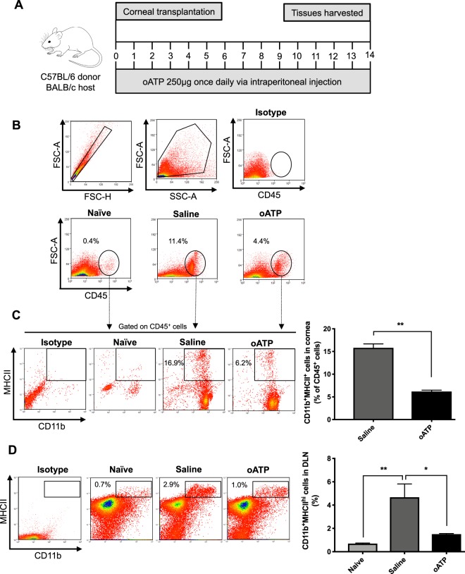
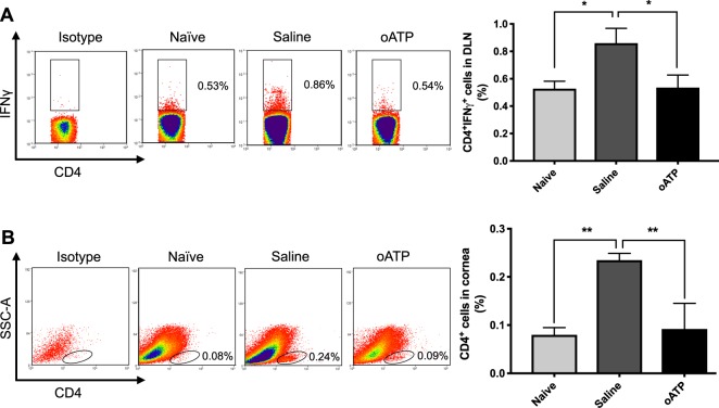
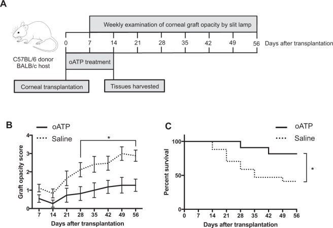
Adenosine triphosphate (ATP) is released into the extracellular environment during transplantation, and acts via purinergic receptors to amplify the alloimmune response. Here, using a well-established murine model of allogeneic corneal transplantation, we investigated the immunomodulatory mechanisms of the purinergic receptor antagonist oxidized ATP (oATP). Corneal transplantation was performed using C57BL/6 donors and BALB/c hosts. oATP or sterile saline was administered via intraperitoneal injection for 2 weeks postoperatively. Frequencies of CD45+ leukocytes, CD11b+MHCII+ antigen presenting cells (APCs), CD4+IFN-γ+ effector Th1 cells and CD4+Foxp3+ regulatory T cells (Tregs) were evaluated by flow cytometry. Slit-lamp microscopy was performed weekly for 8 weeks to evaluate graft opacity and determine transplant rejection. Treatment with oATP was shown to significantly reduce graft infiltration of CD45+ leukocytes, decrease APC maturation and suppress effector Th1 cell generation relative to saline-treated control. No difference in Treg frequencies or Foxp3 expression was observed between the oATP-treated and control groups. Finally, oATP treatment was shown to reduce graft opacity and increase graft survival. This report demonstrates that oATP limits the alloimmune response by regulating APC maturation and suppressing the generation of alloreactive Th1 immunity.
Conflict of interest statement
The authors declare no competing interests.
Figures

Similar articles
-
Inhibition of the purinergic pathway prolongs mouse lung allograft survival.Am J Respir Cell Mol Biol. 2014 Aug;51(2):300-10. doi: 10.1165/rcmb.2013-0362OC. Am J Respir Cell Mol Biol. 2014. PMID: 24661183
-
IFN-γ blocks CD4+CD25+ Tregs and abolishes immune privilege of minor histocompatibility mismatched corneal allografts.Am J Transplant. 2013 Dec;13(12):3076-84. doi: 10.1111/ajt.12466. Epub 2013 Oct 1. Am J Transplant. 2013. PMID: 24119152 Free PMC article.
-
In Vivo Expansion of Regulatory T Cells by Low-Dose Interleukin-2 Treatment Increases Allograft Survival in Corneal Transplantation.Transplantation. 2016 Mar;100(3):525-32. doi: 10.1097/TP.0000000000001044. Transplantation. 2016. PMID: 26881788 Free PMC article.
-
Local Delivery of Regulatory T Cells Promotes Corneal Allograft Survival.Transplantation. 2019 Jan;103(1):182-190. doi: 10.1097/TP.0000000000002442. Transplantation. 2019. PMID: 30247445 Free PMC article.
-
Effect of the purinergic inhibitor oxidized ATP in a model of islet allograft rejection.Diabetes. 2013 May;62(5):1665-75. doi: 10.2337/db12-0242. Epub 2013 Jan 11. Diabetes. 2013. PMID: 23315496 Free PMC article.
Cited by
-
Non-immune and immune functions of interleukin-36γ suppress epithelial repair at the ocular surface.FASEB J. 2022 Aug;36(8):10.1096/fj.202200174RR. doi: 10.1096/fj.202200174RR. FASEB J. 2022. PMID: 35781326 Free PMC article.
-
Lentivirus-mediated IL-10-expressing Bone Marrow Mesenchymal Stem Cells promote corneal allograft survival via upregulating lncRNA 003946 in a rat model of corneal allograft rejection.Theranostics. 2020 Jul 9;10(18):8446-8467. doi: 10.7150/thno.31711. eCollection 2020. Theranostics. 2020. PMID: 32724480 Free PMC article.
-
When rubber meets the road: how innate features of adaptive immune cells play critical roles in transplant alloimmunity.Curr Opin Organ Transplant. 2019 Dec;24(6):659-663. doi: 10.1097/MOT.0000000000000706. Curr Opin Organ Transplant. 2019. PMID: 31577597 Free PMC article. Review.
References
Publication types
MeSH terms
Substances
Grants and funding
LinkOut - more resources
Full Text Sources
Molecular Biology Databases
Research Materials
Miscellaneous
