. 2019 Jun 13;9(1):8625.
doi: 10.1038/s41598-019-44866-0.

Homeostatic pruning and activity of epidermal nerves are dysregulated in barrier-impaired skin during chronic itch development

Affiliations

Homeostatic pruning and activity of epidermal nerves are dysregulated in barrier-impaired skin during chronic itch development

Sonoko Takahashi et al. Sci Rep. .

Abstract

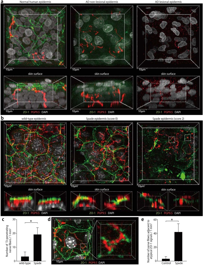
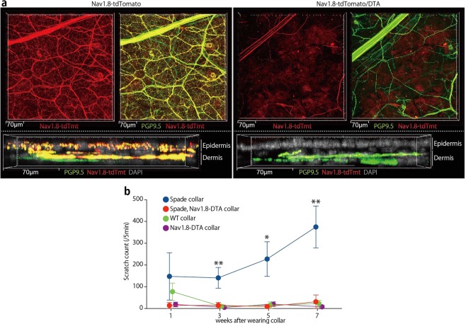
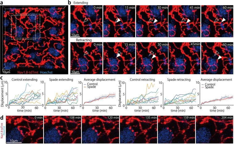
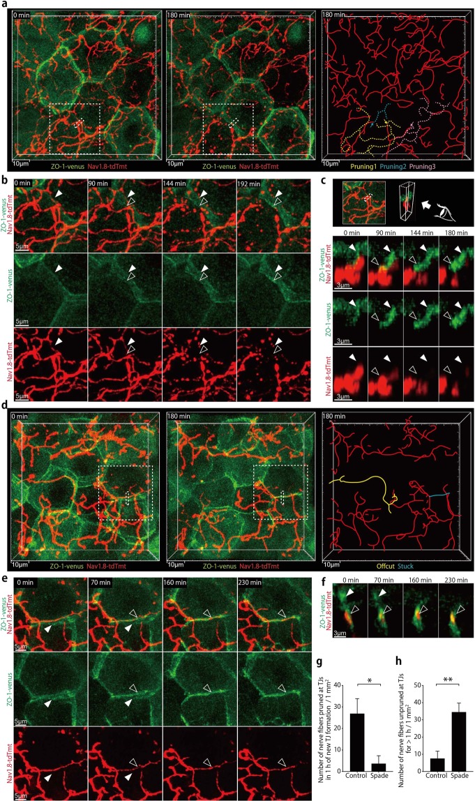
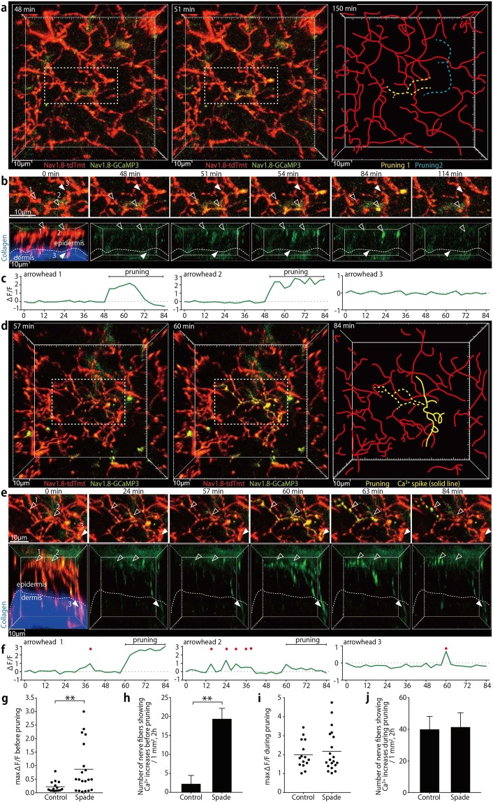
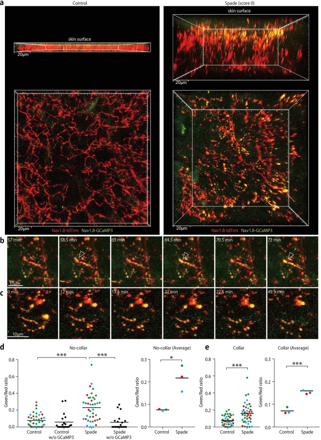
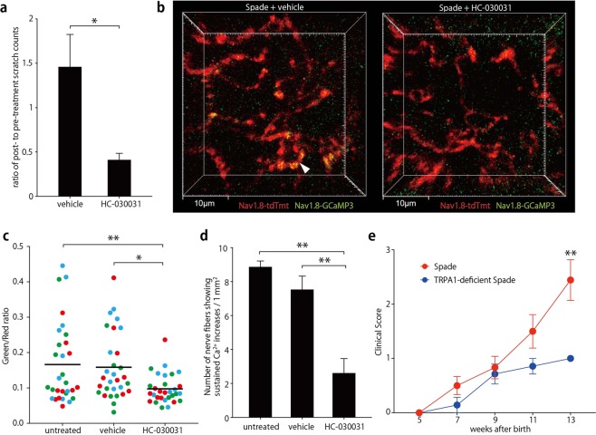
The epidermal barrier is thought to protect sensory nerves from overexposure to environmental stimuli, and barrier impairment leads to pathological conditions associated with itch, such as atopic dermatitis (AD). However, it is not known how the epidermal barrier continuously protects nerves for the sensory homeostasis during turnover of the epidermis. Here we show that epidermal nerves are contained underneath keratinocyte tight junctions (TJs) in normal human and mouse skin, but not in human AD samples or mouse models of chronic itch caused by epidermal barrier impairment. By intravital imaging of the mouse skin, we found that epidermal nerve endings were frequently extended and retracted, and occasionally underwent local pruning. Importantly, the epidermal nerve pruning took place rapidly at intersections with newly forming TJs in the normal skin, whereas this process was disturbed during chronic itch development. Furthermore, aberrant Ca2+ increases in epidermal nerves were induced in association with the disturbed pruning. Finally, TRPA1 inhibition suppressed aberrant Ca2+ increases in epidermal nerves and itch. These results suggest that epidermal nerve endings are pruned through interactions with keratinocytes to stay below the TJ barrier, and that disruption of this mechanism may lead to aberrant activation of epidermal nerves and pathological itch.


Conflict of interest statement

The authors declare no competing interests.

Figures

Figure 1
Figure 1
Epidermal nerve endings are contained under TJs in the normal human and mouse skin but not in the human AD or Spade mouse skin. (a) Whole-mount confocal fluorescence images of the healthy human epidermis and the epidermis of AD patients. PGP9.5+ nerve fibers and TJs visualized as ZO-1 localization are shown in vertical (upper, 44 μm projection depth) and horizontal (lower, 61.5 μm projection depth) projection images. See also Supplementary Movie 1. (b) Whole-mount confocal fluorescence images of the ear epidermis of wild-type and Spade mice without (score 0) or with lesions (score 2). The upper images are the vertical projection (12.5 and 21–22 μm projection depth for the wild-type and Spade mice, respectively). The lower images show horizontal views of the dashed square regions from the right side in the vertical projection images. See also Supplementary Movie 2. (c) The number of nerve fibers penetrating TJs, normalized by the epidermis area. (d) Whole-mount confocal fluorescence images of the SG of the Spade epidermis showing atypical ZO-1 accumulations around a nerve fiber. (e) the area-normalized number of nerve fibers surrounded by atypical ZO-1 accumulations. The data are shown as the mean ± s.e.m. in c and e (WT: n = 9, Spade: n = 20). *p < 0.05.
Figure 2
Figure 2
Epidermal nerve fibers are of Nav1.8-tdTomato+ sensory neurons, which are necessary for itch in Spade mice. (a) Whole-mount confocal fluorescence images of the ear skin from Nav1.8-tdTomato and Nav1.8-tdTomato/DTA mice. Nav1.8-tdTomato+ PGP9.5+ nerves and Nav1.8-tdTomato PGP9.5+ nerves are shown in vertical (top, 33 μm projection depth) and horizontal (bottom, 28.4 μm projection depth) projection images. See also Supplementary Fig. 2. (b) The number of scratching strokes by the hind paws of the indicated mice after wearing Elizabethan collars. The data are shown as the mean ± s.e.m. (n = 3–4). *p < 0.05, **p < 0.01.
Figure 3
Figure 3
Epidermal nerve endings are frequently extended, retracted, and occasionally pruned. (a) An intravital multiphoton image (16 μm projection depth) of nerves in the SG of an Nav1.8-tdTomato mouse. (b) Extending and retracting nerve endings in the dashed rectangles in A. Open and filled arrowheads indicate the positions of nerve endings at 0 min and each later time point, respectively. See also Supplementary Movie 3. (c) Displacement of extending and retracting nerve endings. In the “Average displacement” graphs, data are shown as the mean ± s.e.m. (n = 10). (d) Time-lapse images (14 μm projection depth) of epidermal nerve pruning. See also Supplementary Movie 4.
Figure 4
Figure 4
Epidermal nerve endings are rapidly pruned at newly formed TJs in the normal skin but not in the Spade skin around the disease onset. (a) In vivo time-lapse confocal images (9.5 μm projection depth) of epidermal nerves and TJs in an Nav1.8-tdTomato ZO-1-Venus mouse (far-left and middle panels). The far-right drawing indicates the locations of nerve branches that underwent fragmentation (yellow, blue, and pink dashed lines). The yellow and blue ones were pruned at the newly forming TJ. The pink one was pruned at a non-TJ site (see Supplementary Fig. 4). The sites of nerve pruning are indicated by the arrows. See also Supplementary Movie 5. (b) Time-lapse images of the new TJ formation and nerve pruning in the region demarcated by the dashed squares in (a) See also Supplementary Movie 6. (c) Time-lapse, horizontal views of the region demarcated by the dashed rectangles in a (3.1 μm projection depth). (d) An intravital confocal image of the ear epidermis from a Spade Nav1.8-tdTomato ZO-1-Venus mouse (20 μm projection depth). See also Supplementary Movie 7. (e) Time-lapse images of the region demarcated by the dashed square in (d) (11 μm projection depth). See also Supplementary Movie 8. (f) Time-lapse, horizontal views of the region demarcated by the dashed rectangle in (d) (3.7 μm projection depth). Open and filled arrowheads indicate the new and old TJs, respectively. (g) The area-normalized number of nerve fibers pruned at new TJs in an hour of the new TJ formation. (h) The area-normalized number of nerve fibers unpruned at TJs without being pruned for more than an hour. The data are shown as the mean ± s.e.m. (WT: n = 4, Spade: n = 3). *p < 0.05, **p < 0.01.
Figure 5
Figure 5
Aberrant nerve Ca2+ increases are observed in association with epidermal nerve pruning in Spade mice around the disease onset. (a) In vivo time-lapse multiphoton images (19 μm projection depth) of epidermal and dermal nerves in an Nav1.8-tdTomato/GCaMP3 mouse (far-left and middle). The dashed rectangles indicate the region shown in. (b) The far-right drawing indicates the locations of nerve branches that underwent fragmentation (yellow and blue dashed lines). (b) Local Ca2+ increases in epidermal nerve branches that underwent pruning (open arrowheads). Filled arrowheads indicate one of dermal fibers in which Ca2+ increases were not observed. The upper sequence shows vertical projection views. In the lower sequence of horizontal views, tdTomato and dermal second harmonic signals are shown only in the 0-min time-point image. See also Supplementary Movie 12. (c) Relative fluorescence intensity changes (∆F/F) in the GCaMP3 channel in 23 μm3 regions indicated by the arrowheads in (b). (d) In vivo time-lapse multiphoton images (47 μm projection depth) of epidermal and dermal nerves in a Spade Nav1.8-tdTomato/GCaMP3 mouse (far-left and middle). The dashed rectangles indicate the region shown in (e) The yellow dashed line in the far-right drawing indicates an epidermal nerve branch that showed repetitive Ca2+ increases and underwent fragmentation. The yellow solid line indicates a nerve part that showed a Ca2+ spike without undergoing fragmentation. (e) Repetitive Ca2+ increases in an epidermal nerve branch that underwent pruning (open arrowheads) and a Ca2+ spike that reached a dermal nerve fiber (filled arrowheads). The upper sequence shows vertical projection views. In the lower sequence of horizontal views, tdTomato and dermal second harmonic signals are shown only in the 0-min time-point image. See also Supplementary Movie 13. (f) Relative fluorescence intensity changes (∆F/F) in the GCaMP3 channel in 23 μm3 cubic regions indicated by the arrowheads in (e) Red dots indicate time points when ∆F/F was > 0.5. (g) Maximum fluorescence intensity changes in the GCaMP3 channel before pruning. (h) The area- and time-normalized number of nerve fibers showing Ca2+ increases (max ∆F/F >0.5) before pruning. (i) Maximum fluorescence intensity changes in the GCaMP3 channel during pruning. (j) The area- and time-normalized number of nerve fibers showing Ca2+ increases (max ∆F/F >0.5) during pruning. The data are shown as the mean ± s.e.m. in (h) and (j) (WT: n = 3, Spade: n = 3). *p < 0.01.
Figure 6
Figure 6
Sustained Ca2+ increases are induced in epidermal nerve fibers in the itching skin of Spade mice independently of scratching. (a) Intravital multiphoton images of the ear epidermis from control Nav1.8-tdTomato/GCaMP3 and Spade (score 0) Nav1.8-tdTomato/GCaMP3 mice. Upper and lower images are horizontal and vertical projection views, respectively. See also Supplementary Movie 14. (b,c) Time-lapse images of an epidermal nerve fiber showing repetitive Ca2+ increases (arrowheads in b) and fragmented nerve fibers with increased Ca2+ (c) in the Spade (score 0) mouse. See also Supplementary Movie 15. (d) Fluorescence intensity ratio of GCaMP3 to tdTomato. Shown are the data at the individual positions in the epidermal nerve fibers (left) and the averaged data from each experiment (right). Different colors of the data points indicate different mice in each group. Negative control data from Nav1.8-tdTomato and Spade (score 0) Nav1.8-tdTomato mice without the GCaMP3 allele are also shown in the left graph. (e) Fluorescence intensity ratio of GCaMP3 to tdTomato in epidermal nerves of mice that had worn Elizabethan collars. *p < 0.05, ***p < 0.001.
Figure 7
Figure 7
Inhibition of TRPA1 attenuates epidermal nerve Ca2+ increases and itch in Spade mice. (a) Ratio of ear scratch counts after to before topical treatment of the Spade ear skin with vehicle or HC-030031. (b) Intravital multiphoton images (16 μm projection depth) of the ear epidermis of Spade Nav1.8-tdTomato/GCaMP3 mice after treatment with vehicle or HC-030031. (c) Fluorescence intensity ratio of GCaMP3 to tdTomato. Shown are the data at the individual positions in the epidermal nerve fibers. Different colors of the data points indicate different mice in each group. (d) The area-normalized number of nerve fibers showing Ca2+ increases (fluorescence intensity ratio of GCaMP to tdTomato >0.15) sustained for at least 30 min. (e) Longitudinal analysis of the clinical score of the indicated mouse groups. The data are shown as the mean ± s.e.m. in (a) (n = 5), (d) (n = 3), and (e) (Spade: n = 7–12, TRPA1-deficient Spade: n = 5–7). *p < 0.05, **p < 0.01.

Similar articles

Cited by

References

    1. Kubo A, Nagao K, Amagai M. Epidermal barrier dysfunction and cutaneous sensitization in atopic diseases. J Clin Invest. 2012;122:440–447. doi: 10.1172/JCI57416. - DOI - PMC - PubMed
    1. Misery L, Loser K, Stander S. Sensitive skin. J Eur Acad Dermatol Venereol. 2016;30(Suppl 1):2–8. doi: 10.1111/jdv.13532. - DOI - PubMed
    1. Azimi E, Xia J, Lerner EA. Peripheral Mechanisms of Itch. Curr Probl Dermatol. 2016;50:18–23. doi: 10.1159/000446012. - DOI - PMC - PubMed
    1. De Benedetto A, et al. Tight junction defects in patients with atopic dermatitis. J Allergy Clin Immunol. 2011;127:773–786 e771–777. doi: 10.1016/j.jaci.2010.10.018. - DOI - PMC - PubMed
    1. Yu HS, et al. Claudin-1 polymorphism modifies the effect of mold exposure on the development of atopic dermatitis and production of IgE. J Allergy Clin Immunol. 2015;135:827–830 e825. doi: 10.1016/j.jaci.2014.10.040. - DOI - PubMed

Publication types