. 2019 Jul 9;58(27):2953-2966.
doi: 10.1021/acs.biochem.9b00401. Epub 2019 Jun 25.

Structure and Function of the γ-Secretase Complex

Affiliations

Structure and Function of the γ-Secretase Complex

Michael S Wolfe. Biochemistry. .

Abstract

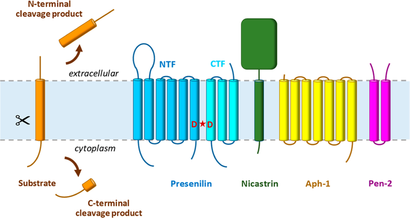
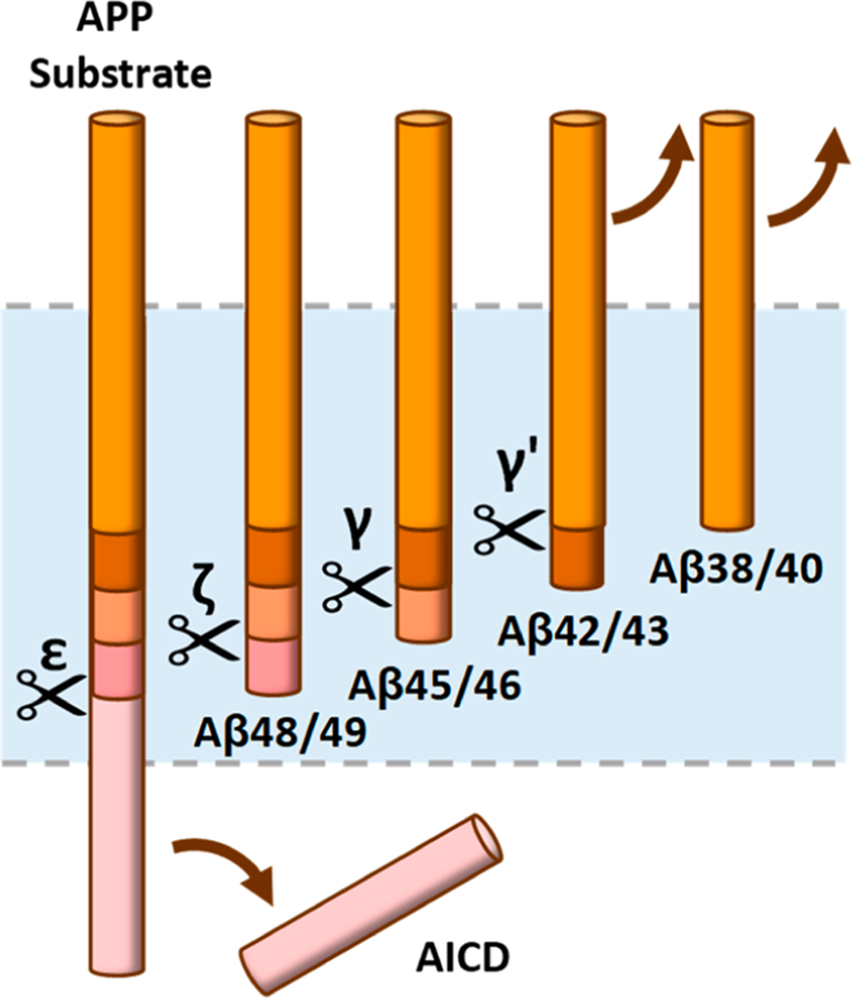
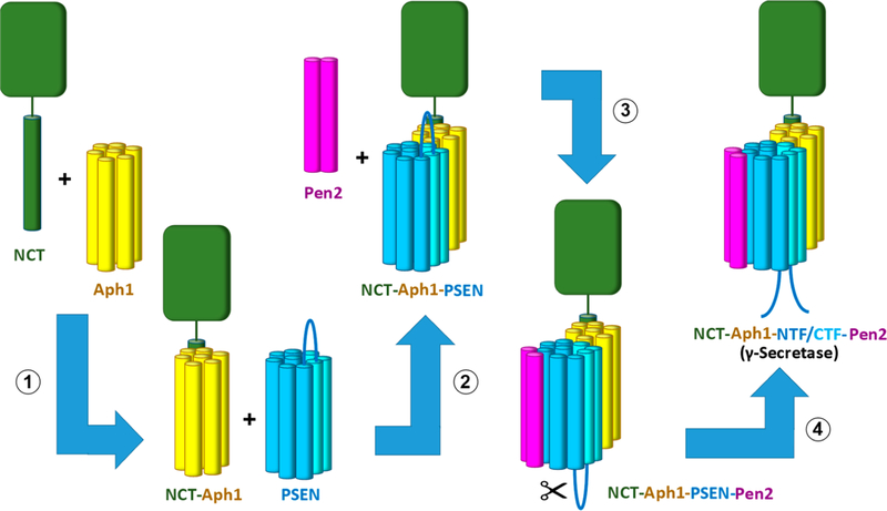
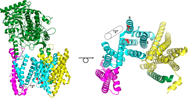
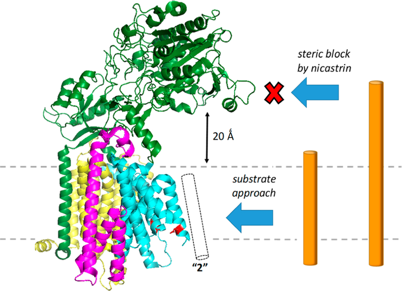
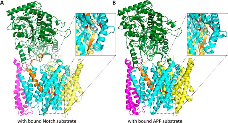
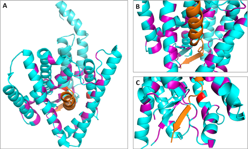
γ-Secretase is a membrane-embedded protease complex, with presenilin as the catalytic component containing two transmembrane aspartates in the active site. With more than 90 known substrates, the γ-secretase complex is considered "the proteasome of the membrane", with central roles in biology and medicine. The protease carries out hydrolysis within the lipid bilayer to cleave the transmembrane domain of the substrate multiple times before releasing secreted products. For many years, elucidation of γ-secretase structure and function largely relied on small-molecule probes and mutagenesis. Recently, however, advances in cryo-electron microscopy have led to the first detailed structures of the protease complex. Two new reports of structures of γ-secretase bound to membrane protein substrates provide great insight into the nature of substrate recognition and how Alzheimer's disease-causing mutations in presenilin might alter substrate binding and processing. These new structures offer a powerful platform for elucidating enzyme mechanisms, deciphering effects of disease-causing mutations, and advancing Alzheimer's disease drug discovery.


Conflict of interest statement

The author declares no competing financial interest.

Figures

Figure 1
Figure 1
Intramembrane proteolysis by the γ-secretase complex. γ-Secretase cleaves >90 different type I integral membrane proteins after ectodomain release by membrane-tethered sheddases. The protease complex carries out proteolysis in the transmembrane domain of these substrates to secrete N-terminal cleavage products into the extracellular milieu and release C-terminal cleavage products into the cytoplasm. The γ-secretase complex is composed of four different multipass membrane proteins, with presenilin as the catalytic component containing two transmembrane aspartates in the active site. Upon assembly with the other subunits (nicastrin, Aph-1, and Pen-2), presenilin undergoes autoproteolysis into an N-terminal fragment (NTF) and C-terminal fragment (CTF) to form active γ-secretase.
Figure 2
Figure 2
Processive proteolysis by γ-secretase. The protease first carries out endoproteolysis (ε) near the cytosolic end of the TMD of the APP substrate, with release of the intracellular domain (AICD). This is followed by carboxypeptidase cleavages (ζ, γ, and γ′) of the remaining long Aβ peptides, in intervals of roughly three amino acids, to secreted peptides that are 38–43 residues in length. There are two general pathways for Aβ generation: Aβ49 → Aβ46 → Aβ43 → Aβ40 and Aβ48 → Aβ45 → Aβ42 → Aβ38.
Figure 3
Figure 3
General mechanism of substrate recognition and processing by γ-secretase. Helical substrate TMD initially binds to presenilin at a docking exosite (where the helix is bound in ES1). This is followed by movement of the substrate in whole or in part (as shown) into the active site, with unwinding of the substrate TMD to set up the transition state (ES1*) for ε cleavage. After release of the intracellular domain, the remaining enzyme-bound product (ES2) again unwinds into the active site for carboxypeptidase cleavage (ES2*). Three pockets in the enzyme active site dictate trimming every three amino acids. Successive carboxypeptidase trimming occurs until short peptide products are released. The inset shows pockets S1′ and S3′ are relatively large and can accommodate bulky aromatic amino acids such as Phe, while S2′ is smaller and cannot accommodate Phe.
Figure 4
Figure 4
Assembly and activation of the γ-secretase complex. Nicastrin and Aph-1 form a stable subcomplex (step 1), followed by addition of presenilin (step 2). Interaction of Pen-2 with TMD4 of presenilin (step 3) triggers autoproteolysis of presenilin into NTF and CTF subunits (step 4) to form γ-secretase capable of cleaving substrates.
Figure 5
Figure 5
First detailed structure of the γ-secretase complex determined by cryo-EM, single-particle analysis, and image reconstruction, with the revised assignment of the presenilin TMDs: green for nicastrin, yellow for Aph-1, magenta for Pen-2, cyan (NTF) and aquamarine (CTF) for PS1, TMDs numbered, and red for catalytic aspartates. TMD2 was not resolved but was modeled using the crystal structure of an archaeal presenilin homologue. This TMD (“2”) is depicted as the dashed outlined cylinder. Looking only at the 19 TMDs of the complex from the cytosolic side (right) illustrates the horseshoe-shaped arrangement, with the active site that can be approached by the substrate from the convex side and flexible TMD2 as the apparent gate. Protein Data Bank entry 5A63.
Figure 6
Figure 6
Nicastrin serves as a gatekeeper of the γ-secretase complex. The large ectodomain of nicastrin juts out over the entryway to the active site, sterically preventing access of substrates with ectodomains that are >20 Å long. Conformationally flexible TMD2 of presenilin is believed to be the gate, allowing substrate entry within the membrane. Protein Data Bank entry 5A63.
Figure 7
Figure 7
Structures of γ-secretase determined using image classification and masked refinement. (A) Class 1 structure of the apoenzyme, with resolution of TMD2 of presenilin and the presence of an unidentified helical density (orange) in presenilin, thought to be a composite of multiple substrates co-purified with the protease complex. This density appears to unwind and disappear as it approaches the active site (catalytic aspartates colored red). TMDs of presenilin are numbered, and loop 1 (L1) is indicated. Protein Data Bank entry 5FN3. (B) Structure of the enzyme bound to small peptide inhibitor DAPT. The protease complex assumes a conformation similar to the class 1 structure but without the unidentified density. A cavity formed where this density would be is indicated by the large purple arrow. Other parts of presenilin become visible or rearrange, most notably the cytoplasmic side of TMD6, which becomes kinked and extended. DAPT, which was only partly resolved binding near the active site, is not shown for the sake of clarity. Protein Data Bank entry 5FN2.
Figure 8
Figure 8
Structures of γ-secretase bound to substrates. (A) γ-Secretase bound to Notch1-derived substrate. Protein Data Bank entry 6IDF. (B) γ-Secretase bound to the APP-derived substrate. Protein Data Bank entry 6IYC. In both structures, the substrate is located inside PSEN1 with the same basic arrangement as that seen in the class 1 apoenzyme with the unidentified density (Figure 7A). Both substrates are now resolved as they unwind and enter the catalytically disabled active site (PSEN1 TMD7 Asp mutated to Ala). Insets show the substrate TMD assumes a β-strand conformation near the cytoplasmic side, stabilized by a β-strand in PSEN1 TMD7, which is in turn stabilized by a short β-strand at the C-terminus of PSEN1 NTF.
Figure 9
Figure 9
Familial Alzheimer’s disease (FAD) hot spots in PSEN1. (A) Sixty-one residues that are each mutated to two or more different amino acids in FAD are colored purple. Only PSEN1 and the APP substrate are shown for the sake of clarity. Note that many of these mutation sites appear to face or interact with the substrate. (B) Mutation sites that interact with the helical region of the substrate. (C) Mutation sites that interact, directly or indirectly, with the substrate in the active site and with the substrate β-strand. Protein Data Bank entry 6IYC.

Similar articles

Cited by

References

    1. Wolfe MS, De Los Angeles J, Miller DD, Xia W, and Selkoe DJ (1999) Are presenilins intramembrane-cleaving proteases? Implications for the molecular mechanism of Alzheimer’s disease. Biochemistry 38, 11223–11230. - PubMed
    1. Wolfe MS, and Kopan R (2004) Intramembrane proteolysis: theme and variations. Science 305, 1119–1123. - PubMed
    1. Hooper NM, and Lendeckel U, Eds. (2007) Intramembrane-Cleaving Proteases (I-CLiPs), Vol. 6, Springer, Dordrecht, The Netherlands.
    1. Freeman M (2014) The rhomboid-like superfamily: molecular mechanisms and biological roles. Annu. Rev. Cell Dev. Biol 30, 235–254. - PubMed
    1. Rawson RB (2013) The site-2 protease. Biochim. Biophys. Acta, Biomembr 1828, 2801–2807. - PubMed

Publication types

MeSH terms