

In Vivo Morphological Changes of the Femoropopliteal Arteries due to Knee Flexion After Endovascular Treatment of Popliteal Aneurysm
- PMID: 31198084
- PMCID: PMC7563842
- DOI: 10.1177/1526602819855441
In Vivo Morphological Changes of the Femoropopliteal Arteries due to Knee Flexion After Endovascular Treatment of Popliteal Aneurysm
Abstract
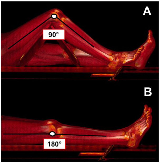
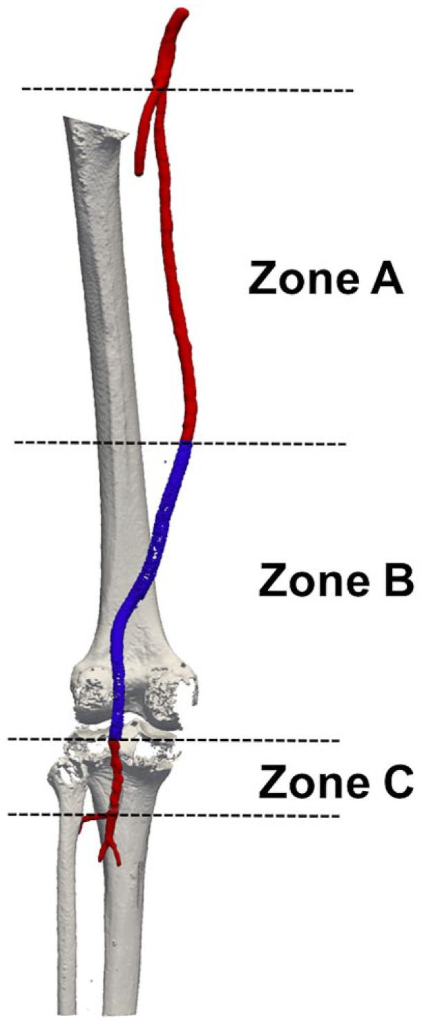
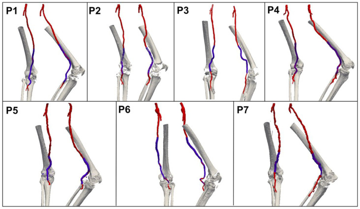
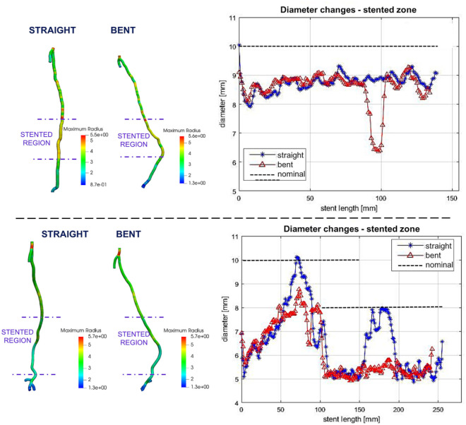
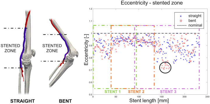
Purpose: To evaluate morphological changes of the femoropopliteal (FP) arteries due to limb flexion in patients undergoing endovascular treatment of popliteal artery aneurysms (PAAs). Materials and Methods: Seven male patients (mean age 68 years) underwent endovascular treatment of PAA with a Viabahn stent-graft between January 2013 and December 2017. During follow-up, one contrast-enhanced computed tomography angiography (CTA) scan of the lower limbs was acquired for each recruited patient. A standardized CTA protocol for acquisitions in both straight-leg and bent-leg positions was used to visualize changes in artery shape due to limb flexion. Three-dimensional reconstruction of the FP segment was performed to compute mean diameter and eccentricity of the vascular lumen and to measure length, tortuosity, and curvature of the vessel centerline in 3 arterial zones: (A) between the origin of the superficial femoral artery and the proximal end of the stent-graft, (B) within the stent-graft, and (C) from the distal end of the stent-graft to the origin of the anterior tibial artery. Results: After limb flexion, all zones of the FP segment foreshortened: 6% in zone A (p=0.001), 4% in zone B (p=0.001), and 8% in zone C (p=0.07), which was the shortest (mean 4.5±3.6 cm compared with 23.8±5.7 cm in zone A and 23.6±7.4 cm in zone B). Tortuosity increased in zone A (mean 0.03 to 0.05, p=0.03), in zone B (0.06 to 0.15, p=0.005), and in zone C (0.027 to 0.031, p=0.1). Mean curvature increased 15% (p=0.05) in zone A, 27% (p=0.005) in zone B, and 95% (p=0.06) in zone C. In all zones, the mean artery diameter and eccentricity were not significantly affected by limb flexion. Conclusion: Limb flexion induces vessel foreshortening and increases mean curvature and tortuosity of the FP segment both within and outside the area of the stent-graft.
Keywords: endovascular treatment; femoropopliteal segment; foreshortening; limb flexion; medical image analysis; morphology; popliteal artery aneurysm; stent-graft; superficial femoral artery; tortuosity.
Conflict of interest statement
Figures
References
-
- Tuveson V, Löfdahl HE, Hultgren R. Patients with abdominal aortic aneurysm have a high prevalence of popliteal artery aneurysms. Vasc Med. 2016;21:369–375. - PubMed
-
- Pulli R, Dorigo W, Castelli P, et al. A multicentric experience with open surgical repair and endovascular exclusion of popliteal artery aneurysms. Eur J Vasc Endovasc Surg. 2013;45:357–363. - PubMed
-
- Gerasimidis T, Sfyroeras G, Papazoglou K, et al. Endovascular treatment of popliteal artery aneurysms. Eur J Vasc Endovasc Surg. 2003;26:506–511. - PubMed
-
- Wrede A, Wiberg F, Acosta S. Increasing the elective endovascular to open repair ratio of popliteal artery aneurysm. Vasc Endovasc Surg. 2018;52:115–123. - PubMed
-
- Lovegrove RE, Javid M, Magee TR. Endovascular and open approaches to non-thrombosed popliteal aneurysm repair: a meta-analysis. Eur J Vasc Endovasc Surg. 2008;36:96–100. - PubMed
Publication types
MeSH terms
Grants and funding
LinkOut - more resources
Full Text Sources
Medical
