. 2019 May 6:2019:3259295.
doi: 10.1155/2019/3259295. eCollection 2019.

MMP-9 Contributes to Dendritic Spine Remodeling Following Traumatic Brain Injury

Affiliations

MMP-9 Contributes to Dendritic Spine Remodeling Following Traumatic Brain Injury

Barbara Pijet et al. Neural Plast. .

Abstract

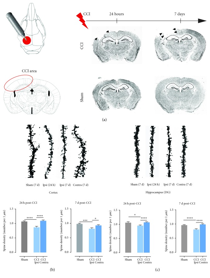
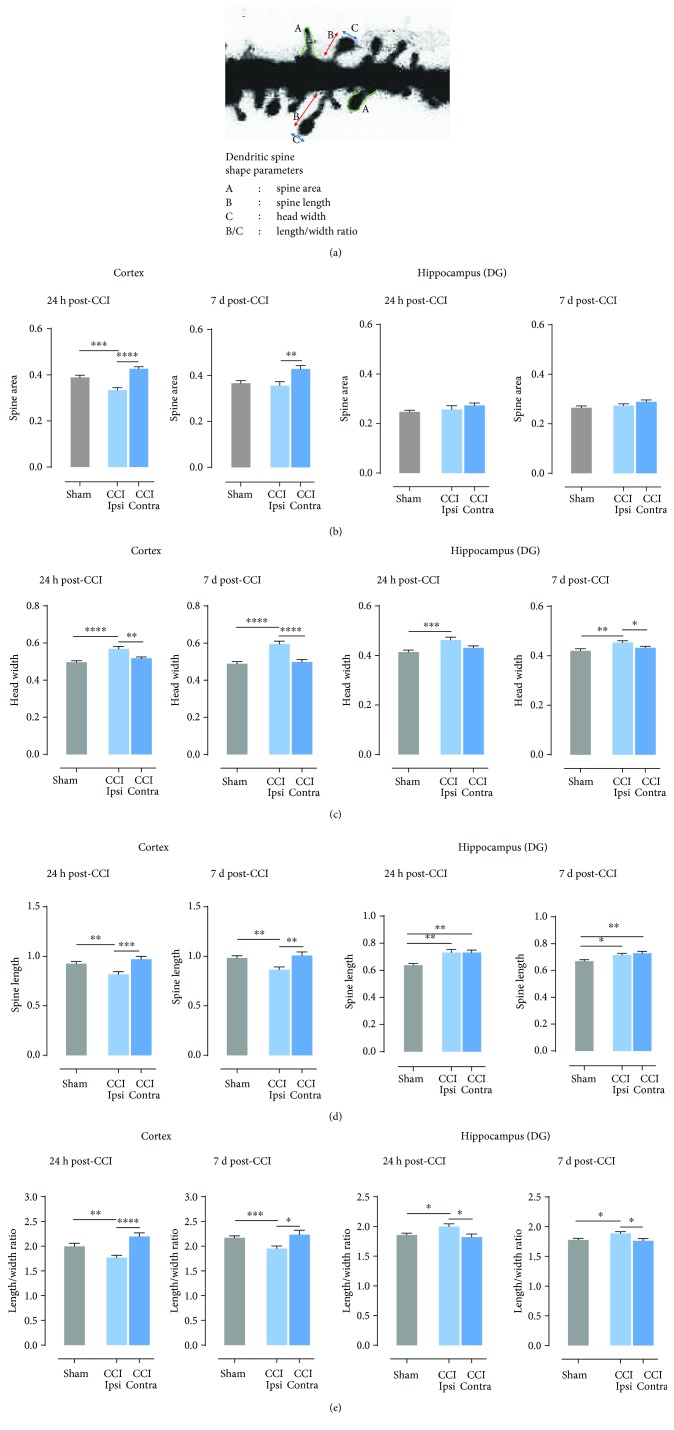
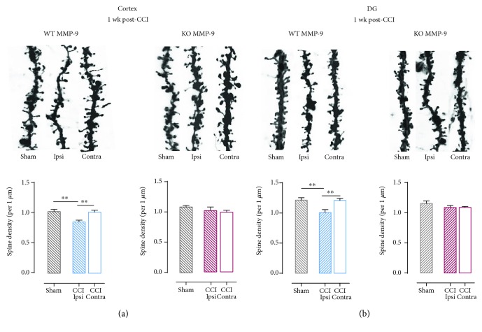
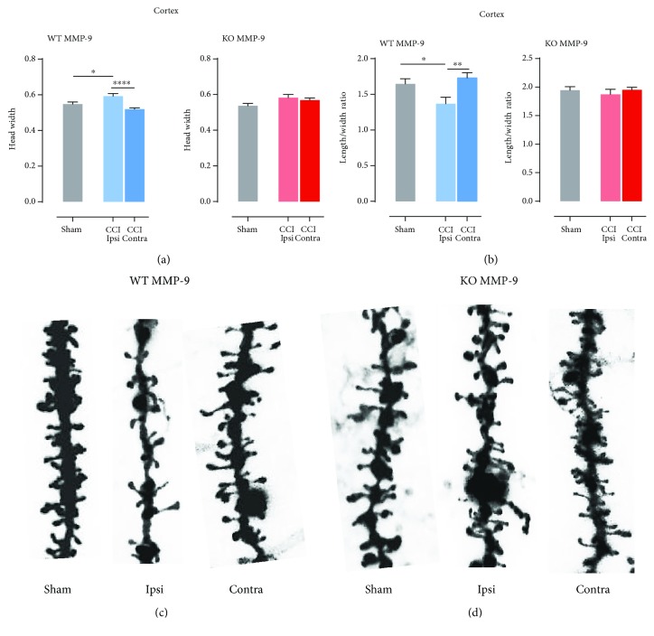
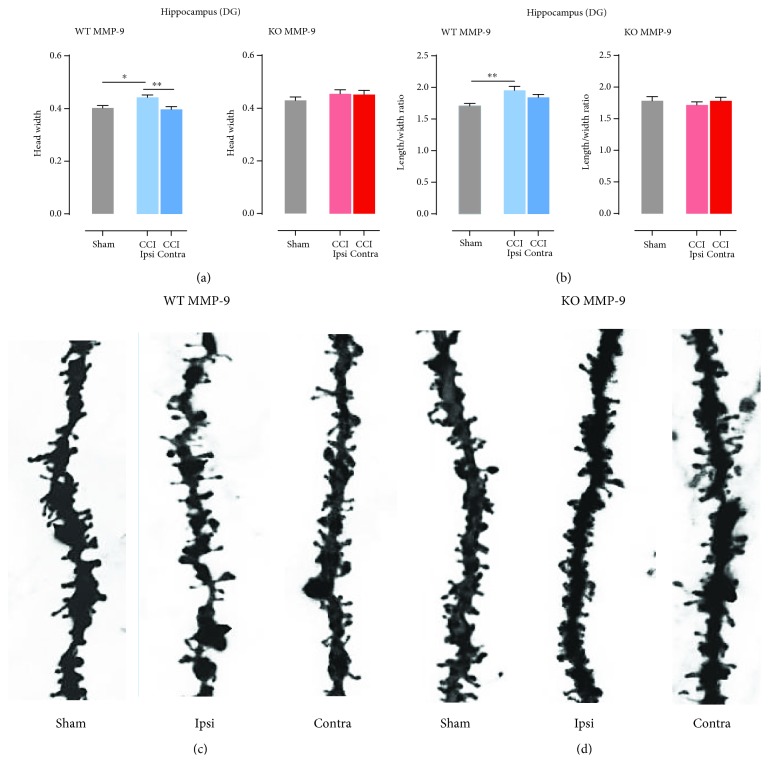
Traumatic brain injury (TBI) occurs when a blow to the head causes brain damage. Apart from physical trauma, it causes a wide range of cognitive, behavioral, and emotional deficits including impairments in learning and memory. On neuronal level, TBI may lead to circuitry remodeling and in effect imbalance between excitatory and inhibitory neurotransmissions. Such change in brain homeostasis may often lead to brain disorders. The basic units of neuronal connectivity are dendritic spines that are tiny protrusions forming synapses between two cells in a network. Spines are dynamic structures that undergo morphological transformation throughout life. Their shape is strictly related to an on/off state of synapse and the strength of synaptic transmission. Matrix metalloproteinase-9 (MMP-9) is an extrasynaptically operating enzyme that plays a role in spine remodeling and has been reported to be activated upon TBI. The aim of the present study was to evaluate the influence of MMP-9 on dendritic spine density and morphology following controlled cortical impact (CCI) as animal model of TBI. We examined spine density and dendritic spine shape in the cerebral cortex and the hippocampus. CCI caused a marked decrease in spine density as well as spine shrinkage in the cerebral cortex ipsilateral to the injury, when compared to sham animals and contralateral side both 1 day and 1 week after the insult. Decreased spine density was also observed in the dentate gyrus of the hippocampus; however, in contrast to the cerebral cortex, spines in the DG became more filopodia-like. In mice lacking MMP-9, no effects of TBI on spine density and morphology were observed.


Figures

Figure 1
Figure 1
Decrease in spine density after controlled cortical impact (CCI). (a) Schematic representation of injured area. Nissl-stained brain sections from animals at 1 and 7 days after CCI (CCI: animals after controlled cortical impact; sham: animals subjected to craniectomy without cortical injury). (b) Spine density (number of spines per 1 μm of dendrite length) in ipsi- and contralateral 2nd and 3rd cortex layers of C57Bl6/J mice, 1 and 7 days after CCI and sham procedures; right panel shows representative dendrites pictures. (c) Spine density in ipsi- and contralateral dentate gyrus of C57Bl6/J mice, 1 and 7 days after CCI and sham procedures; right panel shows representative dendrite pictures. Data are presented as mean ± SEM. Statistical analysis was carried out using one-way ANOVA followed by Tukey's post hoc test. Asterisks indicate statistical significance from the CCI and sham groups, respectively. P < 0.05; ∗∗∗P ≤ 0.001; ∗∗∗∗P < 0.0001.
Figure 2
Figure 2
Time-dependent changes in dendritic spines shape after controlled cortical impact (CCI). (a) Spine shape parameters: A: spine area; B: spine length; C: head width; B/C: length/width ratio. (b) Spine area calculated in the ipsi- and contralateral cortex and hippocampus of C57Bl6/J mice, 1 and 7 days after CCI and sham procedures. (c) Head width calculated in the ipsi- and contralateral cortex and hippocampus of C57Bl6/J mice, 1 and 7 days after CCI and sham procedures. (d) Spine length calculated in the ipsi- and contralateral cortex and hippocampus of C57Bl6/J mice, 1 and 7 days after CCI and sham procedures. (e) Length/width ratio calculated in the ipsi- and contralateral cortex and hippocampus of C57Bl6/J mice, 1 and 7 days after CCI and sham-operated animals. Data are presented as mean ± SEM. Statistical analysis was carried out using one-way ANOVA followed by Tukey's post hoc test. Asterisks indicate statistical significance from the CCI and sham groups, respectively. P < 0.05; ∗∗P ≤ 0.01; ∗∗∗P ≤ 0.001; ∗∗∗∗P < 0.00013.3.
Figure 3
Figure 3
Effect of lack of functional MMP-9 on spine density in animals after controlled cortical impact (CCI). (a) Spine density in ipsi- and contralateral 2nd and 3rd cortex layers of animals with different mmp-9 gene expression levels 1 week after CCI and sham surgeries; upper panel shows representative dendrites pictures. (b) Spine density in the ipsi- and contralateral dentate gyrus of animals with different mmp-9 gene expression levels 1 week after CCI and sham surgeries; upper panel shows representative dendrite pictures. Data are presented as mean ± SEM. Statistical analysis was carried out using one-way ANOVA followed by Tukey's post hoc test. Asterisks indicate statistical significance from the CCI and sham groups, respectively. ∗∗P ≤ 0.01.
Figure 4
Figure 4
Effects of MMP-9 on spine shape changes in the cerebral cortex layers of animals 1 week post-CCI. (a) Head width calculated in the ipsi- and contralateral cortex and hippocampus of C57Bl6/J mice, 7 days after CCI and sham procedures. (b) Length/width ratio calculated in the ipsi- and contralateral cortex and hippocampus of C57Bl6/J mice, 7 days after CCI and sham-operated animals. (c) Dendrite pictures from the ipsi- and contralateral cortex and sham animals. (d) Representative dendrite pictures from the ipsi- and contralateral cortex and sham animals. Data are presented as mean ± SEM. Statistical analysis was carried out using one-way ANOVA followed by Tukey's post hoc test. Asterisks indicate statistical significance from the CCI and sham groups, respectively. P < 0.05; ∗∗P ≤ 0.01.
Figure 5
Figure 5
Effects of MMP-9 on spine shape changes in the dentate gyrus of animals 1 week post-CCI. (a) Head width calculated in the ipsi- and contralateral hippocampus of C57Bl6/J mice, 7 days after CCI and sham procedures. (b) Length/width ratio calculated in the ipsi- and contralateral hippocampus of C57Bl6/J mice, 7 days after CCI and sham-operated animals. (c) Dendrite pictures from the ipsi- and contralateral dentate gyrus and sham animals. (d) Representative dendrite pictures from the ipsi- and contralateral dentate gyrus and sham animals. Data are presented as mean ± SEM. Statistical analysis was carried out using one-way ANOVA followed by Tukey's post hoc test. Asterisk indicate statistical significance from the CCI and sham groups, respectively. P < 0.05; ∗∗P ≤ 0.01.

Similar articles

Cited by

References

    1. Atkins C. M. Decoding hippocampal signaling deficits after traumatic brain injury. Translational Stroke Research. 2011;2(4):546–555. doi: 10.1007/s12975-011-0123-z. - DOI - PMC - PubMed
    1. Reeves T. M., Prins M. L., Zhu J. P., Povlishock J. T., Phillips L. L. Matrix metalloproteinase inhibition alters functional and structural correlates of deafferentation-induced sprouting in the dentate gyrus. The Journal of Neuroscience. 2003;23(32):10182–10189. doi: 10.1523/JNEUROSCI.23-32-10182.2003. - DOI - PMC - PubMed
    1. Shohami E., Biegon A. Novel approach to the role of NMDA receptors in traumatic brain injury. CNS & Neurological Disorders - Drug Targets. 2014;13(4):567–573. doi: 10.2174/18715273113126660196. - DOI - PubMed
    1. Sala C., Segal M. Dendritic spines: the locus of structural and functional plasticity. Physiological Reviews. 2014;94(1):141–188. doi: 10.1152/physrev.00012.2013. - DOI - PubMed
    1. Gray E. G. Axo-somatic and axo-dendritic synapses of the cerebral cortex: an electron microscope study. Journal of Anatomy. 1959;93:420–433. - PMC - PubMed

Publication types

MeSH terms

Substances