

Case Reports
doi: 10.1016/j.radcr.2019.05.016.
eCollection 2019 Aug.
Multimodality imaging evaluation for iliac crest apophysis avulsion injury
Affiliations
- PMID: 31198480
- PMCID: PMC6556494
- DOI: 10.1016/j.radcr.2019.05.016
Item in Clipboard
Case Reports
Multimodality imaging evaluation for iliac crest apophysis avulsion injury
Radiol Case Rep.
.
Abstract
A young cross-country athlete with left thigh pain, and a recent negative MRI of the left hip was referred to nuclear medicine for bone scan imaging. A 3-phase Tc-99m methylene diphosphonate bone scan was performed and revealed left iliac crest apophysis avulsion. This case illustrates that 3-phase bone scan is a great adjunct in the evaluation of sports injuries especially in athletes presenting with vague nonlocalizing symptoms, and prior negative radiographic or MRI imaging.
Keywords: Bone scan; Iliac crest apophysis injury.
Figures
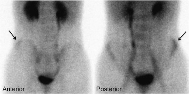
Anterior and posterior blood pool images of a Tc99m-MDP bone scan showing asymmetric increased blood pooling in the region of the left iliac crest apophysis.
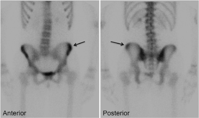
Anterior and posterior 3 hours delayed images of a Tc99m-MDP bone scan showing asymmetric increased radiotracer deposition in the region of the left iliac crest apophysis, better appreciated on the posterior views.
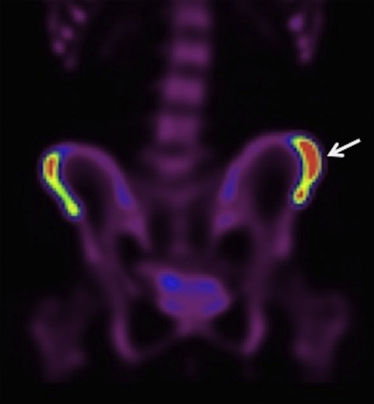
Bone scan SPECT 3D MIP reconstructed image demonstrating asymmetric increased radiotracer deposition in the region of the left anterior iliac crest extending inferiorly to the region of the left anterior iliac spine.
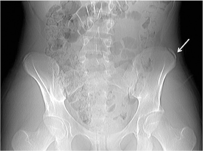
Frontal pelvis radiograph showing a mildly displaced avulsion fracture of the left iliac crest apophysis.
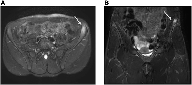
Axial (A) and coronal (B) STIR MRI images showing increased T2 signal in the region of the left iliac crest apophysis.
Similar articles
-
A rare case of avulsion fracture of the iliac crest apophysis in a young female athlete.Trauma Case Rep. 2019 Nov 7;24:100257. doi: 10.1016/j.tcr.2019.100257. eCollection 2019 Dec. Trauma Case Rep. 2019. PMID: 31737774 Free PMC article.
-
Results of operative treatment of avulsion fractures of the iliac crest apophysis in adolescents.Injury. 2014 Apr;45(4):721-4. doi: 10.1016/j.injury.2013.10.005. Epub 2013 Oct 15. Injury. 2014. PMID: 24246879
-
Avulsion of the iliac crest apophysis: a rare fracture in adolescent athletes.Ann Emerg Med. 1993 Jul;22(7):1218-20. doi: 10.1016/s0196-0644(05)80994-1. Ann Emerg Med. 1993. PMID: 8517577
-
U-shaped sacral fracture with iliac crest apophyseal avulsion in a young child.J Pediatr Orthop. 2014 Jul-Aug;34(5):e6-e11. doi: 10.1097/BPO.0000000000000139. J Pediatr Orthop. 2014. PMID: 24327188 Review.
-
A report on the incidence of intestinal 99mTc-methylene diphosphonate uptake of bone scans and a review of the literature.Nucl Med Commun. 2006 Nov;27(11):877-85. doi: 10.1097/01.mnm.0000237991.44948.13. Nucl Med Commun. 2006. PMID: 17021428 Review.
References
-
- Kjellin I., Stadnick M.E., Awh M.H. Orthopaedic magnetic resonance imaging challenge: apophyseal avulsions at the pelvis. Sports Health. 2010;2(3):247–251. - PMC - PubMed
- Kjellin I., Stadnick M.E., Awh M.H.Orthopaedic magnetic resonance imaging challenge: apophyseal avulsions at the pelvis. Sports Health. 2010;2(3):247–51. - PMC - PubMed
-
- Hébert K.J., Laor T., Divine J.G., Emery K.H., Wall E.J. MRI appearance of chronic stress injury of the iliac crest apophysis in adolescent athletes. AJR Am J Roentgenol. 2008;190(6):1487–1491. - PubMed
- Hébert K.J., Laor T., Divine J.G., Emery K.H., Wall E.J.MRI appearance of chronic stress injury of the iliac crest apophysis in adolescent athletes. AJR Am J Roentgenol. 2008;190(6):1487–91. - PubMed
Publication types
LinkOut - more resources
Full Text Sources
