

Antiepileptic Effects of Protein-Rich Extract from Bombyx batryticatus on Mice and Its Protective Effects against H2O2-Induced Oxidative Damage in PC12 Cells via Regulating PI3K/Akt Signaling Pathways
- PMID: 31198493
- PMCID: PMC6526569
- DOI: 10.1155/2019/7897584
Antiepileptic Effects of Protein-Rich Extract from Bombyx batryticatus on Mice and Its Protective Effects against H2O2-Induced Oxidative Damage in PC12 Cells via Regulating PI3K/Akt Signaling Pathways
Abstract
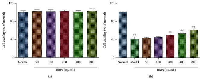
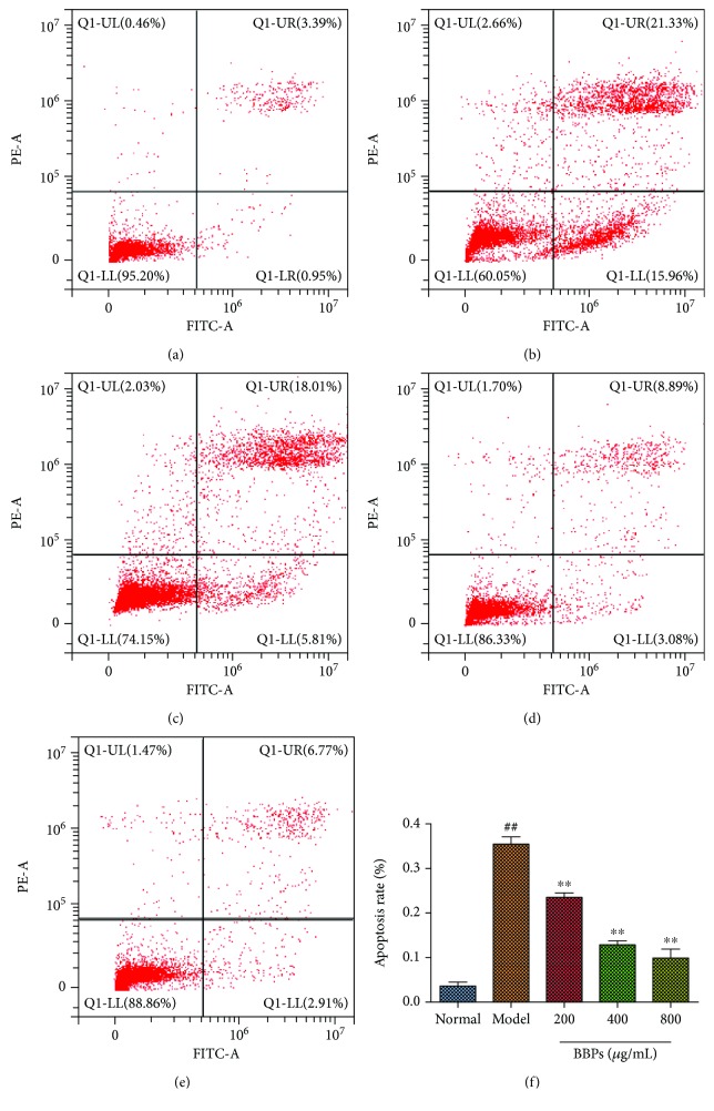
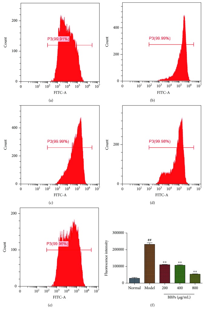
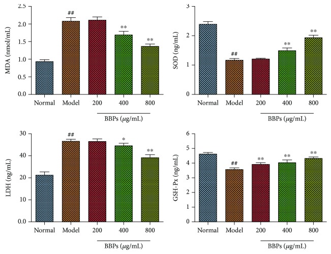
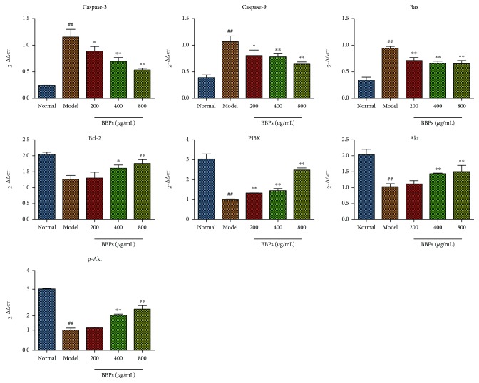
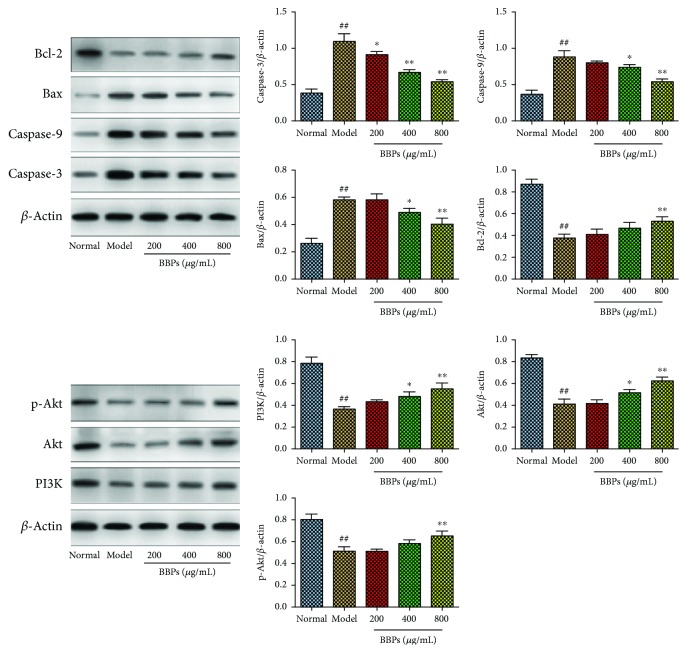
Bombyx batryticatus is a known traditional Chinese medicine (TCM) utilized to treat convulsions, epilepsy, cough, asthma, headaches, and purpura in China for thousands of years. This study is aimed at investigating the antiepileptic effects of protein-rich extracts from Bombyx batryticatus (BBPs) on seizure in mice and exploring the protective effects of BBPs against H2O2-induced oxidative stress in PC12 cells and their underlying mechanisms. Maximal electroshock-induced seizure (MES) and pentylenetetrazole- (PTZ-) induced seizure in mice and the histological analysis were carried out to evaluate the antiepileptic effects of BBPs. The cell viability of PC12 cells stimulated by H2O2 was determined by MTT assay. The apoptosis and ROS levels of H2O2-stimulated PC12 cells were determined by flow cytometry analysis. Furthermore, the levels of malondialdehyde (MDA), superoxide dismutase (SOD), lactate dehydrogenase (LDH), and glutathione (GSH) in PC12 cells were assayed by ELISA and expressions of caspase-3, caspase-9, Bax, Bcl-2, PI3K, Akt, and p-Akt were evaluated by Western blotting and quantitative real-time polymerase chain reaction (RT-qPCR) assays. The results revealed that BBPs exerted significant antiepileptic effects on mice. In addition, BBPs increased the cell viability of H2O2-stimulated PC12 cells and reduced apoptotic cells and ROS levels in H2O2-stimulated PC12 cells. By BBPs treatments, the levels of MDA and LDH were reduced and the levels of SOD and GSH-Px were increased in H2O2-stimulated PC12 cells. Moreover, BBPs upregulated the expressions of PI3K, Akt, p-Akt, and Bcl-2, whereas they downregulated the expressions of caspase-9, caspase-3, and Bax in H2O2-stimulated PC12 cells. These findings suggested that BBPs possessed potential antiepileptic effects on MES and PTZ-induced seizure in mice and protective effects on H2O2-induced oxidative stress in PC12 cells by exerting antioxidative and antiapoptotic effects via PI3K/Akt signaling pathways.
Figures
Similar articles
-
The Effect of Protein-Rich Extract from Bombyx Batryticatus against Glutamate-Damaged PC12 Cells Via Regulating γ-Aminobutyric Acid Signaling Pathway.Molecules. 2020 Jan 28;25(3):553. doi: 10.3390/molecules25030553. Molecules. 2020. PMID: 32012896 Free PMC article.
-
Protection afforded by quercetin against H2O2-induced apoptosis on PC12 cells via activating PI3K/Akt signal pathway.J Recept Signal Transduct Res. 2016;36(1):98-102. doi: 10.3109/10799893.2015.1049363. Epub 2015 Sep 28. J Recept Signal Transduct Res. 2016. PMID: 26414235
-
The Protective Effect of Aspirin Eugenol Ester on Oxidative Stress to PC12 Cells Stimulated with H2O2 through Regulating PI3K/Akt Signal Pathway.Oxid Med Cell Longev. 2021 Jun 24;2021:5527475. doi: 10.1155/2021/5527475. eCollection 2021. Oxid Med Cell Longev. 2021. PMID: 34257805 Free PMC article.
-
N-acetylserotonin protects PC12 cells from hydrogen peroxide induced damage through ROS mediated PI3K / AKT pathway.Cell Cycle. 2022 Nov;21(21):2268-2282. doi: 10.1080/15384101.2022.2092817. Epub 2022 Jun 26. Cell Cycle. 2022. PMID: 35758219 Free PMC article. Review.
-
Neuroprotective effects of natural compounds on neurotoxin-induced oxidative stress and cell apoptosis.Nutr Neurosci. 2022 May;25(5):1078-1099. doi: 10.1080/1028415X.2020.1840035. Epub 2020 Nov 8. Nutr Neurosci. 2022. PMID: 33164705 Review.
Cited by
-
Rhein Ameliorates Cognitive Impairment in an APP/PS1 Transgenic Mouse Model of Alzheimer's Disease by Relieving Oxidative Stress through Activating the SIRT1/PGC-1α Pathway.Oxid Med Cell Longev. 2022 Mar 22;2022:2524832. doi: 10.1155/2022/2524832. eCollection 2022. Oxid Med Cell Longev. 2022. PMID: 35360200 Free PMC article.
-
The Protective Effect of DiDang Tang Against AlCl3-Induced Oxidative Stress and Apoptosis in PC12 Cells Through the Activation of SIRT1-Mediated Akt/Nrf2/HO-1 Pathway.Front Pharmacol. 2020 Apr 15;11:466. doi: 10.3389/fphar.2020.00466. eCollection 2020. Front Pharmacol. 2020. PMID: 32372957 Free PMC article.
-
Antiepileptic Effects of Cicadae Periostracum on Mice and Its Antiapoptotic Effects in H2O2-Stimulated PC12 Cells via Regulation of PI3K/Akt/Nrf2 Signaling Pathways.Oxid Med Cell Longev. 2021 Jul 20;2021:5598818. doi: 10.1155/2021/5598818. eCollection 2021. Oxid Med Cell Longev. 2021. PMID: 34336105 Free PMC article.
-
Hydroxy-α-sanshool Possesses Protective Potentials on H2O2-Stimulated PC12 Cells by Suppression of Oxidative Stress-Induced Apoptosis through Regulation of PI3K/Akt Signal Pathway.Oxid Med Cell Longev. 2020 Jul 9;2020:3481758. doi: 10.1155/2020/3481758. eCollection 2020. Oxid Med Cell Longev. 2020. PMID: 32695254 Free PMC article.
-
Characterization of Polysaccharides from the Pericarp of Zanthoxylum bungeanum Maxim by Saccharide Mapping and Their Neuroprotective Effects.Molecules. 2023 Feb 15;28(4):1813. doi: 10.3390/molecules28041813. Molecules. 2023. PMID: 36838801 Free PMC article.
References
-
- World Health Organization. Epilepsy in the WHO Africa Region, Bridging the Gap: The Global Campaign against Epilepsy, “Out of the Shadows”. Geneva, Switzerland: World Health Organization; 2004.
-
- Thom M. Neuropathology of epilepsy: epilepsy-related deaths and SUDEP. Diagnostic Histopathology. 2019;25(1):23–33. doi: 10.1016/j.mpdhp.2018.11.003. - DOI
MeSH terms
Substances
LinkOut - more resources
Full Text Sources
Medical
Research Materials
Miscellaneous
