Case Reports
. 2019 May 31;2019(5):omz032.
doi: 10.1093/omcr/omz032. eCollection 2019 May.

Antibiotic-resistant hypervirulent Klebsiella pneumoniae causing community- acquired liver abscess: an emerging disease

Affiliations
Case Reports

Antibiotic-resistant hypervirulent Klebsiella pneumoniae causing community- acquired liver abscess: an emerging disease

S Rodriguez-Villar et al. Oxf Med Case Reports. .

Abstract

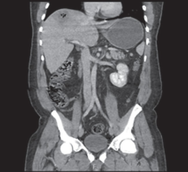
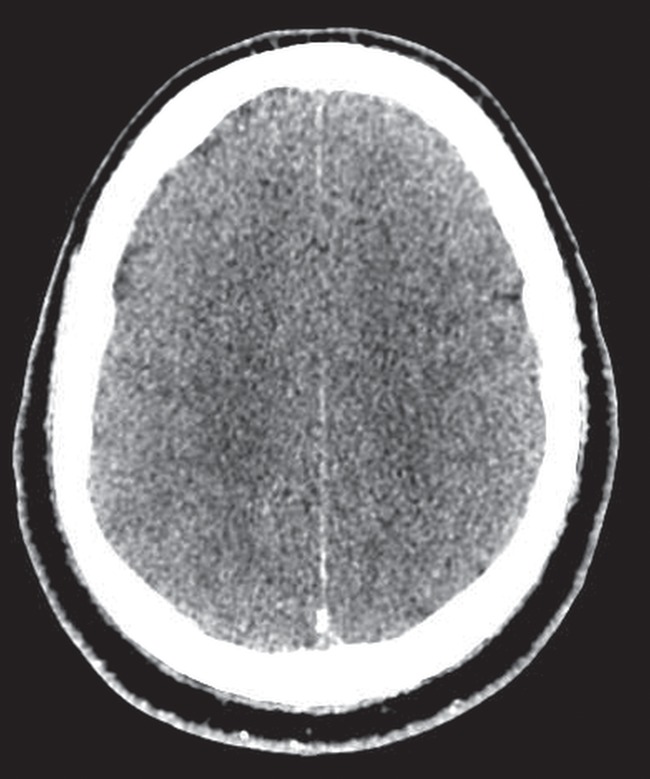
We report a case of a patient with fatal community-acquired pyogenic liver abscess (CA-PLA) caused by multi drug-resistant, hypervirulent, Klebsiella pneumoniae (mdrhvKP). HvKP causing PLA has been described in East and South East Asia and it is recognized as an emerging infection worldwide. The syndrome is characterized by cryptogenic liver abscess formation without a previous history of hepatobiliary or colonic disease and metastatic spread of infection via the bloodstream to distant sites, including lungs, central nervous system and other organ systems. Diabetes mellitus is a recognized risk factor. Most previously reported cases have involved antibiotic susceptible strains of hvKP although reports of bloodstream infections caused by resistant strains, including carbapenemase producers, are increasing. Our report highlights the need for awareness of this devastating infection in patients presenting with sepsis and liver abscess without underlying hepatobiliary or colonic disease.


Figures

Figure 1
Figure 1
Frontal Chest Radiograph (on presentation): The lungs are clear. No pleural effusion. Cardiomediastinal contour are unremarkable.
Figure 2
Figure 2
CT Abdomen (Portal Venous phase). Axial image (Figure 2) and Coronal (Figure 3): there is a 6 cm lobulated mass located in the right lobe of the liver with peripheral enhancement and areas of central necrosis (gas) consistent with abscess formation. The remainder of the liver was normal, in particular, no portal or hepatic vein thrombosis.
Figure 3
Figure 3
CT Abdomen (Portal Venous phase). Axial image (Figure 2) and Coronal (Figure 3): there is a 6 cm lobulated mass located in the right lobe of the liver with peripheral enhancement and areas of central necrosis (gas) consistent with abscess formation. The remainder of the liver was normal, in particular, no portal or hepatic vein thrombosis.
Figure 4
Figure 4
CT pulmonary angiogram. Axial image (lung windows): there is nodular right upper lobe air space opacification with surrounding ground glass nodules noted. No lung abscess demonstrated.
Figure 5
Figure 5
Frontal chest radiograph. Central lines and tubes appropriately sited. There is loss of volume of the right lung with elevation of the right hemi-diaphragm and air space opacification of the lateral aspect of the right mid zone. Left lung is clear.
Figure 6
Figure 6
CT head (non-contrast). Axial image: There is cerebral sulcal effacement of both hemispheres at the convexity in keeping with cerebral oedema.
Figure 7
Figure 7
CT head (non-contrast). Sagittal Image: there is preservation of the suprasellar and pre pontine/pre medullary cisterns. The cerebellar tonsils are above foramen magnum.
Figure 8
Figure 8
CT head (contrast). Axial Image: there is lateral ventricular dilatation, asymmetric to the right with evidence of debri in the occipital horn of the right lateral ventricle.
Figure 9
Figure 9
CT Head (Post Contrast). Sagittal image: There is progressive effacement of the suprasellar, pre pontine/ pre medullary cisterns with inferior cerebellar tonsillar descent below foramen magnum.

Similar articles

Cited by

References

    1. Rossi B, Gasperini ML, Leflon-Guibout V, Gioanni A, de Lastours V, Rossi G, et al. . Hypervirulent Klebsiella pneumoniae in cryptogenic liver abscesses, Paris, France. Emerg Infect Dis 2018;24:221–229. - PMC - PubMed
    1. Siu LK, Yeh KM, Lin JC, Fung CP, Chang FY. Klebsiella pneumoniae liver abscess: a new invasive syndrome. Lancet Infect Dis 2012;12:881–887. - PubMed
    1. Catalan-Najera JC, Garza-Ramos U, Barrios-Camacho H. Hypervirulence and hypermucoviscosity: two different but complementary Klebsiella spp. phenotypes? Virulence 2017;8:1111–1123. - PMC - PubMed
    1. Decré D, Verdet C, Emirian A, Le Gourrierec Petit JC, Offenstadt G, et al. . Emerging severe and fatal infections due to Klebsiella pneumoniae in two university hospitals in France. J Clin Microbiol 2011;49:3012–3014. - PMC - PubMed
    1. Zhang Y, Zhao C, Wang Q, Wang X, Chen H, Li H, et al. . High prevalence of hypervirulent Klebsiella pneumoniae infection in China geographic distribution, clinical characteristics, and antimicrobial resistance. Anitmicrob Agents Chemother 2016;160:6115–6120. - PMC - PubMed

Publication types