Pleural-Based Intrathoracic Cystic Lymphangioma in an Infant Mimicking a Pneumonia
- PMID: 31198614
- PMCID: PMC6526578
- DOI: 10.1155/2019/7920591
Pleural-Based Intrathoracic Cystic Lymphangioma in an Infant Mimicking a Pneumonia
Abstract
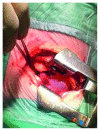
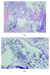
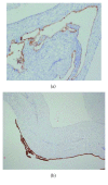
Cystic lymphangioma is an uncommon benign tumor that occurs primarily in children in the cervical region. We report the first case of a pleural-based cystic lymphangioma in an infant. The patient was admitted for RUL pneumonia. Because of the persistence of the radiographic findings despite clinical improvement, a computed tomography (CT) and a magnetic resonance imaging (MRI) scan were performed. They showed a multiloculated cystic lesion in the superior posterior right hemithorax. A surgical procedure was performed with complete resection of the tumor. Histopathological examination showed a pleural-based intrathoracic multicystic lymphangioma. One year after the surgery, the patient feels well without any sign of recurrence.
Figures


Similar articles
-
A huge lymphangioma mimicking pleural effusion with extension to both chest cavities: a case report and review of literature.Iran J Med Sci. 2015 Mar;40(2):181-4. Iran J Med Sci. 2015. PMID: 25821300 Free PMC article.
-
[Cervical lymphangioma in the adult. A report of 2 cases].Cir Cir. 2016 Jul-Aug;84(4):313-7. doi: 10.1016/j.circir.2015.06.028. Epub 2015 Aug 7. Cir Cir. 2016. PMID: 26259743 Review. Spanish.
-
Lymphangioma of the kidney.Int J Urol. 2002 Mar;9(3):178-82. doi: 10.1046/j.1442-2042.2002.00437.x. Int J Urol. 2002. PMID: 12010331 Review.
-
Intrathoracic lymphangioma.Mayo Clin Proc. 1986 Nov;61(11):882-92. doi: 10.1016/s0025-6196(12)62609-3. Mayo Clin Proc. 1986. PMID: 3762227
-
[CASE REPORT: COMPLETE RESECTION OF RETROPERITONEAL CYSTIC LYMPHANGIOMA AND SURROUNDING ORGANS].Nihon Hinyokika Gakkai Zasshi. 2019;110(1):52-55. doi: 10.5980/jpnjurol.110.52. Nihon Hinyokika Gakkai Zasshi. 2019. PMID: 31956220 Japanese.
References
-
- Benninghoff M. G., Todd W. U., Bascom R. Incidental pleural-based pulmonary lymphangioma. Journal of the American Osteopathic Association. 2008;108(9):525–528. - PubMed
Publication types
LinkOut - more resources
Full Text Sources

