A study of endothelial cell count pre- and post-neodymium-doped yttrium aluminum garnet laser iridotomy in subacute angle closure using specular microscope
- PMID: 31198669
- PMCID: PMC6557069
- DOI: 10.4103/tjo.tjo_38_18
A study of endothelial cell count pre- and post-neodymium-doped yttrium aluminum garnet laser iridotomy in subacute angle closure using specular microscope
Abstract
Aim: The aim of this study was to study the effect of neodymium-doped yttrium aluminum garnet (Nd:YAG) laser iridotomy on corneal endothelial cell count in patients with subacute angle closure using specular microscope.
Materials and methods: In this prospective study, 50 cases of narrow-angle Grade 1 and Grade 2 (Shaffer gonioscopic grading) visiting the Regional Institute of Ophthalmology, Government Medical College, Amritsar underwent Nd:YAG laser peripheral iridotomy. After obtaining informed written consent, specular microscopy was performed before and after iridotomy at 1 week, 1 month, 3rd month, and at 6th-month follow-up visits. Central, nasal, and temporal endothelial cell counts were evaluated through noncontact specular microscopy.
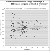
Results: The mean participant age was 51.52 ± 7.9 years, and majority of the participants were females (76%). The mean IOP before the laser was 19.25 ± 1.914 mmHg and it varied from 18.50 ± 1.647 to 18.25 ± 1.699 mmHg (day 1, p = 0.06 and at 6 months, p = 0.04) following laser procedure. The mean corneal endothelial cell count at superotemporal site before laser peripheral iridotomy was 2844 ± 260, and this value decreased to 2807 ± 263, 2699 ± 267, 2656 ± 270, and 2591 ± 275 cells/mm2 at postiridotomy, 1, 3, and 6 months' follow-up visits, respectively; these differences were statistically significant (p < 0.05). The mean total energy required to produce iridotomy was 14.88 ± 6.71 mJ, ranging from 5 to 37 mJ. The linear regression analysis indicated no statistical correlation between change in endothelial cell count at the treated site and total mean energy used. No significant difference was found between preiridotomy and postiridotomy corneal thickness at any site.
Conclusion: This study demonstrated a significant endothelial cell loss at the treated site in 6 months' follow-up and suggested that Nd:YAG laser iridotomy may pose hazard to the corneal endothelium, although corneal decompensation at the treated site or as a whole was not seen.
Keywords: Corneal endothelium; neodymium-doped yttrium aluminum garnet laser; specular microscope.
Conflict of interest statement
The authors declare that there are no conflicts of interests of this paper.
Figures


Similar articles
-
Early intraocular pressure change after peripheral iridotomy with ultralow fluence pattern scanning laser and Nd:YAG laser in primary angle-closure suspect: Kowloon East Pattern Scanning Laser Study Report No. 3.Graefes Arch Clin Exp Ophthalmol. 2018 Feb;256(2):363-369. doi: 10.1007/s00417-017-3860-1. Epub 2017 Dec 7. Graefes Arch Clin Exp Ophthalmol. 2018. PMID: 29218423
-
Effect of laser peripheral iridotomy using argon and neodymium-YAG lasers on corneal endothelial cell density: 7-year longitudinal evaluation.Jpn J Ophthalmol. 2018 Mar;62(2):216-220. doi: 10.1007/s10384-018-0569-6. Epub 2018 Feb 6. Jpn J Ophthalmol. 2018. PMID: 29411172
-
Evaluation of Intraocular Pressure, Refraction, Anterior Chamber Depth, Macular Thickness, and Specular Microscopy Post-Neodymium-Doped Yttrium-Aluminum-Garnet Laser in Patients With Posterior Capsular Opacification.Cureus. 2024 Oct 7;16(10):e70987. doi: 10.7759/cureus.70987. eCollection 2024 Oct. Cureus. 2024. PMID: 39507175 Free PMC article.
-
Neodymium-doped yttrium aluminum garnet (Nd: YAG) laser treatment in ophthalmology: a review of the most common procedures Capsulotomy and Iridotomy.Lasers Med Sci. 2024 Jul 2;39(1):167. doi: 10.1007/s10103-024-04118-8. Lasers Med Sci. 2024. PMID: 38954050 Review.
-
[Early endothelial complications after treatment using a neodymium-Yag laser].J Fr Ophtalmol. 1989;12(1):17-23. J Fr Ophtalmol. 1989. PMID: 2668394 Review. French.
Cited by
-
Association Between Anterior Chamber Angle and Corneal Endothelial Cell Density in Chronic Angle Closure.Clin Ophthalmol. 2021 May 10;15:1957-1964. doi: 10.2147/OPTH.S309005. eCollection 2021. Clin Ophthalmol. 2021. PMID: 34007148 Free PMC article.
References
-
- Stamper RL, Lieberman MF, Drake MV. Becker-Shaffer's Diagnosis and Therapy of the Glaucomas. 8th ed. St. Louis: Mosby; 2009. p. 47. 70, 71, 74, 78, 80, 81.
-
- Wang YX, Xu L, Yang H, Jonas JB. Prevalence of glaucoma in North China: The Beijing eye study. Am J Ophthalmol. 2010;150:917–24. - PubMed
-
- Wu SC, Jeng S, Huang SC, Lin SM. Corneal endothelial damage after neodymium:YAG laser iridotomy. Ophthalmic Surg Lasers. 2000;31:411–6. - PubMed
LinkOut - more resources
Full Text Sources
Miscellaneous

