

Case Reports
Important functional distress in a teenager with optic nerve drusen
Affiliations
- PMID: 31198901
- PMCID: PMC6531770
Item in Clipboard
Case Reports
Important functional distress in a teenager with optic nerve drusen
Rom J Ophthalmol.
2019 Jan-Mar.
Abstract
We present a case of bilateral optic disc drusen and severe visual field loss in a female patient diagnosed at a very young age.
Keywords: optic nerve drusen; optic nerve pathology; perimetry; pseudopapilledema; visual field defects.
Figures
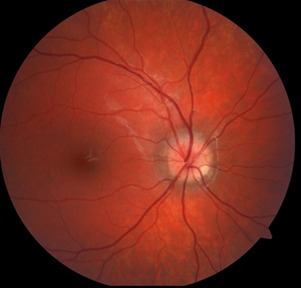
Fundus photography of the right eye
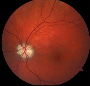
Fundus photography of the left eye
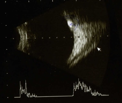
Ultrasonography right eye
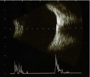
Ultrasonography left eye
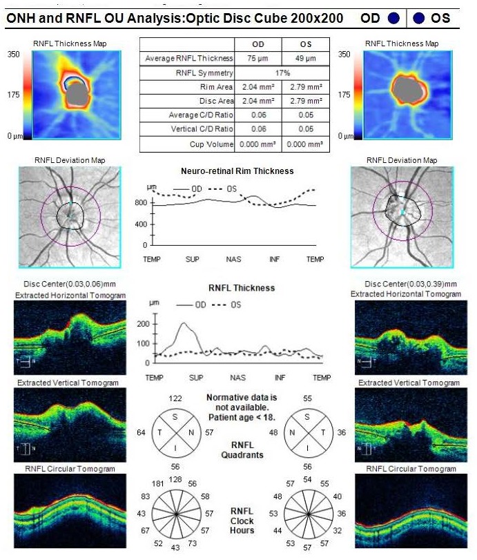
Optical coherence tomography of the optic nerve in both eyes
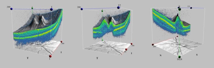
Optical coherence tomography 3D Visualization of the Optic Disc Cube of the right eye
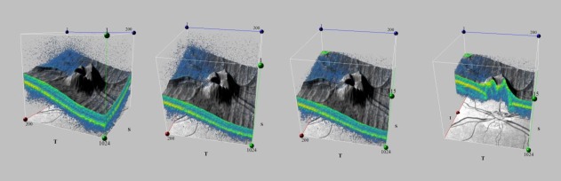
Optical coherence tomography 3D Visualization of the Optic Disc Cube of the left eye
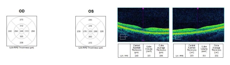
Optical coherence tomography macular thickness analysis for the right and left eye
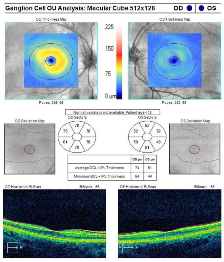
Optical coherence tomography ganglion cell layer analysis
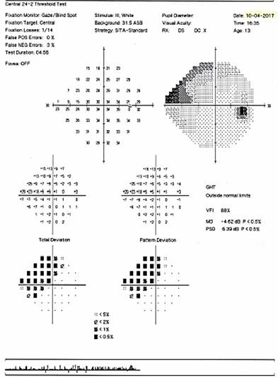
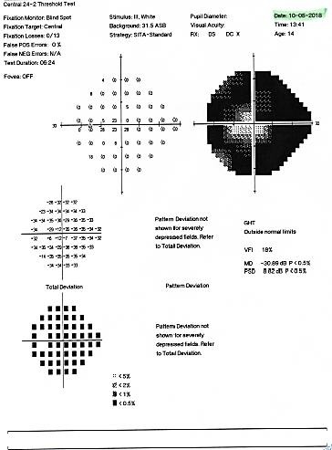
Humphrey visual field of the right eye at presentation showing a superior nasal altitudinal scotoma
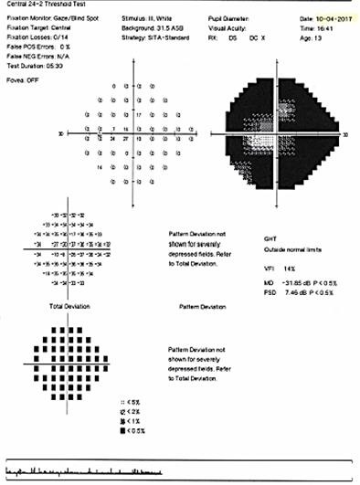
Humphrey visual field of the left eye at presentation showing an important constriction of the visual field, with the preservation of an island of central vision
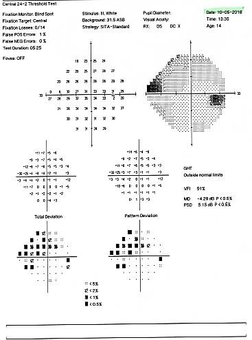
Humphrey visual field of the right eye at follow-up one year later
Humphrey visual field of the left eye at follow up one year later
Similar articles
-
Focal Capillary Dropout Associated With Optic Disc Drusen Using Optical Coherence Tomographic Angiography.J Neuroophthalmol. 2017 Dec;37(4):405-410. doi: 10.1097/WNO.0000000000000502. J Neuroophthalmol. 2017. PMID: 28520583
-
Anatomic and visual function outcomes in paediatric idiopathic intracranial hypertension.Br J Ophthalmol. 2016 Apr;100(4):505-9. doi: 10.1136/bjophthalmol-2015-307043. Epub 2015 Aug 12. Br J Ophthalmol. 2016. PMID: 26269534
-
Optic disc drusen: longitudinal aspects, with emphasis on visual field constriction and enlarged blind spot: a retrospective hospital-based clinical series.Eur J Ophthalmol. 2017 May 11;27(3):372-378. doi: 10.5301/ejo.5000864. Epub 2016 Aug 24. Eur J Ophthalmol. 2017. PMID: 27646322
-
Diagnostic dilemma of papilledema and pseudopapilledema.Int Ophthalmol. 2024 Jun 25;44(1):272. doi: 10.1007/s10792-024-03215-5. Int Ophthalmol. 2024. PMID: 38916684 Review.
-
Optic disk drusen in children.Surv Ophthalmol. 2016 Nov-Dec;61(6):745-758. doi: 10.1016/j.survophthal.2016.03.007. Epub 2016 Mar 29. Surv Ophthalmol. 2016. PMID: 27033945 Free PMC article. Review.
References
-
- Miller NR, Newman NJ, Biousse V, Kerisson JB. Walsh & Hoyt’s Clinical Neuro-Opthalmology. 6th edition. Lippincott Williams & Wilkins; 2005. pp. 177–187.
-
- Antcliff RJ, Spalton DJ. Are optic disc drusen inherited? Ophthalmology. 1999 Jul;106:1278–1281. - PubMed
-
- Kovarik JJ, Doshi PN, Collinge JE, Plager DA. Outcome of pediatric patients referred for papilledema. J AAPOS. 2015;19:344–348. - PubMed
-
- Bowling B. Kanski’s Clinical Opthalmology. A Systematic Approach. eighth edition. Elsevier Limited; 2016. pp. 798–800.
-
- Auw-Haedrich C, Staubach F, Witschel H. Optic disk drusen. Surv Ophthalmol. 2002;47(6):515–532. - PubMed
Publication types
MeSH terms
LinkOut - more resources
Full Text Sources