. 2019 Aug;44(2):630-642.
doi: 10.3892/ijmm.2019.4240. Epub 2019 Jun 12.

Long non-coding RNA XIST promotes extracellular matrix degradation by functioning as a competing endogenous RNA of miR-1277-5p in osteoarthritis

Affiliations

Long non-coding RNA XIST promotes extracellular matrix degradation by functioning as a competing endogenous RNA of miR-1277-5p in osteoarthritis

Tao Wang et al. Int J Mol Med. 2019 Aug.

Abstract

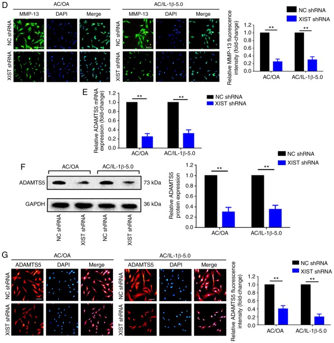
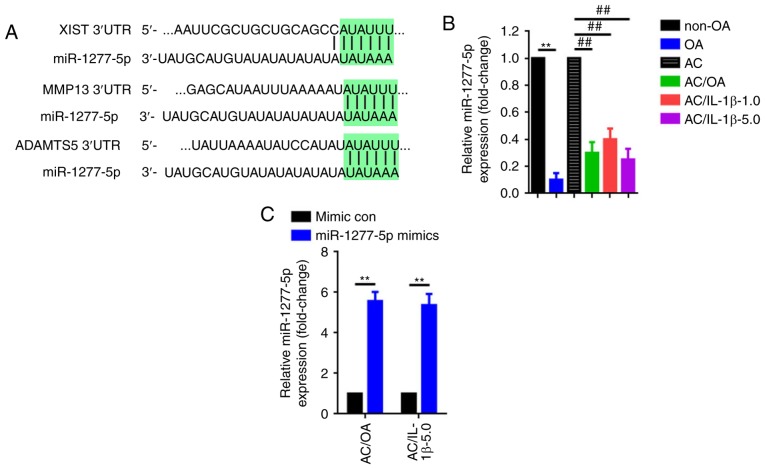
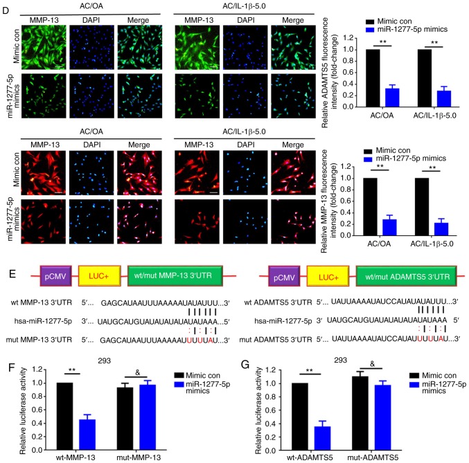
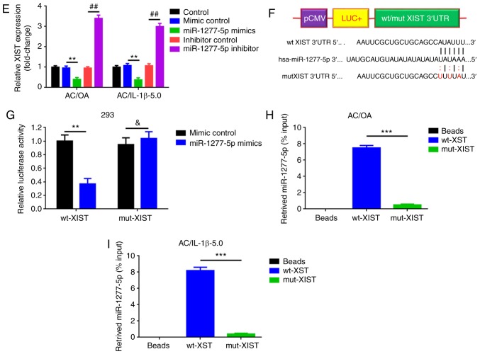
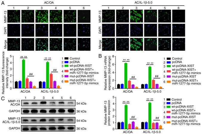
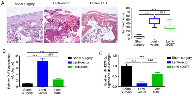
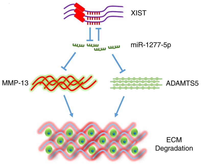
Osteoarthritis (OA) is a common and troublesome disease among the elderly, and is characterized by extracellular matrix (ECM) degradation. The function of the long non‑coding RNA X‑inactive‑specific transcript (XIST) and its working mechanism in ECM degradation remains unclear. In the present study, XIST was revealed to be upregulated in OA specimens and in articular chondrocytes (ACs) derived from OA tissue (AC/OA) and interleukin‑1β (IL‑1β)‑treated ACs. Loss‑of‑function experiments demonstrated that downregulation of XIST suppressed the degradation of the ECM in AC/OA and AC/IL‑1β‑5.0 cells. Furthermore, XIST, matrix metalloproteinase 13 (MMP‑13) and ADAM metallopeptidase with thrombospondin type 1 motif 5 (ADAMTS5) were identified as targets of microRNA (miR)‑1277‑5p, and the reciprocal inhibitive effect between XIST and miR‑1277‑5p was elucidated. Furthermore, the role of XIST in ECM degradation was confirmed to be functioning as a competing endogenous RNA (ceRNA) of miR‑1277‑5p. Finally, the protective effect of the downregulation of XIST on ECM degradation was verified in an OA rat model. In conclusion, the present study suggests that XIST promotes MMP‑13 and ADAMTS5 expression, indicating ECM degradation, by functioning as a ceRNA of miR‑1277‑5p in OA. The present study proposed a novel potential target with a new working mechanism in molecular treating of OA.


Figures

Figure 1
Figure 1
Expression of XIST in OA tissues, OA chondrocytes and IL-1β-treated ACs. (A) Expression of XIST in the collected non-OA (n=20) and OA (n=40) tissue specimens was determined by RT-qPCR using GAPDH as an internal control. (B) Expression of XIST in ACs, AC/OA, and ACs treated with 1.0 and 5.0 ng/ml IL-1β (AC/IL-1β-1.0 and AC/IL-1β-5.0) was also detected by RT-qPCR. All data are normalized to the AC group. The error bars represent standard deviation, (n=3). ***P<0.001. XIST, X-inactive-specific transcript; OA, osteoarthritis; IL-1β, interleukin-1β; RT-qPCR, reverse transcription-quantitative polymerase chain reaction; AC, articular chondrocytes.
Figure 2
Figure 2
Downregulation of XIST decreases extracellular matrix degradation in AC/OA and AC/IL-1β-5.0 cells. (A) Downregulation of XIST by an RNA interference assay was confirmed by RT-qPCR. MMP-13 expression decreased following knockdown of XIST, as determined by (B) RT-qPCR, (C) western blot analysis (lysates were analyzed by immunoblotting with MMP-13 and GAPDH antibodies; the left panel presents data from 3 independent experiments) and (D) immunofluorescence analysis. The knockdown of XIST also downregulated the expression of ADAMTS5 as determined by (E) RT-qPCR, (F) western blot analysis (lysates were analyzed by immunoblotting with ADAMTS5 and GAPDH antibodies; the left panel presents data from 3 independent experiments) and (G) immunofluorescence analysis. All data were normalized to the negative control shRNA group. Magnification, ×100; scale bar, 100 µm. The error bars represent standard deviation (n=3). **P<0.01. XIST, X-inactive-specific transcript; AC, articular chondrocyte; OA, osteoarthritis; IL-1β, interleukin-1β; RT-qPCR, reverse transcription-quantitative polymerase chain reaction; MMP-13, matrix metalloproteinase 13; ADAMTS5, ADAM metallopeptidase with thrombospondin type 1 motif 5; NC, negative control; sh, short hairpin.
Figure 2
Figure 2
Downregulation of XIST decreases extracellular matrix degradation in AC/OA and AC/IL-1β-5.0 cells. (A) Downregulation of XIST by an RNA interference assay was confirmed by RT-qPCR. MMP-13 expression decreased following knockdown of XIST, as determined by (B) RT-qPCR, (C) western blot analysis (lysates were analyzed by immunoblotting with MMP-13 and GAPDH antibodies; the left panel presents data from 3 independent experiments) and (D) immunofluorescence analysis. The knockdown of XIST also downregulated the expression of ADAMTS5 as determined by (E) RT-qPCR, (F) western blot analysis (lysates were analyzed by immunoblotting with ADAMTS5 and GAPDH antibodies; the left panel presents data from 3 independent experiments) and (G) immunofluorescence analysis. All data were normalized to the negative control shRNA group. Magnification, ×100; scale bar, 100 µm. The error bars represent standard deviation (n=3). **P<0.01. XIST, X-inactive-specific transcript; AC, articular chondrocyte; OA, osteoarthritis; IL-1β, interleukin-1β; RT-qPCR, reverse transcription-quantitative polymerase chain reaction; MMP-13, matrix metalloproteinase 13; ADAMTS5, ADAM metallopeptidase with thrombospondin type 1 motif 5; NC, negative control; sh, short hairpin.
Figure 3
Figure 3
miR-1277-5p attenuates extracellular matrix degradation by directly targeting MMP-13 and ADAMTS5 in AC/OA and AC/IL-1β-5.0 cells. (A) XIST, MMP-13 and ADAMTS5 share the same miR-1277-5p binding site within their 3′UTRs. (B) Downregulation of miR-1277-5p in OA tissue specimens and in AC/OA, AC/IL-1β-1.0 and AC/IL-1β-5.0 cells, as confirmed by reverse transcription-quantitative polymerase chain reaction. **P<0.01 vs. non-OA group; ##P<0.01 vs. AC group. (C) miR-1277-5p was upregulated by transfection of miR-1277-5p mimics as determined by reverse transcription-quantitative polymerase chain reaction. **P<0.01 vs. mimic con group. (D) Upregulation of miR-1277-5p (transfection of miR-1277-5p mimics) led to a notable decrease in MMP-13 and ADAMTS5 expression, as measured by immunofluorescence analysis. **P<0.01 vs. mimic control group; magnification, ×100; scale bar, 100 µm. (E) Diagram of the luciferase reporter plasmids with the wt- or mut-MMP-13 and -ADAMTS5 3′UTRs. (F) Compared with the effect of the mimic con, co-transfection of miR-1277-5p mimic and wt-MMP-13 led to a distinct decrease in fluorescence, but the suppressive effect was rescued by a mutation in the putative miR-1277-5p binding sites in the MMP-13 3′UTR (co-transfection of mut-MMP-13 and miR-1277-5p mimic), as verified by dual luciferase assays. **P<0.01 and &P>0.05 vs. mimic con group. (G) The same effects were observed during the verification of the interaction between miR-1277-5p and ADAMTS5 3′UTR. **P<0.01 and &P>0.05 vs. mimic con group. All error bars represented standard deviation (n=3). miR, microRNA; MMP-13, matrix metallopro-teinase 13; ADAMTS5, ADAM metallopeptidase with thrombospondin type 1 motif 5; AC, articular chondrocyte; OA, osteoarthritis; IL-1β, interleukin-1β; UTR, untranslated region; wt, wild type; mut, mutant; con, control; LUC+, luciferase.
Figure 3
Figure 3
miR-1277-5p attenuates extracellular matrix degradation by directly targeting MMP-13 and ADAMTS5 in AC/OA and AC/IL-1β-5.0 cells. (A) XIST, MMP-13 and ADAMTS5 share the same miR-1277-5p binding site within their 3′UTRs. (B) Downregulation of miR-1277-5p in OA tissue specimens and in AC/OA, AC/IL-1β-1.0 and AC/IL-1β-5.0 cells, as confirmed by reverse transcription-quantitative polymerase chain reaction. **P<0.01 vs. non-OA group; ##P<0.01 vs. AC group. (C) miR-1277-5p was upregulated by transfection of miR-1277-5p mimics as determined by reverse transcription-quantitative polymerase chain reaction. **P<0.01 vs. mimic con group. (D) Upregulation of miR-1277-5p (transfection of miR-1277-5p mimics) led to a notable decrease in MMP-13 and ADAMTS5 expression, as measured by immunofluorescence analysis. **P<0.01 vs. mimic control group; magnification, ×100; scale bar, 100 µm. (E) Diagram of the luciferase reporter plasmids with the wt- or mut-MMP-13 and -ADAMTS5 3′UTRs. (F) Compared with the effect of the mimic con, co-transfection of miR-1277-5p mimic and wt-MMP-13 led to a distinct decrease in fluorescence, but the suppressive effect was rescued by a mutation in the putative miR-1277-5p binding sites in the MMP-13 3′UTR (co-transfection of mut-MMP-13 and miR-1277-5p mimic), as verified by dual luciferase assays. **P<0.01 and &P>0.05 vs. mimic con group. (G) The same effects were observed during the verification of the interaction between miR-1277-5p and ADAMTS5 3′UTR. **P<0.01 and &P>0.05 vs. mimic con group. All error bars represented standard deviation (n=3). miR, microRNA; MMP-13, matrix metallopro-teinase 13; ADAMTS5, ADAM metallopeptidase with thrombospondin type 1 motif 5; AC, articular chondrocyte; OA, osteoarthritis; IL-1β, interleukin-1β; UTR, untranslated region; wt, wild type; mut, mutant; con, control; LUC+, luciferase.
Figure 4
Figure 4
XIST and miR-1277-5p repress each other and XIST is a target of miR-1277-5p. (A) The expression of XIST was inversely correlated with that of miR-1277-5p. The overexpression and downregulation of (B) XIST negatively regulated (C) miR-1277-5p expression, as determined by RT-qPCR. **P<0.01 and ##P<0.01 vs. the corresponding control group (n=3). Increasing and decreasing (D) miR-1277-5p levels negatively affected (E) XIST expression, as indicated by RT-qPCR. **P<0.01 and ##P<0.01 vs. control group (n=3). (F) Diagram of the luciferase reporter plasmids with the wt- or mut-XIST 3′UTR. (G) The targeted binding between XIST 3′UTR and miR-1277-5p was verified by a dual luciferase assay. **P<0.01 and &P>0.05 vs. mimic control group. (H) wt-XIST, but not mut-XIST, could pull down miR-1277-5p, as determined by an RNA pull-down assay in AC/OA and (I) AC/IL-1b-5.0. ***P<0.001 vs. the wt-XIST group (normalized to the beads group). The error bars represent standard deviation. (n=3). XIST, X-inactive-specific transcript; miR, microRNA; RT-qPCR, reverse transcription-quantitative polymerase chain reaction; wt, wild type; mut, mutant; UTR, untranslated region.
Figure 4
Figure 4
XIST and miR-1277-5p repress each other and XIST is a target of miR-1277-5p. (A) The expression of XIST was inversely correlated with that of miR-1277-5p. The overexpression and downregulation of (B) XIST negatively regulated (C) miR-1277-5p expression, as determined by RT-qPCR. **P<0.01 and ##P<0.01 vs. the corresponding control group (n=3). Increasing and decreasing (D) miR-1277-5p levels negatively affected (E) XIST expression, as indicated by RT-qPCR. **P<0.01 and ##P<0.01 vs. control group (n=3). (F) Diagram of the luciferase reporter plasmids with the wt- or mut-XIST 3′UTR. (G) The targeted binding between XIST 3′UTR and miR-1277-5p was verified by a dual luciferase assay. **P<0.01 and &P>0.05 vs. mimic control group. (H) wt-XIST, but not mut-XIST, could pull down miR-1277-5p, as determined by an RNA pull-down assay in AC/OA and (I) AC/IL-1b-5.0. ***P<0.001 vs. the wt-XIST group (normalized to the beads group). The error bars represent standard deviation. (n=3). XIST, X-inactive-specific transcript; miR, microRNA; RT-qPCR, reverse transcription-quantitative polymerase chain reaction; wt, wild type; mut, mutant; UTR, untranslated region.
Figure 5
Figure 5
XIST promotes extracellular matrix degradation by acting as a competing endogenous RNA of miR-1277-5p in AC/OA and AC/IL-1β-5.0 cells. Normal XIST overexpression plasmids (wt-pcDNA-XIST) led to a notable upregulation of MMP-13, but the effect was attenuated by the overexpression of miR-1277-5p (co-transfection of wt-pcDNA-XIST and miR-1277-5p mimics). When the putative miR-1277-5p binding site in XIST was mutated (transfection of mut-pcDNA-XIST), the facilitative effect was eliminated. The phenomenon was detected by (A) immunofluorescence analysis, (B) RT-qPCR and (C) western blot analysis (lysates were analyzed by immunoblotting with MMP-13 and GAPDH antibodies; the left panel presents data from 3 independent experiments). **P<0.01 vs. wt-pcDNA-XIST; ##P<0.01 vs. wt-pcDNA-XIST + miR-1277-5p mimic group. wt-pcDNA-XIST, but not mut-pcDNA-XIST, promoted ADAMTS5 expression, and the effect was repressed when the putative miR-1277-5p binding site in XIST was mutated (transfection of mut-pcDNA-XIST). As with MMP-13, elevation of miR-1277-5p also reversed the facilitative effect of wt-pcDNA-XIST on ADAMTS5 expression, as determined by (D) immunofluorescence analysis, (E) RT-qPCR and (F) western blot analysis (lysates were analyzed by immunoblotting with ADAMTS5 and GAPDH antibodies; the left panel presents data from 5 independent experiments). **P<0.01 vs. wt-pcDNA-XIST; ##P<0.01 vs. wt-pcDNA-XIST + miR-1277-5p mimic group. All data were normalized to the control group, and all error bars represent standard deviation (n=3). XIST, X-inactive-specific transcript; miR, microRNA; AC, articular chondrocyte; OA, osteoarthritis; IL-1β, interleukin-1β; wt, wild type; mut, mutant; MMP-13, matrix metalloproteinase 13; ADAMTS5, ADAM metallopeptidase with thrombospondin type 1 motif 5; RT-qPCR, reverse transcription-quantitative polymerase chain reaction.
Figure 5
Figure 5
XIST promotes extracellular matrix degradation by acting as a competing endogenous RNA of miR-1277-5p in AC/OA and AC/IL-1β-5.0 cells. Normal XIST overexpression plasmids (wt-pcDNA-XIST) led to a notable upregulation of MMP-13, but the effect was attenuated by the overexpression of miR-1277-5p (co-transfection of wt-pcDNA-XIST and miR-1277-5p mimics). When the putative miR-1277-5p binding site in XIST was mutated (transfection of mut-pcDNA-XIST), the facilitative effect was eliminated. The phenomenon was detected by (A) immunofluorescence analysis, (B) RT-qPCR and (C) western blot analysis (lysates were analyzed by immunoblotting with MMP-13 and GAPDH antibodies; the left panel presents data from 3 independent experiments). **P<0.01 vs. wt-pcDNA-XIST; ##P<0.01 vs. wt-pcDNA-XIST + miR-1277-5p mimic group. wt-pcDNA-XIST, but not mut-pcDNA-XIST, promoted ADAMTS5 expression, and the effect was repressed when the putative miR-1277-5p binding site in XIST was mutated (transfection of mut-pcDNA-XIST). As with MMP-13, elevation of miR-1277-5p also reversed the facilitative effect of wt-pcDNA-XIST on ADAMTS5 expression, as determined by (D) immunofluorescence analysis, (E) RT-qPCR and (F) western blot analysis (lysates were analyzed by immunoblotting with ADAMTS5 and GAPDH antibodies; the left panel presents data from 5 independent experiments). **P<0.01 vs. wt-pcDNA-XIST; ##P<0.01 vs. wt-pcDNA-XIST + miR-1277-5p mimic group. All data were normalized to the control group, and all error bars represent standard deviation (n=3). XIST, X-inactive-specific transcript; miR, microRNA; AC, articular chondrocyte; OA, osteoarthritis; IL-1β, interleukin-1β; wt, wild type; mut, mutant; MMP-13, matrix metalloproteinase 13; ADAMTS5, ADAM metallopeptidase with thrombospondin type 1 motif 5; RT-qPCR, reverse transcription-quantitative polymerase chain reaction.
Figure 6
Figure 6
Depression of XIST inhibited extracellular matrix degradation in vivo. (A) DMM (lenti-vector and lenti-siXIST group) resulted in increased Safranin-O staining and an elevated summed score compared with the sham surgery group, whereas a certain cartilage-protective effect was presented in the lenti-siXIST group compared with the lenti-vector group. (B) Elevated XIST and (C) depressed miR-1277-5p levels were attributed to DMM, and the injection of lenti-siXIST led to the inhibition of XIST and the promotion of miR-1277-5p compared with the lenti-vector group, as determined by RT-qPCR. (D) In contrast with the sham surgery group, increased MMP-13 expression was detected in the DMM groups (lenti-vector and lenti-siXIST group). In addition, downregulation of XIST (injection of lenti-siXIST) inhibited MMP-13 expression, as detected by IHC, RT-qPCR and western blot analysis (lysates were analyzed by immunoblotting with MMP-13 and GAPDH antibodies; the left panel presents data from 5 independent experiments). (E) Upregulated ADAMTS5 expression was also observed in the DMM groups (lenti-vector and lenti-siXIST group) compared with the surgery group, whereas, compared with the lenti-vector group, the injection of lenti-siXIST led to a decrease in ADAMTS5 expression, as confirmed by IHC, RT-qPCR and western blot analysis (lysates were analyzed by immunoblotting with ADAMTS5 and GAPDH antibodies; the left panel presents data from 5 independent experiments). ***P<0.001 vs. the sham surgery group; ###P<0.001 vs. the lenti-vector group. All data were normalized to the control group, and all error bars represent standard deviation (n=3). XIST, X-inactive-specific transcript; DMM, destabilization of the medial meniscus; siXIST, XIST silencing; RT-qPCR, reverse transcription-quantitative polymerase chain reaction; ADAMTS5, ADAM metallopeptidase with thrombospondin type 1 motif 5; MMP-13, matrix metalloproteinase 13; IHC, immu-nohistochemistry.
Figure 6
Figure 6
Depression of XIST inhibited extracellular matrix degradation in vivo. (A) DMM (lenti-vector and lenti-siXIST group) resulted in increased Safranin-O staining and an elevated summed score compared with the sham surgery group, whereas a certain cartilage-protective effect was presented in the lenti-siXIST group compared with the lenti-vector group. (B) Elevated XIST and (C) depressed miR-1277-5p levels were attributed to DMM, and the injection of lenti-siXIST led to the inhibition of XIST and the promotion of miR-1277-5p compared with the lenti-vector group, as determined by RT-qPCR. (D) In contrast with the sham surgery group, increased MMP-13 expression was detected in the DMM groups (lenti-vector and lenti-siXIST group). In addition, downregulation of XIST (injection of lenti-siXIST) inhibited MMP-13 expression, as detected by IHC, RT-qPCR and western blot analysis (lysates were analyzed by immunoblotting with MMP-13 and GAPDH antibodies; the left panel presents data from 5 independent experiments). (E) Upregulated ADAMTS5 expression was also observed in the DMM groups (lenti-vector and lenti-siXIST group) compared with the surgery group, whereas, compared with the lenti-vector group, the injection of lenti-siXIST led to a decrease in ADAMTS5 expression, as confirmed by IHC, RT-qPCR and western blot analysis (lysates were analyzed by immunoblotting with ADAMTS5 and GAPDH antibodies; the left panel presents data from 5 independent experiments). ***P<0.001 vs. the sham surgery group; ###P<0.001 vs. the lenti-vector group. All data were normalized to the control group, and all error bars represent standard deviation (n=3). XIST, X-inactive-specific transcript; DMM, destabilization of the medial meniscus; siXIST, XIST silencing; RT-qPCR, reverse transcription-quantitative polymerase chain reaction; ADAMTS5, ADAM metallopeptidase with thrombospondin type 1 motif 5; MMP-13, matrix metalloproteinase 13; IHC, immu-nohistochemistry.
Figure 7
Figure 7
Schematic diagram of the mechanism concluded from the present study. XIST, X-inactive-specific transcript; ADAMTS5, ADAM metallopeptidase with thrombospondin type 1 motif 5; MMP-13, matrix metalloproteinase 13; ECM< extracellular matrix; miR, microRNA.

Similar articles

Cited by

References

    1. van der Kraan PM. Osteoarthritis year 2012 in review: Biology. Osteoarthritis Cartilage. 2012;20:1447–1450. doi: 10.1016/j.joca.2012.07.010. - DOI - PubMed
    1. Mobasheri A. Osteoarthritis year 2012 in review: Biomarkers. Osteoarthritis Cartilage. 2012;20:1451–1464. doi: 10.1016/j.joca.2012.07.009. - DOI - PubMed
    1. Lee AS, Ellman MB, Yan D, Kroin JS, Cole BJ, van Wijnen AJ, Im HJ. A current review of molecular mechanisms regarding osteoarthritis and pain. Gene. 2013;527:440–447. doi: 10.1016/j.gene.2013.05.069. - DOI - PMC - PubMed
    1. Sofat N. Analysing the role of endogenous matrix molecules in the development of osteoarthritis. Int J Exp Pathol. 2009;90:463–479. doi: 10.1111/j.1365-2613.2009.00676.x. - DOI - PMC - PubMed
    1. Verma P, Dalal K. ADAMTS-4 and ADAMTS-5: Key enzymes in osteoarthritis. J Cell Biochem. 2011;112:3507–3514. doi: 10.1002/jcb.23298. - DOI - PubMed