. 2019 Jul 2;58(26):2875-2882.
doi: 10.1021/acs.biochem.9b00389. Epub 2019 Jun 19.

Arabinose Alters Both Local and Distal H-D Exchange Rates in the Escherichia coli AraC Transcriptional Regulator

Affiliations

Arabinose Alters Both Local and Distal H-D Exchange Rates in the Escherichia coli AraC Transcriptional Regulator

Alexander Tischer et al. Biochemistry. .

Abstract

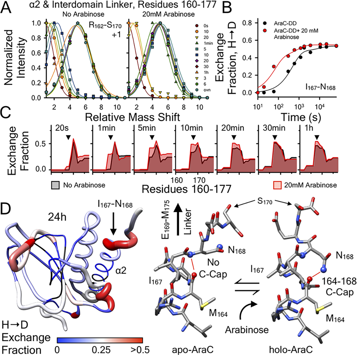
In the absence of arabinose, the dimeric Escherichia coli regulatory protein of the l-arabinose operon, AraC, represses expression by looping the DNA between distant half-sites. Binding of arabinose to the dimerization domains forces AraC to preferentially bind two adjacent DNA half-sites, which stimulates RNA polymerase transcription of the araBAD catabolism genes. Prior genetic and biochemical studies hypothesized that arabinose allosterically induces a helix-coil transition of a linker between the dimerization and DNA binding domains that switches the AraC conformation to an inducing state [Brown, M. J., and Schleif, R. F. (2019) Biochemistry, preceding paper in this issue (DOI: 10.1021/acs.biochem.9b00234)]. To test this hypothesis, hydrogen-deuterium exchange mass spectrometry was utilized to identify structural regions involved in the conformational activation of AraC by arabinose. Comparison of the hydrogen-deuterium exchange kinetics of individual dimeric dimerization domains and the full-length dimeric AraC protein in the presence and absence of arabinose reveals a prominent arabinose-induced destabilization of the amide hydrogen-bonded structure of linker residues (I167 and N168). This destabilization is demonstrated to result from an increased probability to form a helix capping motif at the C-terminal end of the dimerizing α-helix of the dimerization domain that preceeds the interdomain linker. These conformational changes could allow for quaternary repositioning of the DNA binding domains required for induction of the araBAD promoter through rotation of peptide backbone dihedral angles of just a couple of residues. Subtle changes in exchange rates are also visible around the arabinose binding pocket and in the DNA binding domain.


Conflict of interest statement

The authors declare no competing financial interest.

Figures

Figure 1.
Figure 1.
Thermodynamic cycle illustrating the binding of arabinose to AraC in DNA-bound and free states. (A) Arabinose-induced light switch mechanism between the DNA looped repressed state and the unlooped activated state. (B) Arabinose-induced conformational selection in the absence of DNA. HXMS is applied to these conformational states.
Figure 2.
Figure 2.
(A) Structure of the AraC dimerization domain (PDB entry 2ARC) highlighting the secondary structures that respond to arabinose binding. Rendered using UCSF Chimera. (B) Comparison of the hydrogen exchange of the AraC dimerization domain in the presence (red) and absence (black) of 20 mM arabinose as a function of residue number throughout the protein at three exchange time points, 1 min (top), 1 h (middle), and 24 h (bottom). HXMS was quantified in triplicate at each exchange time point. The majority of AraC hydrogen exchange is unchanged upon binding arabinose. Structural regions that show a kinetic structural effect of binding (see Figures 3–5) are indicated by horizontal bars. The exchange kinetics are quenched by arrabinose binding in the arabinose binding barrel containing the β4–β5 hairpin loop. The exchange kinetics are enhanced in the β8–310 loop of the arabinose binding barrel connected through space to A17 in the N-terminal arm. The exchange kinetics are also enhanced in the allosteric C-terminal interdomain linker containing residues I167 and N168.
Figure 3.
Figure 3.
Arabinose binding quenches hydrogen exchange within the β4–β5 loop of the dimerization domain near the arabinose binding pocket. HXMS was quantified in triplicate at times between 10 s to 6 h and 24 h. (A) Peptide envelopes (normalized intensity vs mass shift relative to the “all H” peak) of three peptides covering residues 50–63: top, T50–E63, charge state of +2; middle, G53–E63, charge state of +1; bottom, G55–E63, charge state of +1. Relative to the “all H” peptide at time zero, the level of HX is observed to continually increase as a function of time in the absence of arabinose, whereas the overall level of HX of these peptides remains fairly constant as a function of time in the presence of arabinose. (B) Kinetics of HX (exchange fraction vs time) within site-resolved regions of the β4–β5 loop: top, I51; middle, G55 and V56; bottom, Q60–E63. (C) Site resolution of the HX exchange fraction vs residue number. Quenching of HX (in panels B and C) is primarily observed at longer incubation times between 30 min and 24 h. (D) Hydrogen–deuterium exchange fraction mapped onto the structure of apo-AraC (PDB entry 1XJA) and holo-AraC (PDB entry 2ARC): black, not resolved; blue, 0; white, 0.25; red, ≥0.5. Rendered using UCSF Chimera. Arrows indicate the structural location of the β4–β5 loop.
Figure 4.
Figure 4.
Arabinose binding enhances the hydrogen exchange in residues Y97–R99 and A17 connected through space in the β8–310 loop and the N-terminus, respectively. HXMS was quantified in triplicate at times between 10 s to 6 h and 24 h. (A) Peptide envelopes (normalized intensity vs mass shift relative to the “all H” peak) of three peptides covering the region: top, F98–E106, charge state of +2; middle, V96–W107, charge state of +2; bottom, F15–L19, charge state of +1. (B) Hydrogen–deuterium exchange fraction mapped onto the structure of AraC (PDB entry 2ARC): black, not resolved; blue, 0; white, 0.25; red, ≥0.5. Rendered using UCSF Chimera. Arrows indicate the structural location of this region. (C) HX kinetics of site-resolved Y97–R99 and A17 residues are observed to be faster in the presence of arabinose than in its absence. (D) HX kinetics of the region including and surrounding Y97–R99 between 1 min and 3 h and A17 between 10 s and 30 min illustrate localized HX kinetics. Arrowheads indicate the residues involved in the altered kinetics.
Figure 5.
Figure 5.
Arabinose binding enhances hydrogen exchange in residues I167 and N168 of the C-terminal linker of the dimerization domain. HXMS was quantified in triplicate at times between 10 s to 6 h and 24 h. (A) Peptide envelopes (normalized intensity vs mass shift relative to the “all H” peak) of the R161–S169 peptide at a charge state of +1. (B) HX kinetics of site-resolved residues I167 and N168 of the C-terminal linker are observed to be faster in the presence of arabinose than in its absence. (C) HX kinetics of the region including and surrounding I167 and N168 between 20 s and 1 h illustrate the difference in HX kinetics is localized to I167 and N168 with only moderate differences in the surrounding residues. Arrowheads indicate the residues I167 and N168 involved in the altered kinetics. (D) Hydrogen–deuterium exchange fraction mapped onto the apo structure of AraC (PDB entry 1XJA) and potential C-terminal cap equilibrium of helix α2 containing residues I167 and N168. Structures of the C-termini of apo-AraC (PDB entry 1XJA) and holo-AraC (PDB entry 2ARC) of the α2 helix interdomain linker junction.
Figure 6.
Figure 6.
(A) Comparison of the hydrogen exchange of the full-length AraC protein in the presence (red) and absence (black) of 20 mM arabinose as a function of residue number throughout the protein at three exchange time points: 10 s (top), 1 min (middle), and 10 min (bottom). HXMS was quantified in triplicate at each exchange time point. (B) Hydrogen–deuterium exchange fraction mapped onto the structure of the AraC DNA binding domain (PDB entry 2K9S): black, not resolved; blue, 0; white, 0.25; red, ≥0.5. Rendered using UCSF Chimera. (C) Peptide envelopes (normalized intensity vs mass shift relative to the “all H” peak) of two peptides covering the dimerization domain in full-length AraC that confirm observations in the dimerization domain (Figures 4 and 5): top, F98–E106, charge state of +2; bottom, R162–E169, charge state of +2. HX kinetics of site-resolved Y97–R99 and R162–N168 residues are observed to be faster in the presence of arabinose than in its absence. (D) Peptide envelopes of two peptides covering the DNA binding domain in full-length AraC: top, S225–L237, charge state of +2; bottom, Y260–F276, charge state of +2. HX kinetics of site-resolved residues in α4, A235–L237 and R227–N234, are observed to be slower in the presence of arabinose than in its absence. S262–L276 have fully exchanged in 1 min.

Similar articles

Cited by

References

    1. Brown M, and Schleif RF (2019) Helical Behavior of the Interdomain Linker of the Escherichia coli AraC Protein. Biochemistry, DOI: 10.1021/acs.biochem.9b00234. - DOI - PubMed
    1. Steffen D, and Schleif R (1977) In vitro construction of plasmids which result in overproduction of the protein product of the araC gene of Escherichia coli. Mol. Gen. Genet. 157, 341–344. - PubMed
    1. Hendrickson W, and Schleif R (1985) A dimer of AraC protein contacts three adjacent major groove regions of the araI DNA site. Proc. Natl. Acad. Sci. U. S. A. 82, 3129–3133. - PMC - PubMed
    1. Bustos SA, and Schleif RF (1993) Functional domains of the AraC protein. Proc. Natl. Acad. Sci. U. S. A. 90, 5638–5642. - PMC - PubMed
    1. Sheppard DE, and Englesberg E (1967) Further evidence for positive control of the L-arabinose system by gene araC. J. Mol. Biol. 25, 443–454. - PubMed

MeSH terms

LinkOut - more resources