

White matter hyperintensities: relationship to amyloid and tau burden
- PMID: 31199475
- PMCID: PMC6658846
- DOI: 10.1093/brain/awz162
White matter hyperintensities: relationship to amyloid and tau burden
Abstract
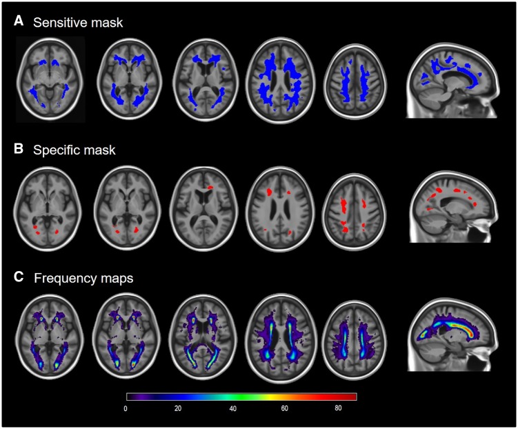
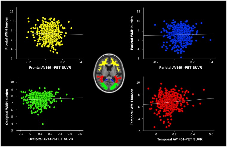
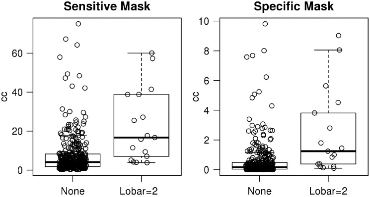
Although white matter hyperintensities have traditionally been viewed as a marker of vascular disease, recent pathology studies have found an association between white matter hyperintensities and Alzheimer's disease pathologies. The objectives of this study were to investigate the topographic patterns of white matter hyperintensities associated with Alzheimer's disease biomarkers measured using PET. From the population-based Mayo Clinic Study of Aging, 434 participants without dementia (55% male) with FLAIR and gradient recall echo MRI, tau-PET (AV-1451) and amyloid-PET scans were identified. A subset had cerebral microbleeds detected on T2* gradient recall echo scans. White matter hyperintensities were semi-automatically segmented using FLAIR MRI in participant space and normalized to a custom template. We used statistical parametric mapping 12-based, voxel-wise, multiple-regression analyses to detect white matter hyperintense regions associated with Alzheimer's biomarkers (global amyloid from amyloid-PET and meta-regions of interest tau uptake from tau-PET) after adjusting for age, sex and hypertension. For amyloid associations, we additionally adjusted for tau and vice versa. Topographic patterns of amyloid-associated white matter hyperintensities included periventricular white matter hyperintensities (frontal and parietal lobes). White matter hyperintense volumes in the detected topographic pattern correlated strongly with lobar cerebral microbleeds (P < 0.001, age and sex adjusted Cohen's d = 0.703). In contrast, there were no white matter hyperintense regions significantly associated with increased tau burden using voxel-based analysis or region-specific analysis. Among non-demented elderly, amyloid load correlated with a topographic pattern of white matter hyperintensities. Further, the amyloid-associated, white matter hyperintense regions strongly correlated with lobar cerebral microbleeds suggesting that cerebral amyloid angiopathy contributes to the relationship between amyloid and white matter hyperintensities. The study did not support an association between increased tau burden and white matter hyperintense burden.
Keywords: cerebral amyloid angiopathy; cerebral microbleeds; tau burden; white matter hyperintensities.
© The Author(s) (2019). Published by Oxford University Press on behalf of the Guarantors of Brain. All rights reserved. For Permissions, please email: journals.permissions@oup.com.
Figures
Similar articles
-
Can white matter hyperintensities based Fazekas visual assessment scales inform about Alzheimer's disease pathology in the population?Alzheimers Res Ther. 2024 Jul 10;16(1):157. doi: 10.1186/s13195-024-01525-5. Alzheimers Res Ther. 2024. PMID: 38987827 Free PMC article.
-
Plasma soluble TREM2 is associated with white matter lesions independent of amyloid and tau.Brain. 2021 Dec 16;144(11):3371-3380. doi: 10.1093/brain/awab332. Brain. 2021. PMID: 34515756
-
Hypertension and cerebral blood flow in the development of Alzheimer's disease.Alzheimers Dement. 2024 Nov;20(11):7729-7744. doi: 10.1002/alz.14233. Epub 2024 Sep 10. Alzheimers Dement. 2024. PMID: 39254220 Free PMC article.
-
White matter hyperintensities are higher among early-onset Alzheimer's disease participants than their cognitively normal and early-onset nonAD peers: Longitudinal Early-onset Alzheimer's Disease Study (LEADS).Alzheimers Dement. 2023 Nov;19 Suppl 9(Suppl 9):S89-S97. doi: 10.1002/alz.13402. Epub 2023 Jul 25. Alzheimers Dement. 2023. PMID: 37491599 Free PMC article. Review.
-
Are the brain's vascular and Alzheimer pathologies additive or interactive?Curr Opin Psychiatry. 2018 Mar;31(2):147-152. doi: 10.1097/YCO.0000000000000395. Curr Opin Psychiatry. 2018. PMID: 29232251 Review.
Cited by
-
[Association between Cerebral Small Vessel and Alzheimer's Disease].Taehan Yongsang Uihakhoe Chi. 2022 May;83(3):486-507. doi: 10.3348/jksr.2022.0041. Epub 2022 May 25. Taehan Yongsang Uihakhoe Chi. 2022. PMID: 36238505 Free PMC article. Review. Korean.
-
The many faces of globular glial tauopathy: A clinical and imaging study.Eur J Neurol. 2023 Feb;30(2):321-333. doi: 10.1111/ene.15603. Epub 2022 Nov 1. Eur J Neurol. 2023. PMID: 36256511 Free PMC article.
-
Can integration of Alzheimer's plasma biomarkers with MRI, cardiovascular, genetics, and lifestyle measures improve cognition prediction?Brain Commun. 2024 Sep 4;6(5):fcae300. doi: 10.1093/braincomms/fcae300. eCollection 2024. Brain Commun. 2024. PMID: 39291164 Free PMC article.
-
Regional white matter hyperintensities in posterior cortical atrophy and logopenic progressive aphasia.Neurobiol Aging. 2022 Nov;119:46-55. doi: 10.1016/j.neurobiolaging.2022.07.008. Epub 2022 Jul 28. Neurobiol Aging. 2022. PMID: 35970009 Free PMC article.
-
Association between migraine and cognitive impairment.J Headache Pain. 2022 Jul 26;23(1):88. doi: 10.1186/s10194-022-01462-4. J Headache Pain. 2022. PMID: 35883043 Free PMC article.
References
Publication types
MeSH terms
Substances
Grants and funding
LinkOut - more resources
Full Text Sources
Medical
