. 2019 Aug 1;142(8):2492-2509.
doi: 10.1093/brain/awz154.

Alzheimer's pathology targets distinct memory networks in the ageing brain

Affiliations

Alzheimer's pathology targets distinct memory networks in the ageing brain

Anne Maass et al. Brain. .

Abstract

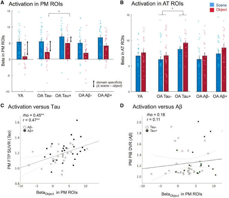
Alzheimer's disease researchers have been intrigued by the selective regional vulnerability of the brain to amyloid-β plaques and tau neurofibrillary tangles. Post-mortem studies indicate that in ageing and Alzheimer's disease tau tangles deposit early in the transentorhinal cortex, a region located in the anterior-temporal lobe that is critical for object memory. In contrast, amyloid-β pathology seems to target a posterior-medial network that subserves spatial memory. In the current study, we tested whether anterior-temporal and posterior-medial brain regions are selectively vulnerable to tau and amyloid-β deposition in the progression from ageing to Alzheimer's disease and whether this is reflected in domain-specific behavioural deficits and neural dysfunction. 11C-PiB PET and 18F-flortaucipir uptake was quantified in a sample of 131 cognitively normal adults (age: 20-93 years; 47 amyloid-β-positive) and 20 amyloid-β-positive patients with mild cognitive impairment or Alzheimer's disease dementia (65-95 years). Tau burden was relatively higher in anterior-temporal regions in normal ageing and this difference was further pronounced in the presence of amyloid-β and cognitive impairment, indicating exacerbation of ageing-related processes in Alzheimer's disease. In contrast, amyloid-β deposition dominated in posterior-medial regions. A subsample of 50 cognitively normal older (26 amyloid-β-positive) and 25 young adults performed an object and scene memory task while functional MRI data were acquired. Group comparisons showed that tau-positive (n = 18) compared to tau-negative (n = 32) older adults showed lower mnemonic discrimination of object relative to scene images [t(48) = -3.2, P = 0.002]. In a multiple regression model including regional measures of both pathologies, higher anterior-temporal flortaucipir (tau) was related to relatively worse object performance (P = 0.010, r = -0.376), whereas higher posterior-medial PiB (amyloid-β) was related to worse scene performance (P = 0.037, r = 0.309). The functional MRI data revealed that tau burden (but not amyloid-β) was associated with increased task activation in both systems and a loss of functional specificity, or dedifferentiation, in posterior-medial regions. The loss of functional specificity was related to worse memory. Our study shows a regional dissociation of Alzheimer's disease pathologies to distinct memory networks. While our data are cross-sectional, they indicate that with ageing, tau deposits mainly in the anterior-temporal system, which results in deficits in mnemonic object discrimination. As Alzheimer's disease develops, amyloid-β deposits preferentially in posterior-medial regions additionally compromising scene discrimination and anterior-temporal tau deposition worsens further. Finally, our findings propose that the progression of tau pathology is linked to aberrant activation and dedifferentiation of specialized memory networks that is detrimental to memory function.

Keywords: anterior-temporal (AT); hyperactivation; memory; posterior-medial (PM); tau.


Figures

Figure 1
Figure 1
Overview of data and main processing steps. A group of 151 subjects spanning from youth through normal ageing to Alzheimer’s disease was examined with PIB-PET (amyloid-β), FTP-PET (tau) and MRI. A subsample of 50 cognitively normal older subjects (OA) and 25 young adults (YA) (including additional subjects without PET) also underwent functional MRI while performing a mnemonic discrimination task on object and scene images (Berron et al., 2018). Our main analyses on anterior-temporal (AT, in red) and posterior-medial (PM, in blue) regions were all performed in subject space using subject-specific regions of interest (ROIs) to extract measures of amyloid-β (PiB DVR), tau burden (FTP SUVR) or task activation (beta) during object or scene discrimination. Anterior-temporal and posterior-medial a priori regions of interest are shown in Fig. 2B. We tested effects of tau and amyloid-β by group-wise comparisons (Tau+/Tau−, amyloid-β+/amyloid-β−) and by regression analyses using continuous PET measures. MA = middle-aged adults.
Figure 2
Figure 2
Tau and amyloid-β accumulation dominates in anterior-temporal and posterior-medial regions in ageing and Alzheimer’s disease. (A) Age-related regional patterns of tau accumulation revealed by a voxelwise two-sample t-test on FTP tau PET scans from 113 older adults (OA) versus 18 young/middle-aged adults (YA/MA). Results are FDR corrected at cluster-level [Pcluster (FDR) < 0.05, Pvoxel (uncorr) < 0.001]. No explicit mask was used. (B) A priori defined anterior-temporal (AT) regions in red comprised fusiform gyrus (FuG; including perirhinal cortex), amygdala, and inferior temporal gyrus (ITG). Posterior-medial (PM) regions in blue included parahippocampal cortex (PHC), retrosplenial cortex (RSC), and precuneus. (C) Mean tau PET measures in anterior-temporal and posterior-medial regions (left) and the anterior-temporal-posterior-medial difference in SUVR (right) across disease progression from 18 young adults/middle-aged, 66 amyloid-β− cognitively normal older adults, 47 amyloid-β+ cognitively normal older adults and 20 amyloid-β+ patients. FTP SUVRs were derived from FreeSurfer regions of interest after partial volume correction and averaged across regions of interest (ROIs). (D) Amyloid-β measures in anterior-temporal and posterior-medial regions (left) and the anterior-temporal-posterior-medial difference (right) assessed by PiB PET. **P < 0.01; *P < 0.05 for paired or two-sample t-test (uncorrected, 2-tailed); error bars denote standard error of the mean (SEM).
Figure 3
Figure 3
Tau and amyloid-β differentially affect object versus scene lure discrimination. (A) Trials consisted of two new stimuli that were subsequently either identically repeated (correct response: ‘old’; hit) or followed by a lure, which is a very similar new version (correct response: ‘new’; correct rejection). (B) Proportion correct for lure stimuli for objects and scenes separated by groups. Tau+ cognitively normal older adults (OA) performed relatively worse on object lures (relative to scenes) compared to Tau− cognitively normal older adults (Tau × Domain interaction). See Supplementary Fig. 3 for response to repeats. YA = young adults (n = 25); OA = older adults (n = 50); **P < 0.01; *P < 0.05 for two-sample t-tests (uncorrected, 2-tailed); error bars denote SEM. (C) A multiple regression revealed that higher anterior-temporal (AT) FTP SUVR was related to worse object performance relative to scenes, whereas in the same model higher posterior-medial (PM) PiB DVR was related to worse scene performance relative to objects (n = 49; one cognitively normal older adult had only PiB SUVR). Adjusted (=residual) behavioural data are plotted adjusting for age, gender and the other PET modality. For bivariate correlations of PET measures and lure performance for objects and scenes, see Supplementary Fig. 4. Aβ = amyloid-β.
Figure 4
Figure 4
Whole brain domain-specific activation for young and older adults. Object versus scene conditions engage anterior-temporal (AT) versus posterior-medial (PM) systems. Scene > object (blue) and object > scene (red) one-sample t-test contrasts in (A) young (n = 23) and (B) older adults (n = 49). Gender was included as covariate. Results are FDR-corrected at cluster-level (Pcluster < 0.05, Pvoxel < 0.001 uncorrected). Scaled t-values are shown overlaid on the T1-group template. Whole-brain functional MRI data were acquired with 1.5 mm3 isotropic resolution using multiband imaging (factor 4). FuG = fusiform gyrus; ITG = inferior temporal gyrus; LOC = lateral occipital cortex; PHC = parahippocampal cortex; PRC = perirhinal cortex; RSC = retrosplenial cortex.
Figure 5
Figure 5
Higher tau measures relate to increased task activation and reduced functional domain-specificity. Mean activation (beta derived from regions of interest in subject space) for scene and object processing relative to a perceptual baseline (i.e. scrambled images) in posterior-medial (PM) (A) and anterior-temporal (AT) (B) regions for young adults as reference and cognitively normal older adults stratified by tau or amyloid-β group. See Fig. 2B for anterior-temporal and posterior-medial regions of interest. Tau+ cognitively normal older adults (OA) showed object-specific over-activation in posterior-medial regions (A) and general over-activation in anterior-temporal regions (B). *P < 0.05 for two-sample t-tests. Paired t-test results are not denoted. Error bars are SEM. (C) Higher tau measures were related to higher activation—here shown for posterior-medial regions during object processing. (D) No such association was seen between posterior-medial amyloid-β PET measures and object activation. Both Spearman rank coefficient (rho) and Pearson coefficient (r) are reported controlling for age and gender. Raw data are plotted (not residuals). **P < 0.01 (uncorrected, 2-tailed). Aβ = amyloid-β; ROIs = regions of interest.
Figure 6
Figure 6
Lower domain-specific activation in posterior-medial regions of interest relates to worse memory in cognitively normal older adults. (A) Partial correlations controlling for age and gender were run within older adults (n = 49). Higher domain-specific activation (ΔScene − Object) in posterior-medial regions of interest was related to better functional MRI task performance (corrected hit rates) for scenes and objects. For simplicity average performance across domains is plotted here. (B) Higher domain-specific activation was also positively related to an independent measure of visual memory (Wechsler Memory Scale) assessed during the neuropsychological testing session. Both Spearman rank coefficient (rho) and Pearson coefficient (r) are reported. Raw data are plotted (not residuals). Mean betas were derived from FreeSurfer regions of interest in subject space. *P < 0.05, **P < 0.01 (uncorrected, 2-tailed).

Similar articles

Cited by

References

    1. Albert MS, DeKosky ST, Dickson D, Dubois B, Feldman HH, Fox NC et al. . The diagnosis of mild cognitive impairment due to Alzheimer’s disease: recommendations from the National Institute on Aging-Alzheimer’s Association workgroups on diagnostic guidelines for Alzheimer’s disease. Alzheimers Dement 2011; 7: 270–9. - PMC - PubMed
    1. Andrews-Hanna JR, Reidler JS, Sepulcre J, Poulin R, Buckner RL. Functional-anatomic fractionation of the brain’s default network. Neuron 2010; 65: 550–62. - PMC - PubMed
    1. Baker SL, Maass A, Jagust WJ. Considerations and code for partial volume correcting [18F]-AV-1451 tau PET data. Data Brief 2017; 15: 648–57. - PMC - PubMed
    1. Bakker A, Albert MS, Krauss G, Speck CL, Gallagher M. Response of the medial temporal lobe network in amnestic mild cognitive impairment to therapeutic intervention assessed by fMRI and memory task performance. NeuroImage Clin 2015; 7: 688–98. - PMC - PubMed
    1. Bakker A, Krauss GL, Albert MS, Speck CL, Jones LR, Stark CE et al. . Reduction of hippocampal hyperactivity improves cognition in amnestic mild cognitive impairment. Neuron 2012; 74: 467–74. - PMC - PubMed

Publication types

MeSH terms