. 2019 Oct 15;200(8):1045-1056.
doi: 10.1164/rccm.201810-1897OC.

The S52F FOXF1 Mutation Inhibits STAT3 Signaling and Causes Alveolar Capillary Dysplasia

Affiliations

The S52F FOXF1 Mutation Inhibits STAT3 Signaling and Causes Alveolar Capillary Dysplasia

Arun Pradhan et al. Am J Respir Crit Care Med. .

Abstract

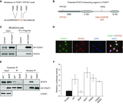
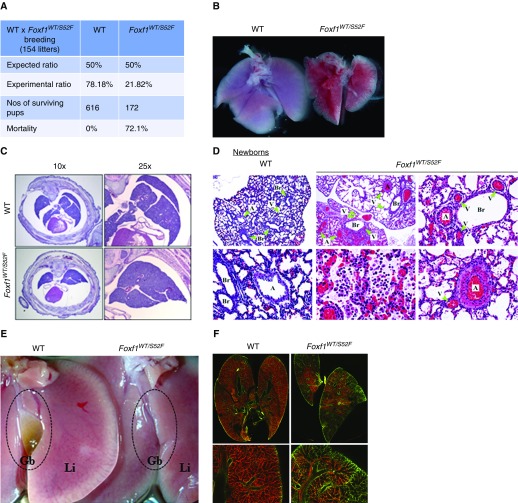
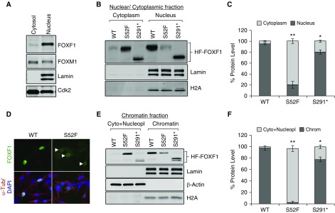
Rationale: Alveolar capillary dysplasia with misalignment of pulmonary veins (ACDMPV) is a lethal congenital disorder causing respiratory failure and pulmonary hypertension shortly after birth. There are no effective treatments for ACDMPV other than lung transplant, and new therapeutic approaches are urgently needed. Although ACDMPV is linked to mutations in the FOXF1 gene, molecular mechanisms through which FOXF1 mutations cause ACDMPV are unknown.Objectives: To identify molecular mechanisms by which S52F FOXF1 mutations cause ACDMPV.Methods: We generated a clinically relevant mouse model of ACDMPV by introducing the S52F FOXF1 mutation into the mouse Foxf1 gene locus using CRISPR/Cas9 technology. Immunohistochemistry, whole-lung imaging, and biochemical methods were used to examine vasculature in Foxf1WT/S52F lungs and identify molecular mechanisms regulated by FOXF1.Measurements and Main Results: FOXF1 mutations were identified in 28 subjects with ACDMPV. Foxf1WT/S52F knock-in mice recapitulated histopathologic findings in ACDMPV infants. The S52F FOXF1 mutation disrupted STAT3-FOXF1 protein-protein interactions and inhibited transcription of Stat3, a critical transcriptional regulator of angiogenesis. STAT3 signaling and endothelial proliferation were reduced in Foxf1WT/S52F mice and human ACDMPV lungs. S52F FOXF1 mutant protein did not bind chromatin and was transcriptionally inactive. Furthermore, we have developed a novel formulation of highly efficient nanoparticles and demonstrated that nanoparticle delivery of STAT3 cDNA into the neonatal circulation restored endothelial proliferation and stimulated lung angiogenesis in Foxf1WT/S52F mice.Conclusions: FOXF1 acts through STAT3 to stimulate neonatal lung angiogenesis. Nanoparticle delivery of STAT3 is a promising strategy to treat ACDMPV associated with decreased STAT3 signaling.

Keywords: ACDMPV; FOXF1 transcription factor; STAT3; alveolar capillary dysplasia with misalignment of pulmonary veins; neonatal pulmonary angiogenesis.


Figures

Figure 1.
Figure 1.
S52F mutation disrupts FOXF1–STAT3 protein–protein interaction and decreases FOXF1 transcriptional activity. (A) Protein sequence shows a frequently mutated region, PPYSY (P49-Y53), located in DNA-binding domain of FOXF1. (B) Schematic diagram shows two potential SH2 domain-binding regions in FOXF1. Consensus STAT3-binding sequences are shown in red. (C) Immunoblots show STAT3 and FOXF1 proteins in immunoprecipitation fractions of MFLM-91 U cells after HF-FOXF1 purification. Cells stably expressing HF-Foxf1 were subjected to two-step affinity purification using anti-Flag Ab followed by nickel affinity columns. Vector alone–transduced cells were used as a control. (D) Immunofluorescent images show colocalization of FOXF1 and STAT3 proteins in nuclei of MFLM-91 U cells. Magnification ×400. (E) Immunoblots show that transfected S52F-FOXF1 protein does not interact with STAT3. Exogenous HF-FOXF1 mutant proteins were detected by α-Flag antibody. (F) S52F FOXF1 mutation inhibits transcriptional activity. Dual-luciferase assay was performed using 6× FOXF1-LUC reporter plasmid (n = 5 in each group). *P< 0.05. IP = immunoprecipitation; WT = wild type.
Figure 2.
Figure 2.
Heterozygous S52F-Foxf1+/− mutation causes increased mortality and pulmonary hypoplasia. (A) Table shows the survival data of Foxf1WT/S52F pups (n = 618) from 154 litters after breeding of Foxf1WT/S52F males and wild-type females. Survival was assessed 1 month after birth. (B) Image shows pulmonary hypoplasia and diffuse hemorrhage in a Foxf1WT/S52F newborn. (C) Hematoxylin and eosin–stained sections of E18.5 embryos show fusion of lung lobes in Foxf1WT/S52F mice. (D) Hematoxylin and eosin staining shows lung inflammation, hemorrhage, hypertrophy of pulmonary arteries, and misalignment of pulmonary veins in Foxf1WT/S52F newborn mice. Green arrowheads show veins, arteries, and bronchioles. (E) Image shows absence of gallbladder in adult Foxf1WT/S52F mouse. (F) P2 pups were intravenously injected with isolectin B4. Perfused lung vasculature was imaged using confocal microscopy (red). Green shows autofluorescence. Magnification: top left and middle panels in D, ×50; bottom middle panel in D, ×400; remaining panels in D, ×200; top panels in F, ×10; bottom panels in F, ×50. A = artery; Br = bronchiole; Gb = gallbladder; Li = liver; V = vein; WT = wild type.
Figure 3.
Figure 3.
Decreased endothelial cell proliferation and STAT3 signaling in Foxf1WT/S52F mice. (A) PECAM1 and FLK1 staining was decreased in lungs of E15.5 Foxf1WT/S52F embryos. Magnification ×200, inserts ×400. (B) Protein and mRNA of Flk1 and Pecam1 were reduced in lungs from E15.5 Foxf1WT/S52F mice as shown by Western blot (top) and qRT-PCR (bottom) (n = 5 embryos in each group were used for qRT-PCR). (C) Decreased pulmonary endothelial cell proliferation in the Foxf1WT/S52F mice is shown using Ki-67 and BrdU immunostaining. Magnification ×400, inserts ×1,600. (D) Graphical representation of cell proliferation by Ki-67 and BrdU staining. Percentage of Ki-67-positive and BrdU-positive cells was counted in 10 random microscope fields (n = 3–6 mice in each group). (E and F) Immunoblots (top) and qRT-PCR (bottom) show gene expression in lungs of Foxf1WT/S52F and Foxf1+/− E18.5 embryos. mRNA was normalized to β-actin mRNA (n = 5 embryos in each group were used for qRT-PCR). *P< 0.05. WT = wild type.
Figure 4.
Figure 4.
FOXF1 stimulates STAT3 expression. (A) Immunostaining shows decreased pSTAT3, Ki-67, FLK1, and Cyclin D1 in human alveolar capillary dysplasia with misalignment of pulmonary veins (ACDMPV) compared with donor lungs (n = 3). Sections show ACDMPV lung associated with A231fs FOXF1 mutation. Green lines show arteries. Black arrowheads show Ki-67+ and Cyclin D1+ cells. Magnification: left, ×200; remaining panels; ×400, inserts ×1,000. A = artery; Br = bronchiole; V = vein. (B) Immunoblots show decreased FOXF1, phospho-STAT3 (Y705), and total STAT3 in FOXF1-depleted MFLM-91 U endothelial cells. FOXF1 levels were decreased by siRNA (siFoxf1). (C) FOXF1 depletion decreases Foxf1, Flk1, Pdgfb, Pecam1, Stat3, Bax, Bcl2, Mmp2, Mmp9, and Ccnd1 mRNAs. Transfection of WT FOXF1 (FOXF1-OE) but not S52F FOXF1 increased gene expression in FOXF1-depleted MFLM-91 U cells. mRNAs were normalized to β-actin (n = 5 cell cultures in each group), *P< 0.05. H&E = hematoxylin and eosin; UTR = untranslated region.
Figure 5.
Figure 5.
S52F-FOXF1 exhibits diminished nuclear localization and decreased binding to chromatin. (A) Immunoblot shows the nucleocytoplasmic distribution of endogenous FOXF1 in MFLM-91 U endothelial cells. Lamin A/C was used to assess nuclear purification. (B and C) Immunoblots show the nucleocytoplasmic distribution of exogenous HF-WT, HF-S52F, and HF-S291* FOXF1 proteins after stable transfection into MFLM-91 U cells. Data were quantified using densitometry (n = 3 immunoblots). (D) Immunofluorescent images show the subcellular distribution of HF-tagged FOXF1 and S52F-FOXF1 proteins in MFLM-91 U cells. Arrowheads show decreased FOXF1 staining in cell nuclei. Magnification ×400. (E and F) Immunoblots show the chromatin association of WT HF-tagged FOXF1 and mutant FOXF1 proteins. Lamin A/C, H2A served as loading controls for the chromatin fractions (n = 3 immunoblots). *P< 0.05 and **P< 0.01. WT = wild type.
Figure 6.
Figure 6.
Nanoparticle-mediated delivery of STAT3 restores endothelial cell proliferation and angiogenesis in Foxf1WT/S52F newborn mice. (A) Fluorescence-activated cell sorter analysis gating strategy for the hematopoietic (a), endothelial (b), epithelial (c), and lineage negative cells (d) with histograms highlighting respective cell-selective targeting (n = 3 mice). (B and C) Immunoblots show the levels of STAT3, pSTAT3, FLK-1, PECAM-1, and PDGFb in lung extracts after nanoparticle-mediated delivery of CMV-STAT3 via facial vein. CMV-empty was used as a control. Nanoparticle/DNA complexes were injected at P2 and mice were harvested at P7. Images were quantified using densitometry (n = 3–6 mice per group). *P< 0.05. (D) qRT-PCR shows the expression of Flk1 and Pecam1 mRNAs in P7 lungs after nanoparticle-mediated delivery of CMV-STAT3 (n = 3–6 mice in each group). (E) Images show the Ki-67 (arrowheads), isolectin B4, and endomucin staining of P7 lungs after nanoparticle-mediated delivery of STAT3. Magnification ×400. (F) Percentage of Ki-67–positive endothelial cells was determined using 10 random Ki-67–stained lung images (n = 3–6 mice in each group). (G) Quantification of endomucin staining was performed using ImageJ software in 10 random images from three to six mouse lungs in each group. **P< 0.01. CMV = cytomegalovirus; NS = not significant; WT = wild type.
Figure 7.
Figure 7.
Schematic diagram shows the proposed molecular mechanisms whereby FOXF1 regulates STAT3 signaling and pulmonary angiogenesis. FOXF1 induces transcription of Stat3 gene leading to increased expression of cell cycle regulatory genes (Ccnb1, Ccnd1, and c-Myc). FOXF1 physically binds the STAT3 protein and these protein-protein interactions are dependent on Serine 52 of FOXF1. Increased STAT3 signaling leads to activation of endothelial cell proliferation and increased angiogenesis. FOXF1 regulates other proangiogenic genes independently of STAT3. S52 = Serine 52.

Comment in

References

    1. Bishop NB, Stankiewicz P, Steinhorn RH. Alveolar capillary dysplasia. Am J Respir Crit Care Med. 2011;184:172–179. - PMC - PubMed
    1. Towe CT, White FV, Grady RM, Sweet SC, Eghtesady P, Wegner DJ, et al. Infants with atypical presentations of alveolar capillary dysplasia with misalignment of the pulmonary veins who underwent bilateral lung transplantation. J Pediatr. 2018;194:158–164. - PMC - PubMed
    1. Stankiewicz P, Sen P, Bhatt SS, Storer M, Xia Z, Bejjani BA, et al. Genomic and genic deletions of the FOX gene cluster on 16q24.1 and inactivating mutations of FOXF1 cause alveolar capillary dysplasia and other malformations Am J Hum Genet 200984780–791.[Published erratum appears in Am J Hum Genet 85:537.] - PMC - PubMed
    1. Sen P, Yang Y, Navarro C, Silva I, Szafranski P, Kolodziejska KE, et al. Novel FOXF1 mutations in sporadic and familial cases of alveolar capillary dysplasia with misaligned pulmonary veins imply a role for its DNA binding domain. Hum Mutat. 2013;34:801–811. - PMC - PubMed
    1. Garabedian MJ, Wallerstein D, Medina N, Byrne J, Wallerstein RJ. Prenatal diagnosis of cystic hygroma related to a deletion of 16q24.1 with haploinsufficiency of FOXF1 and FOXC2 genes. Case Rep Genet. 2012;2012:490408. - PMC - PubMed

Publication types

MeSH terms

Substances

Supplementary concepts