. 2019 Sep;33(9):10049-10064.
doi: 10.1096/fj.201900179RR. Epub 2019 Jun 14.

Timely expression and activation of YAP1 in granulosa cells is essential for ovarian follicle development

Affiliations

Timely expression and activation of YAP1 in granulosa cells is essential for ovarian follicle development

Xiangmin Lv et al. FASEB J. 2019 Sep.

Abstract

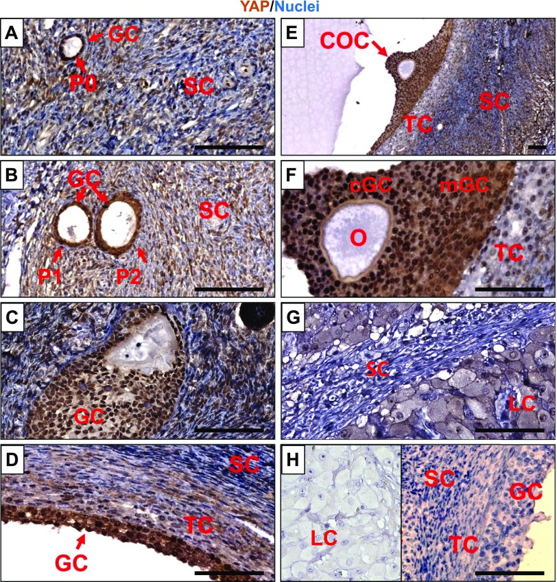
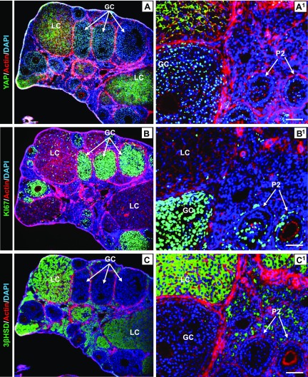
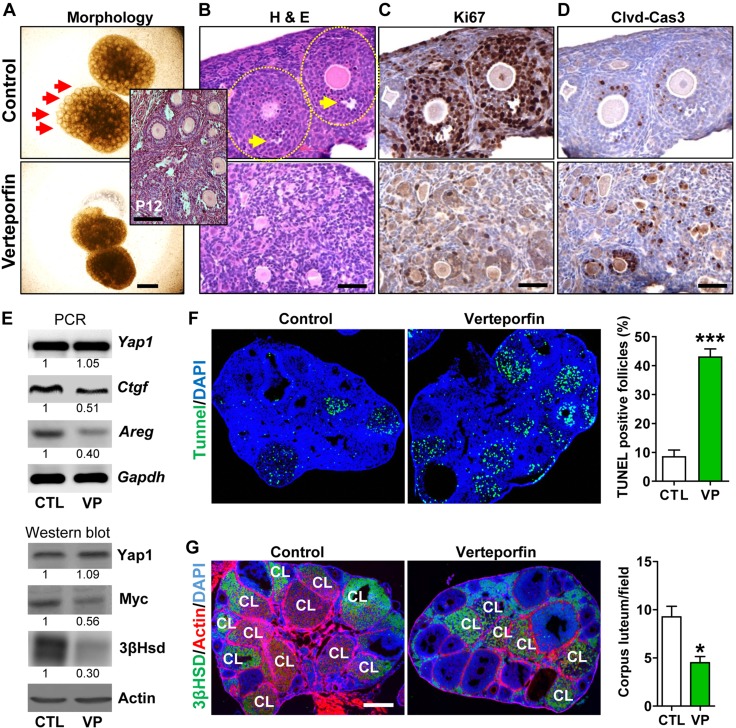
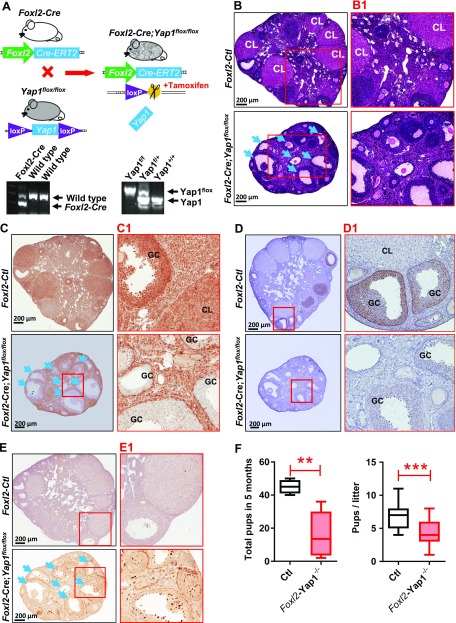
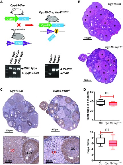
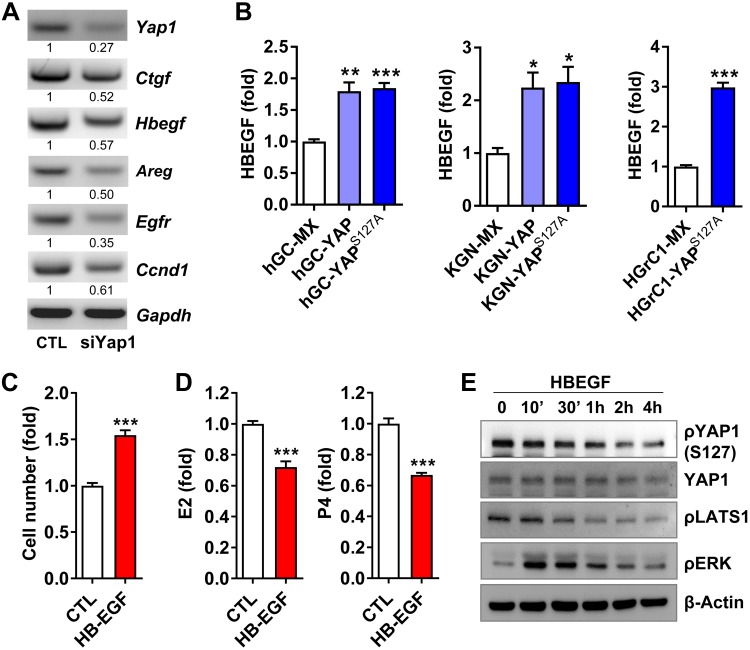
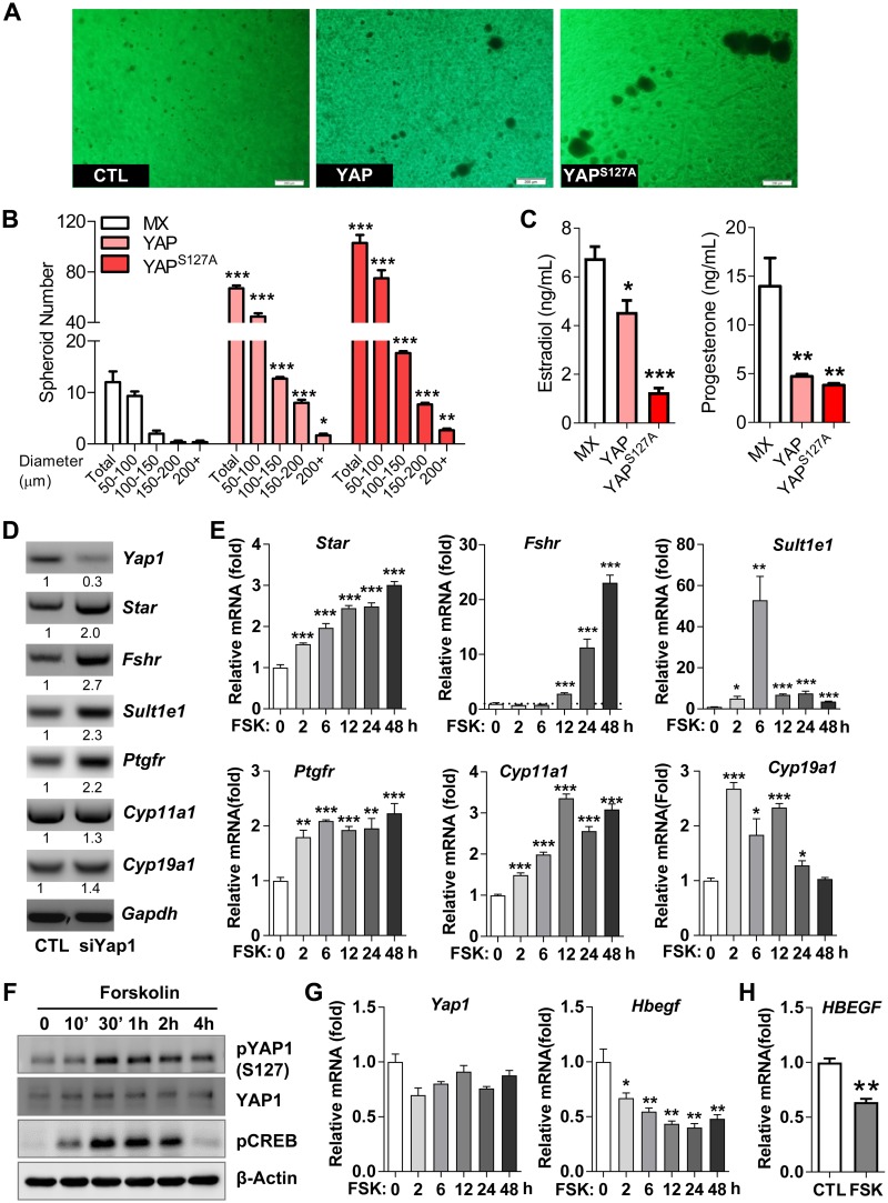
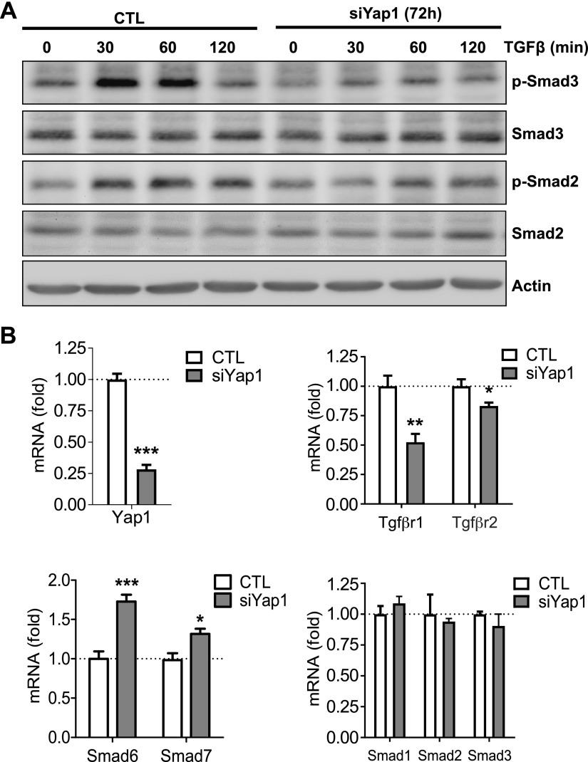
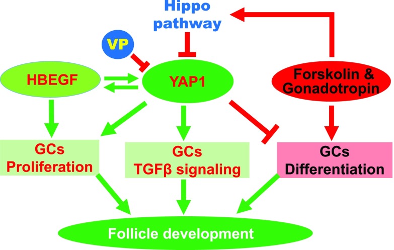
Although the role of the Hippo signaling pathway in development and tumorigenesis has been extensively studied in multiple organs, its role in ovarian follicle development remains largely unknown. Here, we report that Yes-Associated Protein 1 (YAP1), the major effector of Hippo signaling, is spatiotemporally expressed in ovarian granulosa cells and plays a critical role in the regulation of follicle development. We found that the active form of YAP1 (nuclear YAP1) was predominantly expressed in proliferative granulosa cells, whereas the inactive form of YAP1 (cytoplasmic YAP1) was mainly detected in luteal cells (terminally differentiated granulosa cells). Pharmacological inhibition of YAP1 activity disrupted mouse ovarian follicle development in vitro and in vivo. Foxl2 promoter-driven knockout of Yap1 in ovarian granulosa cells resulted in increased apoptosis of granulosa cells, decreased number of corpora lutea, reduced ovarian size, and subfertility in transgenic mice. However, Cyp19a1 promoter-driven knockout of Yap1 in differentiated granulosa cells of preovulatory follicles and luteal cells of corpora lutea had no effect on ovarian morphology and fertility. Mechanistic studies demonstrated that YAP1 interacted with epidermal growth factor receptor and TGF-β signaling pathways to regulate granulosa cell proliferation, differentiation, and survival. Results from this study identify YAP1 as a critical regulator of granulosa cell proliferation and differentiation. Balanced expression and activation of YAP1 is essential for follicle development and successful reproduction. YAP1 is a promising target for treatment of subfertility associated with abnormal granulosa cell function.-Lv, X., He, C., Huang, C., Wang, H., Hua, G., Wang, Z., Zhou, J., Chen, X., Ma, B., Timm, B. K., Maclin, V., Dong, J., Rueda, B. R., Davis, J. S., Wang, C. Timely expression and activation of YAP1 in granulosa cells is essential for ovarian follicle development.

Keywords: Hippo pathway; Steroidogenesis; Yes-Associated Protein 1; fertility.


Conflict of interest statement

The authors thank Eric N. Olson (University of Texas Southwestern Medical Center, Dallas, TX) for providing the Yap1-floxed mouse model. The authors also thank Janice A. Taylor and James R. Talaska of the Advanced Microscopy Core Facility at the University of Nebraska Medical Center for providing assistance with confocal microscopy. This work was supported by a University of Nebraska Medical Center Fellowship; the Eunice Kennedy Shriver National Institute of Child Health and Human Development (5R00HD059985); The National Cancer Institute and U.S. National Institutes of Health (NIH) (1R01CA197976, 1R01CA201500), the Vincent Center for Reproductive Biology, Department of Obstetrics and Gynecology, Massachusetts General Hospital–Harvard Medical School; the Olson Center for Women’s Health at the University of Nebraska Medical Center; The Department of Veteran’s Affairs, and the Colleen’s Dream Foundation. X.L., C. He, and C. Huang are co-first authors. The authors declare no conflicts of interest.

Figures

Figure 1
Figure 1
Expression and localization of YAP1 protein in human ovarian cells. A–G) The chromogenic IHC showing YAP1 protein expression in primordial follicles (A), primary and secondary follicles (B), growing follicles (C), preovulatory follicles (D–F), and corpus luteum (G) of human ovaries. H) Antibody control showing negative staining of YAP1 in granulosa cells, theca cells, and stromal cells in normal ovarian tissues and luteal cells in a corpus luteum (insert). cGC, cumulus granulosa cell; COC, cumulus-oocyte complex; LC, luteal cell; GC, granulosa cell; mGC, mural granulosa cell; O, oocyte; P0, primordial follicle; P1, primary follicle; P2, secondary follicle; SC, stromal cell; TC, theca cell. Scale bars, 100 μm.
Figure 2
Figure 2
Expression and localization of Yap1 protein in mouse ovarian cells. A–C) Fluorescent IHC and confocal microscopy showing expression and localization of Yap1 (A), Ki67 (B), and 3β-Hsd (C) in ovarian cells using series sections of mouse ovarian tissues. The green color in A–C represents expression and localization of Yap1, Ki67, and 3β-HSD in mouse ovarian cells, respectively. A1–C1) Corresponding high-resolution images. Actin filaments were stained with rhodamine-phalloidin. Nuclei were stained with DAPI. GC, granulosa cell; LC, luteal cell; P2, secondary follicle. Scale bars, 100 μm.
Figure 3
Figure 3
Pharmacological inhibition of Yap1 disrupts ovarian follicle development in vitro and in vivo. A) Representative images showing the morphology of postnatal d 12 mouse ovaries incubated for 7 d in the presence or absence of VP (Verteporfin, a selective YAP1 antagonist). Scale bar, 500 μm. B) Hematoxylin and eosin (H&E) stain showing histology of control (CTL) and VP-treated ovaries. Scale bar, 50 μm. The insert image shows the histology of ovaries in postnatal d 12 mice. Please note that only early stage growing follicles (<5 layers of GCs) are present in these ovaries. C, D) Representative IHC images showing expression of Ki67 and cleaved Caspase-3 (Clvd-Cas3) in serial sections of cultured CTL and VP-treated ovarian tissues. Scale bars, 50 μm. E) Representative images showing expression of Yap1, Yap1-targeting genes [connective tissue growth factor (Ctgf), Myc, and Areg], and 3β-Hsd in the CTL and VP-treated ovaries determined by RT-PCR and Western blot. F) TUNEL assay showing the apoptosis of granulosa cells and atresia of follicles in CTL and VP-treated ovarian tissues. Scale bar, 500 μm. The percentages of TUNEL-positive follicles in CTL and VP-treated groups are also presented in a graph next to images. ***P < 0.001 compared with CTL. G) Inhibition of Yap1 decreases the number of corpora lutea in the mouse ovary. 3β-Hsd staining was used to identify the corpus luteum (CL). Scale bar, 500 μm. *P < 0.05 compared with CTL.
Figure 4
Figure 4
Knockout of Yap1 in granulosa cells disrupts ovarian follicle development. A) Top panel: schematic cartoon showing the generation of Foxl2 promoter–driven granulosa cell–specific Yap1-knockout mouse model (Foxl2-CRE;Yap1flox/flox). Lower panel: Representative gel images showing the genotyping PCR products. B) Representative hematoxylin and eosin (H&E) staining images showing the ovarian morphology of control (Ctl) and Foxl2-CRE;Yap1flox/flox mice. Scale bars, 200 μm. C) Representative images showing expression of Yap1 in ovarian cells of the Ctl and Foxl2-CRE;Yap1flox/flox mice. Yap1 protein was determined by IHC staining. Scale bars, 200 μm. D) Representative images showing the phosphorylation of Akt1 determined by IHC in ovarian granulosa cells of Ctl and Foxl2-CRE;Yap1flox/flox mice. Scale bars, 200 μm. E) Representative IHC images showing expression of cleaved caspase-3 in ovarian cells of the Ctl and Foxl2-CRE;Yap1flox/flox mice. Serial sections from ovaries of the Ctl and Foxl2-CRE;Yap1flox/flox mice were used for morphologic and IHC analyses. B1–E1 are high-resolution images of BE, respectively. Arrows indicate follicles with granulosa cell–specific Yap1 gene knockout. Scale bars, 200 μm. F) Accumulated pups and pups/litter in Ctl and Foxl2-CRE;Yap1flox/flox mice. GC, granulosa cell; CL, corpus luteum. **P < 0.01, ***P < 0.001, compared with Ctl.
Figure 5
Figure 5
Knockout of Yap1 in luteinized granulosa cells has limited effect on mouse ovarian follicle development and fertility. A) Top panel: schematic cartoon showing the generation of Cyp19 promoter–driven GC-specific Yap1-knockout mouse model (Cyp19-CRE;Yap1flox/flox). Lower panel: Representative gel images showing the genotyping PCR products. B) Representative images [hematoxylin and eosin (H&E) staining] showing the ovarian morphology of control (Ctl) and Cyp19-CRE;Yap1flox/flox mice. Each astrisk marks a corpus luteum. Scale bars, 500 μm. C) Top panel: representative images showing Yap1 expression in ovarian tissues of Ctl and Cyp19-CRE;Yap1flox/flox mice. Yap1 was stained using IHC. Nuclei were counterstained with hematoxylin. Scale bars, 500 μm. Lower panel: high-resolution images showing expression of Yap1 protein in ovarian tissues of the Ctl and Cyp19-CRE;Yap1flox/flox mice. Scale bars, 100 μm. D) Accumulated pups and litter size in Ctl and Cyp19-CRE;Yap1flox/flox mice. GC, granulosa cell; LC, luteal cell; ns, no significance. P>0.05.
Figure 6
Figure 6
YAP1 interacts with EGFR signaling pathway to drive granulosa cell proliferation. A) Knockdown of Yap1 in mouse granulosa cells reduced mRNA levels of Egfr, EGF-like ligands (Hbegf and Areg), and cyclin D1 (Ccnd1). Relative mRNA levels were determined using RT-PCR. The experiment was repeated ≥3 times, and representative images are presented. B) Expression of HBEGF in hGCs, KGN cells, and HGrC1 cells transfected with empty vectors [MX as control (CTL)], vectors expressing wild-type YAP1 (YAP), or constitutively active YAP1 (YAPS127A). Relative mRNA levels were quantified using real-time PCR. Each bar represents the mean ± sem (n = 4). C, D) HBEGF treatment (50 ng/ml, 3 d) stimulated cell proliferation (C), but suppressed estradiol (E2) and progesterone (P4) production (D) in cultured granulosa cells. Each bar represents the mean ± sem (n = 4). E) HBEGF (50 ng/ml) induced rapid dephosphorylation of LATS1 (S909) and YAP1 (S127) and phosphorylation of ERK1/2 in cultured granulosa cells. β-Actin protein was used as loading CTL. The relative levels of the total proteins and phosphoproteins were determined by Western blot. Ctgf, connective tissue growth factor; Gapdh, glyceraldehyde 3-phosphate dehydrogenase; p, phosphorylated. Experiments were repeated ≥3 times, and representative images are presented. *P < 0.05, **P < 0.01, ***P < 0.001, compared with corresponding CTL groups (MX groups).
Figure 7
Figure 7
YAP1 stimulates proliferation but suppresses differentiation of granulosa cells. A) Representative images showing spheroids formed in KGN-MX, KGN-YAP, and KGN-YAPS127A cells in a 3D culture system. Scale bars, 200 μm. B) Numbers of spheroids with different diameters formed by KGN-MX, KGN-YAP, and KGN-YAPS127A cells in the 3D culture system in A. Each bar represents the mean ± sem (n = 4). *P < 0.05, **P < 0.01, ***P < 0.001 compared with corresponding control (CTL). C) Production of E2 and progesterone in KGN-MX, KGN-YAP, and KGN-YAPS127A cells in the 3D culture system. *P < 0.05, **P < 0.01, ***P < 0.001 compared with MX control. D) Knockdown of Yap1 with Yap1-specific siRNA (siYap) in cultured mouse granulosa cells up-regulated the expression of granulosa cell differentiation–associated genes. The relative mRNA levels of Star, Fshr, Sult1e1, Ptgfr, cyp11A1, cyp19a1, and Gapdh were determined by RT-PCR. E) Forskolin (FSK) induced expression of differentiation-associated genes in cultured primary mouse granulosa cells. The relative mRNA levels of Yap1, Star, Fshr, Sult1e1, Ptgfr, cyp11a1, and cyp19a1 in mouse granulosa cells treated with 10 μM FSK for the indicated time points were determined by quantitative PCR. *P < 0.05, **P < 0.01, ***P < 0.001 compared with CTL. F) FSK (10 μM) induced phosphorylation of YAP1 in cultured HGrC1 cells. Please note that phosphorylation results in inactivation of YAP1 protein. The relative levels of the total proteins and phosphoproteins were determined by Western blot. All experiments were repeated ≥3 times, and representative images were presented. G) FSK (10 μM) suppressed the expression of Hbegf mRNA in cultured primary mouse granulosa cells in a time-dependent manner. Each bar represents the mean ± sem (n = 4). *P < 0.05, **P < 0.01 compared with CTL. H) FSK (10 μM, 72 h) suppressed HBEGF mRNA expression in cultured primary hGCs. CREB, cAMP responsive element binding protein; Gapdh, glyceraldehyde 3-phosphate dehydrogenase; p, phosphorylated. Each bar represents the mean ± sem (n = 4). **P < 0.01 compared with CTL.
Figure 8
Figure 8
Knockdown of Yap1 compromises Tgf-β signaling in mouse ovarian granulosa cells. A) Representative blots showing alteration of TGF-β1–induced phosphorylation of Smad2 and Smad3 in mouse granulosa cells with or without Yap1 knockdown. The relative levels of the total proteins and phosphoproteins were determined by Western blot. Experiments were repeated ≥3 times. B) Relative mRNA levels of Tgfßr1, Tgfßr2, Smad6, Smad7, Smad1, Smad2, and Smad3 in ovarian granulosa cells with or without Yap1 knockout. p, phosphorylated; siYap, Yap1-specific siRNA. Each bar represents the mean ± sem (n = 4). *P < 0.05, **P < 0.01, ***P < 0.001 compared with corresponding control (CTL).
Figure 9
Figure 9
A schematic illustration showing the interactions between YAP1 and HBEGF, gonadotropin signaling transduction, and TGF-β signaling pathways in regulating granulosa cell (GC) proliferation and differentiation and, thereby, follicle development. In the growing follicle, YAP1 and HBEGF form a feedforward loop to drive GC proliferation. Activation of gonadotropin signaling triggers the Hippo pathway and phosphorylates YAP1, leading to differentiation of GCs. YAP1 activity is also essential for appropriate TGF-β signal transduction in GCs, which is critical for follicle development and oocyte maturation. Therefore, our study suggests that timely expression and activation of YAP1 is essential for GC function and ovarian follicle development.

References

    1. Hsueh A. J., Kawamura K., Cheng Y., Fauser B. C. (2015) Intraovarian control of early folliculogenesis. Endocr. Rev. 36, 1–24 - PMC - PubMed
    1. Stocco C., Telleria C., Gibori G. (2007) The molecular control of corpus luteum formation, function, and regression. Endocr. Rev. 28, 117–149 - PubMed
    1. Meignin C., Alvarez-Garcia I., Davis I., Palacios I. M. (2007) The salvador-warts-hippo pathway is required for epithelial proliferation and axis specification in Drosophila. Curr. Biol. 17, 1871–1878 - PMC - PubMed
    1. Polesello C., Tapon N. (2007) Salvador-warts-hippo signaling promotes Drosophila posterior follicle cell maturation downstream of notch. Curr. Biol. 17, 1864–1870 - PubMed
    1. Huang J., Kalderon D. (2014) Coupling of Hedgehog and Hippo pathways promotes stem cell maintenance by stimulating proliferation. J. Cell Biol. 205, 325–338 - PMC - PubMed

Publication types

MeSH terms