Review
. 2019 Sep;92(1101):20170980.
doi: 10.1259/bjr.20170980. Epub 2019 Jun 20.

MDCT prior to median re-sternotomy in cardiovascular surgery: our experiences, infrequent findings and the crucial role of radiological report

Affiliations
Review

MDCT prior to median re-sternotomy in cardiovascular surgery: our experiences, infrequent findings and the crucial role of radiological report

Tullio Valente et al. Br J Radiol. 2019 Sep.

Abstract

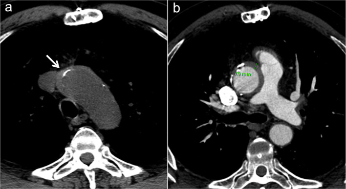
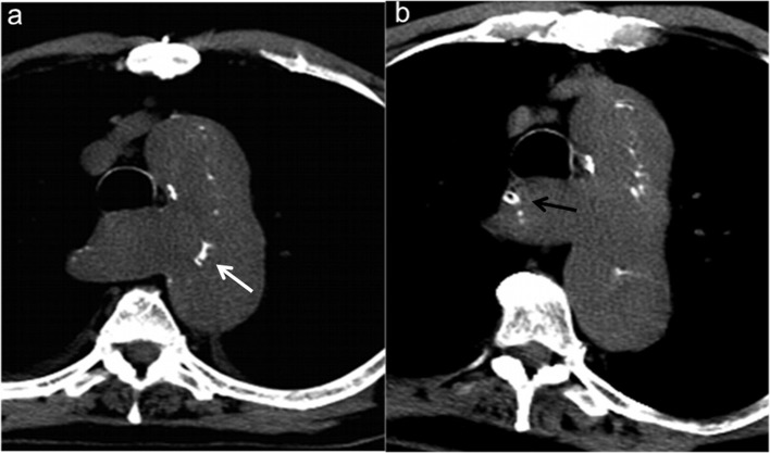
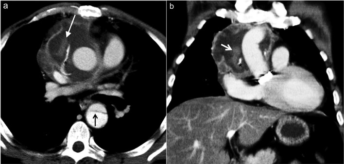
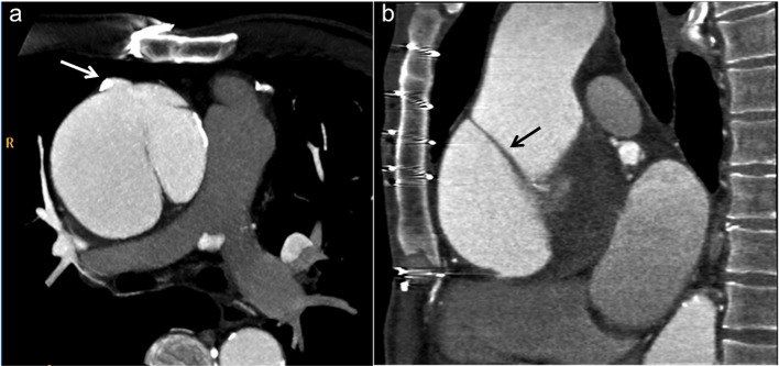
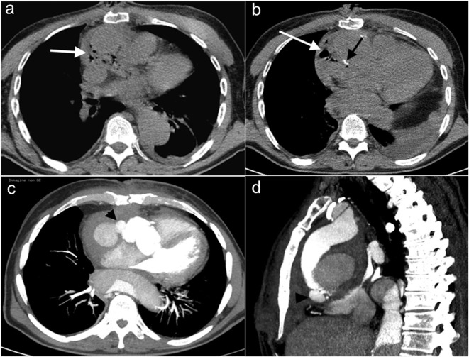
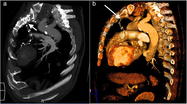
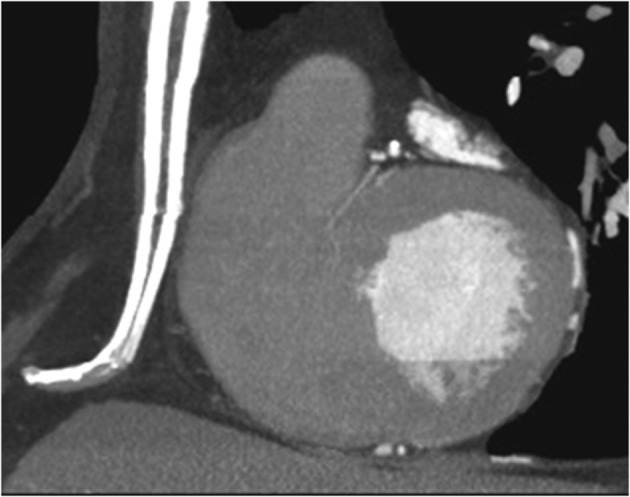
Resternotomy (RS) is a common occurrence in cardiac surgical practice. It is associated with an increased risk of injury to old conduits, cardiac structures, catastrophic hemorrhage and subsequent high morbidity and mortality rate in the operating room or during the recovery period. To mitigate this risk, we evaluated the role of multidetector CT (MDCT) in planning repeat cardiac surgery. We evaluated sternal compartment abnormalities, sternal/ascending aorta distance, pre-reoperative assessment of the aorta (wall, diameters, lumen, valve), sternal/right ventricle distance, diaphragm insertion, pericardium and cardiac chambers, sternal/innominate vein distance, connection of the grafts to the predicted median sternotomy cut, graft patency and anatomic course, possible aortic cannulation and cross-clamping sites and additional non-cardiovascular significant findings. Based on the MDCT findings, surgeons employed tailored operative strategies, including no-touch technique, clamping strategy and cardiopulmonary bypass (CPB) via peripheral cannulation assisted resternotomy. Our experience suggests that MDCT provides information which contributes to the safety of re-operative heart surgery reducing operative mortality and adverse outcomes. The radiologist must be aware of potential surgical options, including in the report any findings relevant to possible resternotomy complications.


Figures

Figure 1a–b.
Figure 1a–b.
Normal sternotomy and midsternal lucent stripe. (a) VR reconstruction shows normal median sternotomy; (b) VR reconstruction shows midsternal lucent stripe >3 mm (arrow) 12 months after undergoing sternotomy. This finding may be indicative of dehiscence only if the stripe is thicker than 3 mm. VR, volumetric rendering.
Figure 2a–b.
Figure 2a–b.
Retrosternal adhesions findings. Axial MIP reconstruction (a) shows an irregularly triangular shape adhesion with anterior large apex between the sternum and the retrosternal structures (“peak sign”); MDCT axial scan (b) demonstrates severe adhesions by retrosternal structures flattened over the sternum ("flat sign“). MDCT, multidetector CT; MIP, maximum intensity projection.
Figure 3a–c.
Figure 3a–c.
Relations between sternum and vascular/cardiac structures. (a) Axial MIP reconstruction shows S/RV critical contiguity (<3 mm). (b) Sagittal MIP reconstruction shows risk of AA damage (S/AA ≤5 mm) (arrow). (c) Axial MIP reconstruction shows sternum/ left innominate vein critical contiguity (<3 mm). AA, ascending aorta; MIP, maximum intensity projection; RV, right ventricle; S, sternal.
Figure 4a–b.
Figure 4a–b.
Diffuse atherosclerotic disease of the aorta. (a) Coronal MPR reconstruction and (b) oblique sagittal MIP reconstruction show areas of ascending aortic heavily calcifications (arrow) as at high risk for embolization with surgical clamping and manipulation. MIP, maximum intensity projection; MPR, Multiplanar reformation.
Figure 5a–b.
Figure 5a–b.
Pre-operative assessment in a 58-year-old male with unstable angina and a history of two prior coronary artery bypass graftings. (a) Unenhanced and (b) enhanced axial MDCT images show a high density aortic wall suggestive for acute intramural hematoma (IMH) associated to displaced calcifications (white arrow). MDCT, multidetector CT.
Figure 6a–b.
Figure 6a–b.
A 71-year-old male who presented with acute chest pain 4 months after undergoing composite graft repair for ascending aortic aneurysm. Unenhanced axial MDCT images (a, b) show displacement of calcifications (white arrow) extending to a dissected lusory artery (black arrow). MDCT, multidetector CT.
Figure 7a–b.
Figure 7a–b.
A 56-year-old Marfan patient contrast-enhanced MDCT after Bentall procedure for A type aortic dissection. Axial (a) an coronal (b) MIP reconstructions show hyperdense dislodged surgical material (asterisk) and a contrast medium leakage (white arrow) surrounded by a organized serosanguineous collection; note dissection flap in descending aorta (black arrow). MDCT, multidetector CT; MIP, maximum intensity projection.
Figure 8a–b.
Figure 8a–b.
Assessment of a 53-year-old male with a history of CABG and subsequent development of a dissecting aneurysm of the ascending aorta who presented to the emergency department with chest pain. Axial (a) and sagittal (b) MIP reconstructions show a Stanford type A chronic slow-growing dissecting aneurysm with aortic valve insufficiency and high mediastinal occupying rate. Note the flat and fixed appearance of the fibrotic immobile and thickened flap (black arrow) characteristic of a chronic dissection and the site of the venous graft in the ascending aorta (white arrow). The patient underwent to resternotomy for button Bentall procedure. CABG, coronary artery bypass grafting; MIP, maximumintensity projection.
Figure 9a–d.
Figure 9a–d.
Infection and graft anastomotic dehiscence in a 70-year-old male who had undergone replacement of the ascending aorta with biological valve for acute type A aortic dissection 7 months earlier. A 2 months follow-up unenhanced axial CT scan examination was performed in another institution to evaluate for the aetiology of night sweats, mild chills, and fever. (a, b) Unenhanced MDCT axial scans show abnormal low-attenuation perigraft material and multiple gas bubbles (white arrows) near the graft anastomosis (black arrow); these findings suggesting purulent fluid due to graft infection were misinterpreted as a normal postoperative pattern. After other 2 months this patient came to our observation for fever and worsening dyspnoea and was performed an emergent MDCT. (c) Axial and (d) sagittal coronal MIP reconstructions show active contrast material extravasation (black arrowhead) arising from proximal graft anastomotic dehiscence. MDCT, multidetector CT; MIP, maximum intensity projection.
Figure 10a–b.
Figure 10a–b.
Pre-operative assessment of a 64-year-old male who had undergone a previous CABG, before repeat CABG and AVR. (a) Sagittal oblique MIP reconstructions and (b) 3D VR sagittal reconstruction shows that the left ITA is adherent to the sternum at the midline behind the manubrium, adjacent to the highest sternal suture (arrow); further down it continues with a torturous way toward its anastomosis with the LAD artery. ITA, internal thoracic artery; CABG, coronary artery bypassgrafting; LAD, left anterior descending;MIP, maximum intensity projection.
Figure 11.
Figure 11.
MIP sagittal reconstruction shows that, due to high insertion of the diaphragm, the sternotomy should spare the distal 2–3 cm of the sternum. MIP, maximum intensity projection.

Similar articles

Cited by

References

    1. Morales DLS, Zafar F, Arrington KA, Gonzalez SM, McKenzie ED, Heinle JS, et al. . Repeat sternotomy in congenital heart surgery: no longer a risk factor. Ann Thorac Surg 2008; 86: 897–902. doi: 10.1016/j.athoracsur.2008.04.044 - DOI - PubMed
    1. Morales D, Williams E, John R. Is resternotomy in cardiac surgery still a problem?☆. Interactive CardioVascular and Thoracic Surgery 2010; 11: 277–86. doi: 10.1510/icvts.2009.232090 - DOI - PubMed
    1. American Heart Association Heart disease and stroke statistics 2009 update. A report from the American Heart Association statistics committee and stroke statistics Committee. Circulation 2009; 119: e21–181. - PubMed
    1. Cremer J, Teebken OE, Simon A, Hutzelmann A, Heller M, Haverich A, et al. . Thoracic computed tomography prior to redo coronary surgery. Eur J Cardiothorac Surg 1998; 13: 650–4. doi: 10.1016/S1010-7940(98)00087-6 - DOI - PubMed
    1. Gilkeson RC, Markowitz AH, Ciancibello L. Multisection CT evaluation of the reoperative cardiac surgery patient. RadioGraphics 2003; 23(suppl_1): S3–S17. doi: 10.1148/rg.23si035505 - DOI - PubMed

MeSH terms