. 2019 Aug;33(8):9030-9043.
doi: 10.1096/fj.201800263RR. Epub 2019 Jun 14.

Liver disease-associated keratin 8 and 18 mutations modulate keratin acetylation and methylation

Affiliations

Liver disease-associated keratin 8 and 18 mutations modulate keratin acetylation and methylation

Kwi-Hoon Jang et al. FASEB J. 2019 Aug.

Abstract

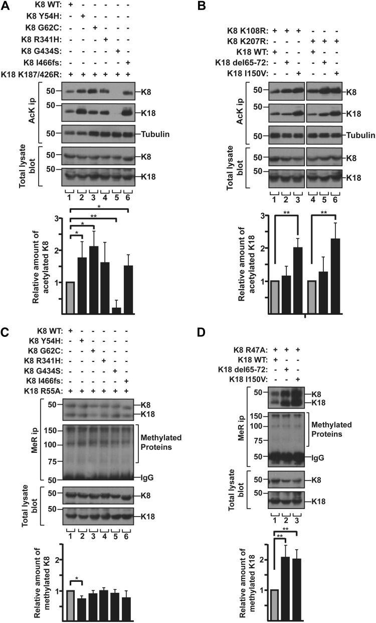
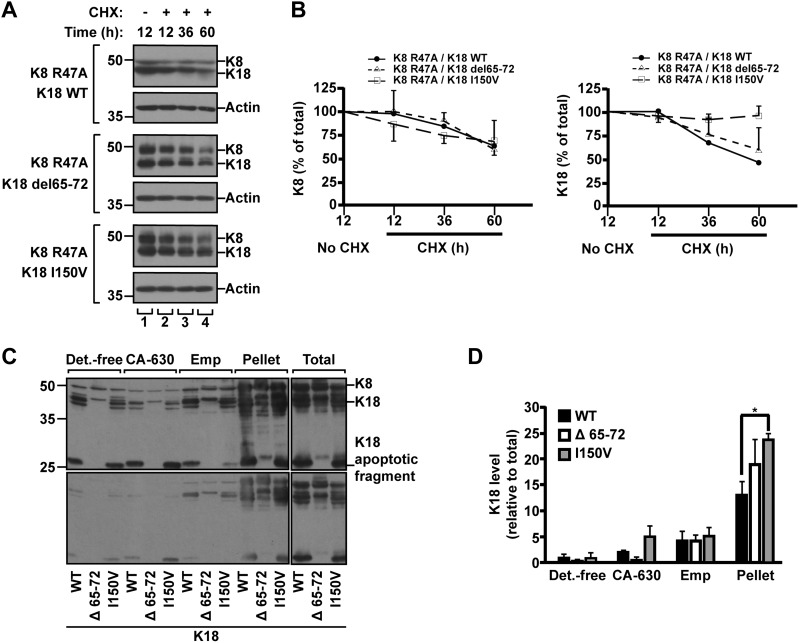
Keratin 8 (K8) and keratin 18 (K18) are the intermediate filament proteins whose phosphorylation/transamidation associate with their aggregation in Mallory-Denk bodies found in patients with various liver diseases. However, the functions of other post-translational modifications in keratins related to liver diseases have not been fully elucidated. Here, using a site-specific mutation assay combined with nano-liquid chromatography-tandem mass spectrometry, we identified K8-Lys108 and K18-Lys187/426 as acetylation sites, and K8-Arg47 and K18-Arg55 as methylation sites. Keratin mutation (Arg-to-Lys/Ala) at the methylation sites, but not the acetylation sites, led to decreased stability of the keratin protein. We compared keratin acetylation/methylation in liver disease-associated keratin variants. The acetylation of K8 variants increased or decreased to various extents, whereas the methylation of K18-del65-72 and K18-I150V variants increased. Notably, the highly acetylated/methylated K18-I150V variant was less soluble and exhibited unusually prolonged protein stability, which suggests that additional acetylation of highly methylated keratins has a synergistic effect on prolonged stability. Therefore, the different levels of acetylation/methylation of the liver disease-associated variants regulate keratin protein stability. These findings extend our understanding of how disease-associated mutations in keratins modulate keratin acetylation and methylation, which may contribute to disease pathogenesis.-Jang, K.-H., Yoon, H.-N., Lee, J., Yi, H., Park, S.-Y., Lee, S.-Y., Lim, Y., Lee, H.-J., Cho, J.-W., Paik, Y.-K., Hancock, W. S., Ku, N.-O. Liver disease-associated keratin 8 and 18 mutations modulate keratin acetylation and methylation.

Keywords: MDB; intermediate filament; post-translational modification; protein stability.


Conflict of interest statement

This work was supported by the Korean Ministry of Education, Science, and Technology Grants 2016R1A2B4012808 and 2018R1D1A1A02086060, the Yonsei University Research Fund 2018-22-0072 (to N.-O.K.), and the National Research Foundation of Korea funded by the Ministry of Science and Information and Communication Technology (ICT) Grant NRF-2016R1A5A1010764 (to J.-W.C.). The authors declare no conflicts of interest.

Figures

Figure 1
Figure 1
Verification of PTMs of K8 and K18 in vivo after okadaic acid (OA), TSA/MS-275/nicotinamide, or CORM/hemin treatment. A) HT29 cells were treated with phosphatase inhibitor (OA; 1 μg/ml, 2 h). K8/K18 were immunoprecipitated using L2A1 Ab. K8/K18 immunoprecipitates were analyzed by SDS-PAGE, followed by Coomassie Blue staining. Hyperphosphorylated keratins were detected as blurred bands just above the major K8 or K18 bands. B) HT29 cells were treated with deacetylase inhibitors (TSA/MS-275/nicotinamide; 2 μM, 5 μM, and 20 mM for 24 h). Acetylated proteins were immunoprecipitated with anti-AcK Ab and separated by SDS-PAGE followed by immunoblotting with anti-K8 or K18 Ab. The level of K8 acetylation was enhanced after inhibitor treatment, whereas the level of K18 acetylation was similar to that of basal conditions. C) HT29 cells were incubated with enhancers of Arg methylation (CORM/hemin; 100 and 10 μM for 10 h). Using anti-mono-MeR antibody, anti-symmetric di-methylated Arg antibody (SYM10) and anti-asymmetric di-methylated Arg antibody (ASYM24), both methylome and methylated keratins were confirmed from total lysate and high salt extracted fraction, respectively. Actin blot for total lysates and Coomassie staining for high salt extracted were used as loading control. HSE, high salt extracted.
Figure 2
Figure 2
Identification of acetylation/methylation sites of K8 and K18 using acetylation and methylation mutant keratins. A) BHK cells were cotransfected with K18 WT and K8 acetylation mutant construct (Lys-to-Arg mutant; K108R or K207R), or with K8 WT and K18 acetylation mutant construct (K187R or K426R). Cells were treated with TSA/MS-275/nicotinamide and harvested after 24 h. Levels of acetylated K8 and K18 were verified by immunoprecipitation using anti-AcK Ab, followed by immunoblotting with anti-K8 and anti-K18 antibodies. Tubulin blot was used as a loading control. B) K8 K108R or K8 K207R together with K18 K187/426R were transfected in BHK cells. Transfected cells were analyzed as described in A. The graph represents means ± sd of 3 independent experiments. C) Comparison of the sequence adjacent to methylation motif (RGG; Arg-Gly-Gly) site of keratins with other proteins known to be methylated. RGG and RG sequences are highlighted in gray. hnRNP A1, heterogeneous nuclear RNP A1; hnRNP K, heterogeneous nuclear RNP K; TAF15, TATA-box binding protein associated factor 15; ERa, estrogen receptor α; p53, tumor protein 53; Smad6, smad family member 6; asterisk, Arg residue modified by methylation; aDMA, asymmetric dimethylated Arg; sDMA, symmetric dimethylated Arg. D) BHK cells were cotransfected with either combination of K18 WT and K8 methylation mutant construct (K8 R47A or R47K) or the combination of K8 WT and K18 methylation mutant construct (K18 R55A or R55K). Levels of MMA K8 and K18 were verified by immunoprecipitation using anti-MeR Ab, followed by immunoblot with anti-K8/K18 Ab. MMA proteins were used as loading control. E) BHK cells were transfected with K8 and 18 WT, or combination of methylation mutant K8 and K18 mutant construct. Transfected cells were analyzed as described in D. The graph represents means ± sd of 3 independent experiments. *P < 0.05, **P < 0.005, ***P < 0.0005.
Figure 3
Figure 3
Mutation of acetylation or methylation sites on keratins alters their protein stability. A) BHK cells were cotransfected with indicated keratin constructs and treated with CHX for 12, 36, or 60 h. Total lysates were separated by SDS-PAGE, followed by immunoblotting with anti-K8/K18 Ab. B, C) Quantity of K8 and K18 normalized by actin quantitatively analyzed by densitometry. Graph represented as means ± sd. Note: stability of acetylation mutant K8 and K18 showed a limited change, compared with WT, but methylation mutants K8 and K18 exhibited significant reduction in both Arg-to-Ala and Arg-to-Lys mutants, compared with WT. D) BHK cells were transfected with K8 and K18 WT and then were treated with DMSO as a solvent control or the indicated methylation inhibitors for 48 h. Levels of methylated K8 and K18 were verified by immunoprecipitation using anti-K8/K18 Ab, followed by immunoblotting with anti-MeR Ab. Note that the level of keratin methylation was dramatically decreased after AdOX treatment. The graph represents means ± sd from 3 independent experiments. *P < 0.05. E) BHK cells were cotransfected with the indicated keratin constructs and were treated with DMSO or AdOX (20 µM) for 24 or 48 h. The cells were then fixed and stained with anti-K8/K18 Ab (red) and DAPI (blue). Images were obtained using fluorescence microscopy equipped with a 40 × /1.3 oil objective lens. Cells with normal (N) filament network and cells with collapsed (C) filament network were counted in 2–3 independent experiments, and at least 55 cells were counted per each experiment. Graph shows the % ratio of N and C cells and represents the means ± sd. Scale bar, 10 µm. *P < 0.05, **P < 0.005. F) Representative confocal images of Fig. 2E. Arrows indicate C cells. Scale bar, 20 µm.
Figure 4
Figure 4
Schematic diagram representing human liver disease-related keratin variants and acetylation/methylation sites in K8 and K18. Keratin proteins are composed of 3 domains: conserved α-helical rod domain flanked by nonhelical head and tail domains. Rod domain is composed of subdomains IA, IB, IIA, and IIB, connected by linker1 (L1), linker12 (L12), and linker2 (L2). Shaded region within IA and IIB indicates highly conserved region. Previously described genetic variants of K8 (A) and K18 (B) related to liver diseases (50, 51). PTM sites examined herein. a, acetylation; m, methylation.
Figure 5
Figure 5
Human liver disease–related keratin mutations alter the acetylation/methylation state. A) BHK cells were cotransfected with the combination of K18 acetylation mutant K187/426R and indicated K8 constructs. The level of acetylated K8 and K18 was verified by immunoprecipitation using anti-AcK Ab, followed by immunoblotting with anti-K8 and anti-K18 antibodies. Acetylated keratins were quantitatively analyzed after 3 independent experiments. Results are represented as means ± sd. B) Acetylation mutant K8 K108R or K207R were cotransfected with the indicated K18 constructs in BHK cells. Acetylated keratins were quantitatively analyzed as described in A. C) BHK cells were cotransfected with K18 methylation mutant R55A and the indicated K8 constructs. Levels of MMA K8 and K18 were verified by immunoprecipitation using anti-MeR Ab, followed by immunoblot with anti-K8/K18 Ab. MMA keratins were quantitatively analyzed after 3 independent experiments. Results are represented as means ± sd. D) K8 methylation mutant R47A were transfected with the indicated K18 constructs in BHK cells. Transfected cells were analyzed as described in C. *P < 0.05, **P < 0.005.
Figure 6
Figure 6
Protein stability and solubility of highly methylated human liver disease–related K18 variants. A, B) BHK cells transfected with the indicated keratin constructs were incubated with CHX for 12, 36, or 60 h. Total lysates were separated by SDS-PAGE, followed by immunoblotting with anti-K8/K18 Ab (A). Amounts of K8 and K18 were quantitatively analyzed by densitometry, and graphs were represented as means ± sd (B). C, D) BHK cells were cotransfected with a combination of indicated K18 constructs together with K8 WT. Transfected cells were used for the preparation of sequential subcellular fractions (15). Detergent-free buffer (Det.-free), nonionic (CA-630), ionic (Emp) detergent, remaining pellet and total lysates sample were prepared. Total lysates and cell fractions were separated by SDS-PAGE, followed by immunoblotting with anti-K8/K18 Ab. C) Upper and lower panels represent long and short exposures of the immunoblotted membrane, respectively. D) Amounts of K8/K18 and K18 human variants were quantitatively analyzed by densitometry. Note: K18 I150V variant showed significant increase in insoluble pellet fraction compared with K18 WT. The graph represents the means ± sd of 3 independent experiments. *P < 0.05.

Similar articles

Cited by

References

    1. Eriksson J. E., Dechat T., Grin B., Helfand B., Mendez M., Pallari H. M., Goldman R. D. (2009) Introducing intermediate filaments: from discovery to disease. J. Clin. Invest. 119, 1763–1771 - PMC - PubMed
    1. Schweizer J., Bowden P. E., Coulombe P. A., Langbein L., Lane E. B., Magin T. M., Maltais L., Omary M. B., Parry D. A., Rogers M. A., Wright M. W. (2006) New consensus nomenclature for mammalian keratins. J. Cell Biol. 174, 169–174 - PMC - PubMed
    1. Ku N. O., Strnad P., Zhong B. H., Tao G. Z., Omary M. B. (2007) Keratins let liver live: mutations predispose to liver disease and crosslinking generates Mallory-Denk bodies. Hepatology 46, 1639–1649 - PubMed
    1. Omary M. B., Ku N. O., Strnad P., Hanada S. (2009) Toward unraveling the complexity of simple epithelial keratins in human disease. J. Clin. Invest. 119, 1794–1805 - PMC - PubMed
    1. Ku N. O., Strnad P., Bantel H., Omary M. B. (2016) Keratins: biomarkers and modulators of apoptotic and necrotic cell death in the liver. Hepatology 64, 966–976 - PMC - PubMed

Publication types

MeSH terms