. 2019 Jun 14;17(6):e3000316.
doi: 10.1371/journal.pbio.3000316. eCollection 2019 Jun.

Protein interactions and consensus clustering analysis uncover insights into herpesvirus virion structure and function relationships

Affiliations

Protein interactions and consensus clustering analysis uncover insights into herpesvirus virion structure and function relationships

Anna Hernández Durán et al. PLoS Biol. .

Abstract

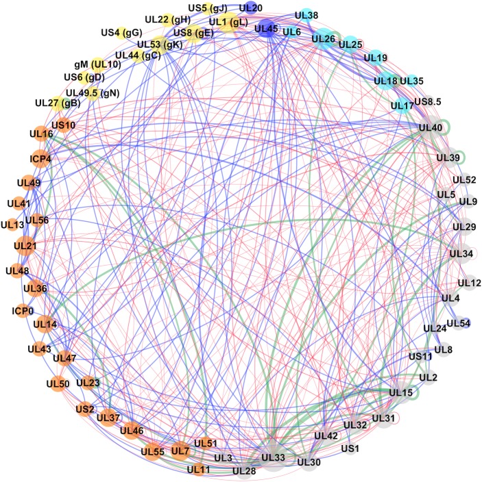
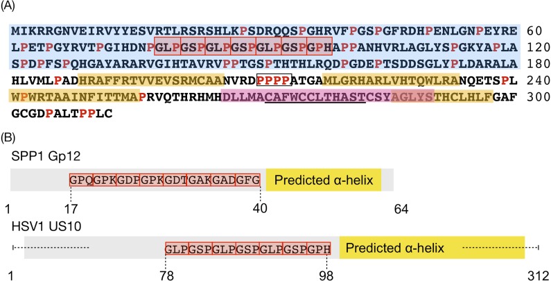
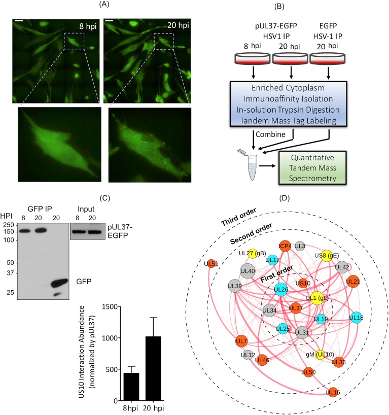
Infections with human herpesviruses are ubiquitous and a public health concern worldwide. Current treatments reduce the severity of some symptoms associated to herpetic infections but neither remove the viral reservoir from the infected host nor protect from the recurrent symptom outbreaks that characterise herpetic infections. The difficulty in therapeutically tackling these viral systems stems in part from their remarkably large proteomes and the complex networks of physical and functional associations that they tailor. This study presents our efforts to unravel the complexity of the interactome of herpes simplex virus type 1 (HSV1), the prototypical herpesvirus species. Inspired by our previous work, we present an improved and more integrative computational pipeline for the protein-protein interaction (PPI) network reconstruction in HSV1, together with a newly developed consensus clustering framework, which allowed us to extend the analysis beyond binary physical interactions and revealed a system-level layout of higher-order functional associations in the virion proteome. Additionally, the analysis provided new functional annotation for the currently undercharacterised protein pUS10. In-depth bioinformatics sequence analysis unravelled structural features in pUS10 reminiscent of those observed in some capsid-associated proteins in tailed bacteriophages, with which herpesviruses are believed to share a common ancestry. Using immunoaffinity purification (IP)-mass spectrometry (MS), we obtained additional support for our bioinformatically predicted interaction between pUS10 and the inner tegument protein pUL37, which binds cytosolic capsids, contributing to initial tegumentation and eventually virion maturation. In summary, this study unveils new, to our knowledge, insights at both the system and molecular levels that can help us better understand the complexity behind herpesvirus infections.


Conflict of interest statement

The authors have declared that no competing interests exist.

Figures

Fig 1
Fig 1. Network assembly framework.
(A and B) PPI data for a total of 12 herpesvirus species (nine human and three nonhuman herpesviruses, together covering members of all three subfamilies, i.e., the Alpha-, Beta-, and Gammaherpesvirinae, S1 and S2 Tables) were collected from seven public resources [21]. (C) PPIs detected in any of its orthologous herpesvirus species (oPPIs) were used to predict PPIs in HSV1 (pPPIs). Predictions were conducted based on a sequence-based interologues mapping [22] approach (green box) and included the following steps: for each protein involved in a binary oPPI, (1) sequence-based homologous sequences in the HSV1 proteome were searched for using HHblits [23]; (2) a conservative homology threshold was applied to filter out potential spurious matches among the list of candidates returned by HHblits. From the remaining matches, the best scoring sequence was selected as the most reliable putative HSV1 homologue; (3) if potential HSV1 homologous sequences were found for both proteins in the initial oPPI, an interaction between the two HSV1 sequences was predicted. (D) PPIs experimentally detected in HSV1 were transferred to its interactome (ePPIs). (E) Predicted and experimentally supported PPIs were joined into a nonredundant data set and scored based on their supporting evidence (see Materials and Methods). DIP, Database of Interacting Proteins; EMDB, Electron Microscopy Data Bank; ePPI, experimentally detected PPI; HHblits, HMM-HMM—based lightning-fast iterative sequence search; HSV1, herpes simplex virus type 1; MuHV1, Murid betaherpesvirus; MuHV4, Murid gammaherpesvirus 4; oPPI, PPI in orthologous species; PDB, Protein Data Bank; PPI, protein–protein interaction; pPPI, computationally predicted PPI; PRV, pseudorabies virus.
Fig 2
Fig 2. Reconstructed PPI network for HSV1.
Nodes represent proteins, and their size is proportional to the number of interactions associated with each protein in the network (degree). Nodes are colour-coded as follows: cyan for capsid and capsid-associated proteins, orange for tegument proteins, yellow for envelope glycoproteins, blue for nonglycosylated envelope proteins, and grey for proteins that are not present in the mature virion particle (i.e., typically only expressed during intracellular stages). Edge (or link) thickness reflects the confidence score for the interaction (the thicker, the higher the confidence). Edges are colour-coded to indicate the type of supporting evidence behind it, i.e., blue for experimentally supported interactions, red for computationally predicted, and green for interactions with both experimental and computational supporting evidence. gB, glycoprotein B; gC, glycoprotein C; gD, glycoprotein D; gE, glycoprotein E; gG, glycoprotein G; gH, glycoprotein H; gK, glycoprotein K; gJ, glycoprotein J; gL, glycoprotein L; gN, glycoprotein N; HSV1, herpes simplex virus type 1; PPI, protein–protein interaction; UL, unique long region; US, unique short region.
Fig 3
Fig 3. Consensus clustering protocol.
(A) Starting from an input network, a series of bootstrap sample graphs are generated. (B) Each sample graph is generated taking 80% of the edges of the input graph G. (C) At each bootstrap iteration, 13 clustering algorithms are applied to the sample graph Gi. From the resulting partitions, those with positive modularity are integrated into a consensus partition for that iteration (ICM). (D) Throughout the bootstrap procedure, the number of times that two nodes appear in the same sample graph is tracked (SCM) to use later when computing p-values. (E) All ICMs are integrated into a BCM. (F) p-values are calculated as indicated in Eq 3, using matrices ICM and BCM. (G) Cells with statistically significant values are used to define the final clusters in the network. BCM, bootstrap co-occurrence matrix; ICM, Iteration co-occurrence matrix; SCM, sampling co-occurrence matrix.
Fig 4
Fig 4. Community structure inferred for the HSV1 virion network.
Different communities are delineated by grey areas and labels. Nodes and edges colour-coding follows the same criteria as described in Fig 2, i.e., blue for experimentally supported interactions, red for computationally predicted, and green for interactions with both experimental and computational supporting evidence. The dashed line in community D indicates the two subsets of proteins observed in the community (see text). The associated pictogram reflects the physical and functional relationships among pUS10, pUL37, and gE in the context of the community. gB, glycoprotein B; gC, glycoprotein C; gD, glycoprotein D; gE, glycoprotein E; gG, glycoprotein G; gH, glycoprotein H; gK, glycoprotein K; gJ, glycoprotein J; gL, glycoprotein L; gM, glycoprotein M; HSV1, herpes simplex virus type 1; ICP, infected cell protein; pUL, protein in unique long region; pUS, protein in unique short region; RL, repeat long; RS, repeat short; UL, unique long region; US, unique short region.
Fig 5
Fig 5. Sequence characterisation of pUS10.
(A) Both previously described and newly identified features are indicated. Predicted disordered regions, α-helices, and transmembrane helices are highlighted in blue, yellow, and pink, respectively. CLRs are framed in red boxes. Prolines are highlighted in red. The 4-residue polyproline sequence is framed in a black box. The previously found consensus zinc finger sequence [68] is underscored. (B) Schematic comparison of the structural features of gp12 from bacteriophage SPP1 and pUS10 of HSV1. Protein sequences are shown in grey, CLRs as red boxes with the involved residues annotated, and predicted α-helical regions are highlighted as yellow boxes. CLR, collagen-like repeat; gp12, glycoprotein 12; HSV1, herpes simplex virus type 1; pUS, protein in unique short region; SPP1, secreted phosphoprotein 1.
Fig 6
Fig 6. Experimental validation of the pUL37–pUS10 interaction in HSV1-infected primary human fibroblasts.
(A) Visualisation of pUL37-EGFP during HSV1 infection of human foreskin fibroblasts using live-cell epifluorescence microscopy. Images show a representative field of infected cells at 8 and 20 HPI. Zoomed images show localisation of pUL37-EGFP (green) in the same cell at 8 and 20 HPI. Scale bar = 50 μm. (B) IP–MS workflow. Human fibroblasts were synchronously infected (multiplicity of infection = 10) with either pUL37-EGFP or EGFP HSV1, with two replicates per condition. HSV1-UL37GFP was collected at 8 and 20 HPI and HSV1-GFP at 20 HPI (HSV1-GFP). pUL37-EGFP and its interactions were isolated from the cytoplasmic cell fraction by IP using anti-GFP antibodies. Proteins were digested with trypsin, and the resulting peptides from each sample were labelled with unique TMT reagents and then combined prior to nanoliquid chromatography–tandem MS analysis. (C) The recovery of pUL37-EGFP and EGFP in the immunoisolates was assessed by western blot with an anti-GFP antibody (S6 Table, columns S-V). 10% of each sample was analysed. The abundance of pUS10 interaction with pUL37 at 8 and 20 HPI (average ± range, N = 2). The relative amount of pUS10 was calculated by TMT–MS quantification and normalised by the respective pUL37 TMT abundance in each IP. (D) PPIs around pUL37 present in the reconstructed HSV1 network (Fig 2) and supported by IP results. EGFP, enhanced green fluorescent protein; gB, glycoprotein B; gE, glycoprotein E; GFP, green fluorescent protein; gL, glycoprotein L; gM, glycoprotein M; HPI, hours postinfection; HSV1, herpes simplex virus type 1; ICP, infected cell protein; IP, immunoaffinity purification; MS, mass spectrometry; PPI, protein–protein interaction; pUL, protein in unique long region; pUS, protein in unique short region; TMT, tandem mass tag; UL, unique long region; US, unique short region.

Similar articles

Cited by

References

    1. Arvin A, Campadelli-Fiume G, Mocarski E, Moore PS, Roizman B, Whitley R, et al. Human Herpesviruses: Biology, Therapy, and Immunoprophylaxis. Cambridge: Cambridge University Press; 2007. - PubMed
    1. Eimer WA, Vijaya Kumar DK, Navalpur Shanmugam NK, Rodriguez AS, Mitchell T, Washicosky KJ, et al. Alzheimer’s Disease-Associated β-Amyloid Is Rapidly Seeded by Herpesviridae to Protect against Brain Infection. Neuron. 2018;99: 56–63.e3. 10.1016/j.neuron.2018.06.030 - DOI - PMC - PubMed
    1. Readhead B, Haure-Mirande J-V, Funk CC, Richards MA, Shannon P, Haroutunian V, et al. Multiscale Analysis of Independent Alzheimer’s Cohorts Finds Disruption of Molecular, Genetic, and Clinical Networks by Human Herpesvirus. Neuron. Cell Press; 2018;99: 64–82.e7. 10.1016/j.neuron.2018.05.023 - DOI - PMC - PubMed
    1. Grünewald K, Desai P, Winkler DC, Heymann JB, Belnap DM, Baumeister W, et al. Three-Dimensional Structure of Herpes Simplex Virus from Cryo-Electron Tomography. Science. American Association for the Advancement of Science; 2003;302: 1396–1398. 10.1126/science.1090284 - DOI - PubMed
    1. Spear PG, Longnecker R. Herpesvirus entry: an update. J Virol. American Society for Microbiology (ASM); 2003;77: 10179–10185. 10.1128/JVI.77.19.10179-10185.2003 - DOI - PMC - PubMed

Publication types