. 2019 Jun 14;14(6):e0217624.
doi: 10.1371/journal.pone.0217624. eCollection 2019.

Immunoglobulin G structure and rheumatoid factor epitopes

Affiliations

Immunoglobulin G structure and rheumatoid factor epitopes

Sheila Lefoli Maibom-Thomsen et al. PLoS One. .

Abstract

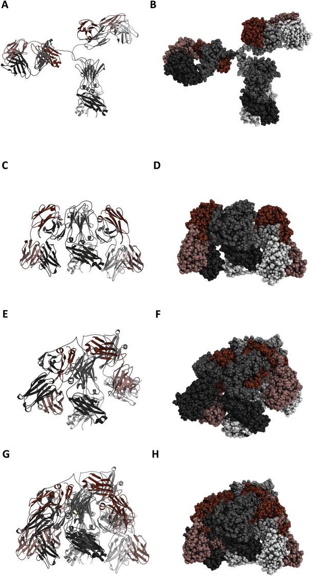
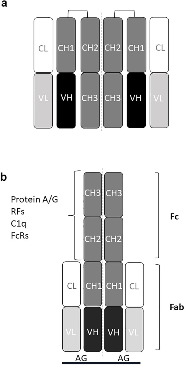
Antibodies are important for immunity and exist in several classes (IgM, IgD, IgA, IgG, IgE). They are composed of symmetric dimeric molecules with two antigen binding regions (Fab) and a constant part (Fc), usually depicted as Y-shaped molecules. Rheumatoid factors found in patients with rheumatoid arthritis are autoantibodies binding to IgG and paradoxically appear to circulate in blood alongside with their antigen (IgG) without reacting with it. Here, it is shown that rheumatoid factors do not react with native IgG in solution, and that their epitopes only become accessible upon certain physico-chemical treatments (e.g. heat treatment at 57 °C), by physical adsorption on a hydrophobic surface or by antigen binding. Moreover, chemical cross-linking in combination with mass spectrometry showed that the native state of IgG is a compact (closed) form and that the Fab parts of IgG shield the Fc region and thereby control access of rheumatoid factors and presumably also some effector functions. It can be inferred that antibody binding to pathogen surfaces induces a conformational change, which exposes the Fc part with its effector sites and rheumatoid factor epitopes. This has strong implications for understanding antibody structure and physiology and necessitates a conceptual reformulation of IgG models.


Conflict of interest statement

Statens Serum Institut has filed a patent application (EP 18169502.4) on the use of heat-treated immunoglobulins in immunoassays with GH as inventor. This does not alter our adherence to PLOS ONE policies on sharing data and materials.

Figures

Fig 1
Fig 1. Immunoglobulin model and immunoassay formats used in this work.
(a). Schematic structure of an immunoglobulin G (IgG) molecule consisting of two heavy chains of γ type and two light chains of either κ or λ type linked by disulphide bridges. The variable part of an heavy chain together with the variable part of a light chain together form the antigen binding site. A light chain together with variable heavy and CH1 domains form a Fab (fragment antigen-binding) part (“arm”) and the CH2 and CH3 domains constitute the Fc (fragment constant or crystallizable) part with effector functions. (b). Direct antigen antibody ELISA, where the antigens are immobilized by non-covalent forces. (c). Rheumatoid factor (RF) ELISA, where the antigen is IgG. (d). Bead-based fluorescent capture sandwich immunoassay, where the capture antibody is immobilized by covalent bonds. (e). RF sandwich/bridging assay. Ab: antibody, Ag: antigen, E: enzyme, b: biotin, S: streptavidin, PE: phycoerythrin, RF: rheumatoid factor. CL: constant light, VL: variable light, VH: variable heavy, CH: constant heavy.
Fig 2
Fig 2. RF reactivity with IgG.
RFs do not react in a bead-based fluorescent sandwich/bridging immunoassay with immobilized IgG and bIgG in solution but do so after exposure of the bIgG to elevated temperature. (a). Reactivity of RF-positive and -negative sera to IgG immobilized on fluorescent beads (positive control: RaHIgG). Individual data points represent single determinations on nine individual sera. (b). RFs react with heat-treated (57 °C, 24 h, heating cabinet) bIgG (***: p = 0.0003 for RF IgA, p = 0.0001 for RF IgM). Individual data points represent single determinations on the same nine sera as used in (a). The figures in (a,b) show one representative experiment of three. c. Temperature dependence of RF reactivity. IgG incubated at the indicated temperatures was tested for reaction with RF-positive or–negative sera using beads with immobilized IgG. The figure shows mean +/- SD of four experiments. (d-f). Native IgG (0 h) and IgG incubated at 57 °C for 4 h or 24 h was covalently immobilized on fluorescent beads and incubated with RF-positive or -negative serum samples in the presence of bIgG, which was either non-heated (d, 0 h) or had been incubated at 57 °C for 4 h (e) or 24 h (f). The panel shows one of two experiments. Individual data points of the time experiment are single determinations on individual sera (4 in all). The positive control was rabbit antibodies to human IgG (RaHIgG).
Fig 3
Fig 3. RF reactivity with IgG subclasses.
RFs do not react with native IgG in a bead-based fluorescent sandwich/bridging immunoassay with immobilized IgG subclasses but does so after exposure of the bIgG to elevated temperature (57 °C, 24 h). (a). IgG1. (b). IgG2. (c). IgG3. (d). IgG4. The figures show one of two experiments.
Fig 4
Fig 4. IgG reactivity with RFs.
(a, b). Reaction of different IgG forms with Rheumatoid factors in inhibition assays. IgG (a) or IFX (b) was coated on the surface of polystyrene ELISA plates and incubated with RF-containing serum or control serum (healthy donor serum) in the absence or presence of the indicated concentrations of inhibitor (native or heat-treated IgG). The experiments were done as titrations and data points are single determinations. (c). Immobilised IFX incubated first at 37 °C—57 °C and then immobilized on beads reacts with RaHIgG and can be bridged to native IFX in solution by TNF but not by RFs. No reaction is seen with IFX incubated at 62 °C or 67 °C due to precipitation of the IFX. (d). IFX immobilized covalently on beads can be bridged by RFs to heat-treated IFX (57 °C) but not by RF-negative sera. The figures show one representative experiment out of two. HD: healthy donors (pool).
Fig 5
Fig 5. Heat-treated IgG retains antigen binding and antigen binding exposes cryptic RF epitopes.
(a). Tetanus toxoid (TT), diphtheria toxoid (DT) and Epstein Barr virus nuclear antigen 1 (EBNA1) were coated in ELISA wells and incubated with bIgG (bIVIG), which had been pre-incubated at the indicated temperatures. (b). TNF was coated in ELISA wells and incubated with bIFX, which had been pre-incubated at the indicated temperatures. (c). TNF was immobilized on the surface of a microtitre plate, incubated with IFX and then incubated with RF-positive or -negative serum. (d). Infliximab was immobilized in ELISA wells and incubated with an RF-positive serum, which had first been incubated with IFX (which had itself been pre-incubated with TNF). The figures show one experiment out of two. HD: healthy donors (pool).
Fig 6
Fig 6. Models of the open and closed IgG (IFX) conformation constructed by Modeller27 and using the PDB structures 5VH4, 5VH5 and 1IGT as templates.
Left side: ribbon (cartoon) presentation, right side: sphere (space-filling) presentation. Light chains are colored in shades of red, heavy chains in shades of grey. (a, b). Model of the open conformation of the IFX antibody. (c, d). Model of the first closed conformation. (e, f). Model of the second closed conformation. (g, h). Superimposition of first and second closed models.
Fig 7
Fig 7. Immunoglobulin G models.
(a). Graphical model of native (closed) IgG molecule consisting of two heavy chains of γ type and two light chains of either κ or λ type linked by disulphide bridges. The variable part of a heavy chain domain together with the variable part of a light chain domain (VL) together form the antigen binding site. A light chain together with variable heavy and CH1 domains form a Fab (fragment antigen-binding) part (“arm”), and the CH2 and CH3 domains constitute the Fc (fragment constant or crystallizable) part with effector functions. RF binding sites and effector sites for C1/C1q and FcRs reside in the Fc (CH2-CH3) domains and are shielded by the Fab “arms”. (b). Model of the conformational change in IgG from closed to open upon antigen binding. Epitopes for RFs, C1q, FcRs and protein A/G are indicated on the Fc. CL: constant light, VL: variable light, VH: variable heavy, CH: constant heavy, Ag: antigen, RF: rheumatoid factor, FcR: Fc receptor.

Similar articles

Cited by

References

    1. Murphy K. Immunobiology. Garland Science, NY, USA; 2016.
    1. Lu LL, Suscovich TJ, Fortune SM, Alter G. Beyond binding: antibody effector functions in infectious diseases. Nat Rev Immunol. 2018; 18: 46–61. 10.1038/nri.2017.106 - DOI - PMC - PubMed
    1. Panda S, Ding JL. Natural antibodies bridge innate and adaptive immunity. J. Immunol. 2015; 194: 13–20. 10.4049/jimmunol.1400844 - DOI - PubMed
    1. Heyman B. Antibodies as natural adjuvants. Curr Top Microbiol Immunol. 2014; 382: 201–219. 10.1007/978-3-319-07911-0_9 - DOI - PubMed
    1. Holm BE, Sandhu N, Tronstrøm J, Lydolph M, Trier NH, Houen G. Species cross-reactivity of rheumatoid factors and implications for immunoassays. Scand J Clin Lab Invest. 2015; 75: 51–63. 10.3109/00365513.2014.965738 - DOI - PubMed

Publication types

MeSH terms