. 2019 Jun 14;14(6):e0218109.
doi: 10.1371/journal.pone.0218109. eCollection 2019.

SLAMF6 clustering is required to augment T cell activation

Affiliations

SLAMF6 clustering is required to augment T cell activation

Matthew A Dragovich et al. PLoS One. .

Abstract

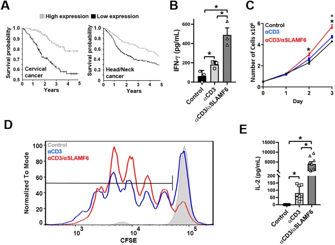
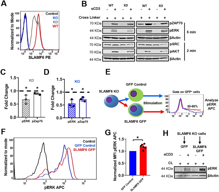
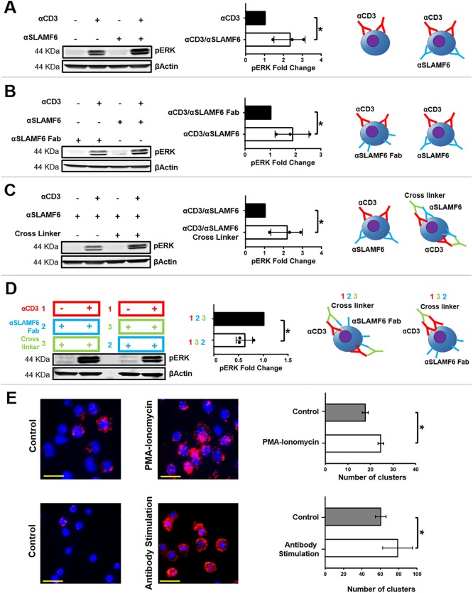
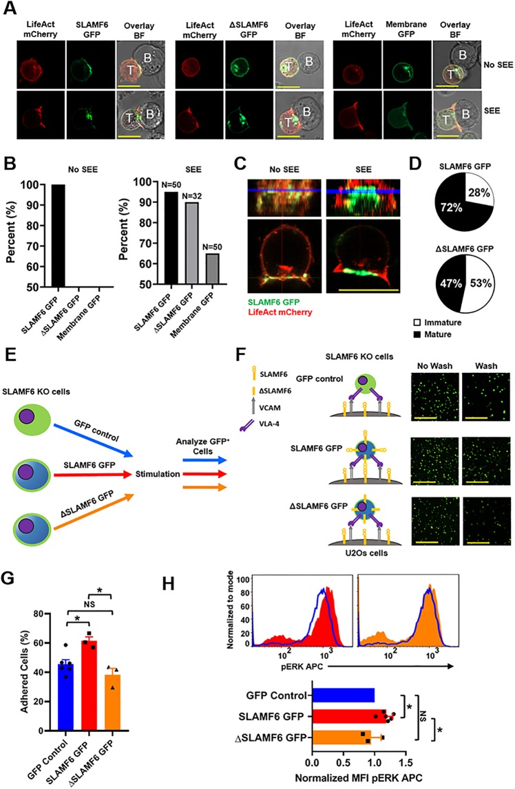
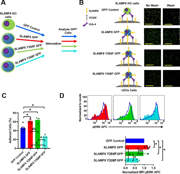
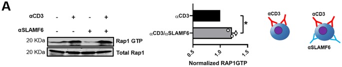
The signaling lymphocytic activation molecule (SLAM) family is comprised of nine distinct receptors that are expressed exclusively on hematopoietic cells. Most of these transmembrane receptors are homotypic by nature and downstream signaling occurs when cells that express the same SLAM receptor interact. Previous studies have determined that anti-SLAMF6 antibodies can have a therapeutic effect in autoimmunity and cancer. However, little is known about the role of SLAMF6 in the adaptive immune responses and in order to utilize SLAMF6 interventional approaches, a better understanding of the biology of this receptor in T cell is warranted. Accordingly, the objective of our study was to investigate both functionally and structurally the role of SLAMF6 in T cell receptor (TCR) mediated responses. Biochemical and genetic experiments revealed that SLAMF6 was required for productive TCR downstream signaling. Interestingly, SLAMF6 ectodomain was required for its function, but not for its recruitment to the immunological synapse. Flow-cytometry analysis demonstrated that tyrosine 308 of the tail of SLAMF6 was crucial for its ability to enhance T cell function. Imaging studies revealed that SLAMF6 clustering, specifically with the TCR, resulted in dramatic increase in downstream signaling. Mechanistically, we showed that SLAMF6 enhanced T cell function by increasing T cell adhesiveness through activation of the small GTPase Rap1. Taken together SLAMF6 is an important regulator of T cell activation where both its ectodomain and its endodomain contribute differentially to T cell functions. Additional studies are underway to better evaluate the role of anti-SLAMF6 approaches in specific human diseases.


Conflict of interest statement

The authors have declared that no competing interests exist.

Figures

Fig 1
Fig 1. SLAMF6 antibody engagement leads to T cell activation.
(A) Survival curves demonstrating that high expression of SLAMF6 in tumors is positively correlated with cancer survival. (B) Freshly isolated primary CD3+ T cells were cultured in the presence of αCD3 or αCD3 + αSLAMF6. After 48 hrs. the supernatant was harvested and IFN-γ levels were analyzed by ELSIA for two independent experiments (n = 2) with 3 technical replicates of one experiment shown. *p<0.05 for an unpaired student t-test (C) Jurkat T cells were cultured in the presence of αCD3 or αCD3 + αSLAMF6 for 72 hrs. Cell number was assessed by automated cell counting every 24 hrs. The experiment was done in triplicate (n = 3). *p<0.05 for αCD3 + αSLAMF6 compared to control and + p<0.05 for αCD3 compared to control for an unpaired student t-test. (D) Freshly isolated primary CD3+ T cells were stained with CFSE then cultured in the presence of immobilized αCD3 or αCD3 + αSLAMF6. After 120 hrs. the cells were assayed for FITC fluorescence for three independent experiments (n = 3). The data was analyzed for percent (%) of proliferating cells as depicted; αCD3 + αSLAMF6 had a 15–25% greater proliferation advantage over αCD3 alone conditions. (E) Also, the supernatant was harvested, and IL-2 levels were analyzed by ELSIA for three independent experiments (n = 3) with 3 technical replicates for each experiment shown together. *p<0.05 for an unpaired student t-test.
Fig 2
Fig 2. SLAMF6 knock down or knock out leads to a decrease in TCR activation.
(A) Flow cytometry verification of SLAMF6 KO via CRISPR-Cas9 and SLAMF6 KD via shRNA. (B) WT Jurkat T cells, SLAMF6 KD and SLAMF6 KO were treated with αCD3 + cross linker and then stimulated at 37° C for 5 min. Blots were generated by lysing the cells, separating the lysates by tris-glycine PAGE and transferring to a nitrocellulose membrane. pZAP70, pERK, pSRC and pAKT were assessed. Quantification was given for three independent experiments (n = 3) for SLAMF6 KD Jurkat T cells (C) and SLAMF6 KO Jurkat T cells (D). *p<0.05 for an unpaired student t-test. Further quantification of pSRC and pAKT can be found in S5 Fig. (E) A schematic of the flow cytometry assay used to rescue SLAMF6 function. SLAMF6 KO Jurkat T cells were transiently transfected with GFP control or SLAMF6 GFP. Cells were treated with αCD3 + cross linker, stimulated at 37° C for 5 min and subjected to Flow cytometry. Cell were gated on GFP positive cells and analyzed for pERK APC. (F) Representative curve for functional recue of SLAMF6 by flow cytometry. (G) Quantification for six independent functional recue of SLAMF6 by flow cytometry experiments (n = 6). *p<0.05 for an unpaired student t-test. (H) Functional recue of SLAMF6 by western blot. SLAMF6 KO cells were transiently transfected with GFP control (pEGFPN1 vector) or SLAMF6 GFP (SLAMF6 pEGFPN1 vector) then treated with αCD3 + cross linker and stimulated at 37° C for 5 min. Blot was generated and pERK were assessed.
Fig 3
Fig 3. SLAMF6 clustering with the TCR increases T cell activation.
(A) Jurkat T cells were treated with αCD3 or αCD3 + αSLAMF6 and then stimulated at 37° C for 5 min. Blots were generated by lysing the cells, separating the lysates by tris-glycine PAGE and transferring to a nitrocellulose membrane. pERK was assessed in three independent experiments (n = 3). *p<0.05 for an unpaired student t-test. (B) Jurkat T cells were treated with αCD3 + αSLAMF6 or αCD3 + αSLAMF6 Fab and then stimulated as above. Blots were generated assessing pERK in three independent experiments (n = 3). *p<0.05 for an unpaired student t-test. (C) Jurkat T cells were treated with αCD3 + αSLAMF6 or αCD3 + αSLAMF6 + cross linker and then stimulated. pERK was assessed in three independent experiments (n = 3). *p<0.05 for an unpaired student t-test. (D) Jurkat T cells were treated with αCD3 + αSLAMF6 Fab + cross linker or αCD3 + cross linker + αSLAMF6 Fab as delineated by color and number (cells were washed in between steps) and then stimulated. Blots were produced for pERK in three independent experiments (n = 3). *p<0.05 for an unpaired student t-test. (E) A proximity ligation assay (PLA) was performed on Jurkat T cells for SLAMF6 and the TCR. Cells were stimulated with PMA ionomycin at 37° C for 5 min, then fixed in 1% PFA for 10 min. Next, the cells were then blocked and treated with αCD3 + αSLAMF6 for 1 hr. at room temperature. This experiment was replicated in freshly isolated primary CD3+ T cells stimulated with immobilized αCD3 + αSLAMF6 for 120 hrs. The PLA assay was then performed as described by the manufactures protocol.
Fig 4
Fig 4. The SLAMF6 ectodomain is not required for SLAMF6 presence immunological synapse, but contributes to its co-receptor function.
(A) Jurkat T cells were transfected with LifeAct mCherry and SLAMF6 GFP, ΔSLAMF6 or membrane GFP. Raji B cells (top row) or Raji B cells loaded with SEE (bottom row) were co-cultured with the transfected Jurkat T cells. Images are representative of at least 32 cells from a least two independent experiments. Scale bar is 10μm. Actin clearance was defined as a mature IS. If actin was not cleared from the IS the synapse was considered immature. Only images with actin clearance were considered for analysis of SLAMF6 presence in the IS when Raji B cells were loaded with SEE. (B) Quantification of SLAMF6 GFP, ΔSLAMF6 or membrane GFP at the point of contact between a Jurkat T cell and Raji B cell (left). Quantification of SLAMF6 GFP, ΔSLAMF6 GFP or membrane GFP at IS between a Jurkat T cell and a SEE loaded Raji B cell (right). (C) SLAMF6 localizes to the PSMAC in mature synapses. (D) The percent of mature and immature synapses for Jurkat T cells transfected with SLAMF6 GFP or ΔSLAMF6 GFP. (E) A schematic of the assay used to analyze SLAMF6 ectodomain function. (F) SLAMF6 KO jurkat T cells were transfected with GFP control, SLAMF6 GFP or ΔSLAMF6 and co-cultured with U2Os cells stably expressing SLAMF6 and VCAM in the presence of αCD3 for 20 min at 37°C. They were then imaged before and after four washes. Scale bar is 100μm. (G) Quantification of number of adhered cells for at least three independent experiments of SLAMF6 KO Jurkat T cells transfected with GFP control, SLAMF6 GFP or ΔSLAMF6 (bottom) (n = 3–6). *p<0.05 for an unpaired student t-test. (H) Representative histograms analyzing pERK APC for SLAMF6 KO Jurkat T cells (top). SLAMF6 KO Jurkat T cells were transiently transfected with GFP control, SLAMF6 GFP or ΔSLAMF6. Cells were treated with αCD3 + cross linker, stimulated at 37° C for 5 min, fixed permeabilized, stained and subjected to flow cytometry. Cells were gated on GFP positive cells and analyzed for pERK APC. Quantification of pERK for at least three independent experiments of SLAMF6 KO Jurkat T cells transfected with GFP control, SLAMF6 GFP or ΔSLAMF6 (bottom) (n = 3–6). *p<0.05 for an unpaired student t-test.
Fig 5
Fig 5. SLAMF6 tyrosine 308 is crucial for its ability to promote adhesion.
(A) A schematic of the assay used to analyze SLAMF6 endodomain function. (B) SLAMF6 KO Jurkat T cells were transfected with GFP control, SLAMF6 GFP, SLAMF6 Y284F GFP and SLAMF6 Y308F GFP and co-cultured with U2Os cells stably expressing SLAMF6 and VCAM in the presence of αCD3 for 20 min at 37°C. They were then imaged before and after four washes. Scale bar is 100μm. (C) Quantification of number of adhered cells for at least three independent experiments of SLAMF6 KO Jurkat T cells transfected with GFP control, SLAMF6 GFP, SLAMF6 Y284F GFP and SLAMF6 Y308F GFP (bottom) (n = 3–6). *p<0.05 for an unpaired student t-test. (D) Representative histograms analyzing pERK APC for SLAMF6 KO Jurkat T cells t (top). SLAMF6 KO Jurkat T cells were transiently transfected with GFP control, SLAMF6 GFP, SLAMF6 Y284F GFP and SLAMF6 Y308F GFP. Cells were treated with αCD3 + cross linker, stimulated at 37° C for 5 min, fixed permeabilized, stained and subjected to flow cytometry. Cells were gated on GFP positive cells and analyzed for pERK APC. Quantification of pERK for at least three independent experiments of SLAMF6 KO Jurkat T cells transfected with GFP control, SLAMF6 GFP, SLAMF6 Y284F GFP and SLAMF6 Y308F GFP (bottom) (n = 3–6). *p<0.05 for an unpaired student t-test.
Fig 6
Fig 6. SLAMF6 clustering increases Rap1 activation.
The amount of activated Rap1 was measured by glutathione S-transferase (GST) pull-down assay. Quantification is shown for at least three independent experiments. *p<0.05 for an unpaired student t-test.

Similar articles

Cited by

References

    1. Jain A, Pasare C: Innate Control of Adaptive Immunity: Beyond the Three-Signal Paradigm. J Immunol 2017, 198(10):3791–3800. 10.4049/jimmunol.1602000 - DOI - PMC - PubMed
    1. Wakamatsu E, Omori H, Ohtsuka S, Ogawa S, Green JM, Abe R: Regulatory T cell subsets are differentially dependent on CD28 for their proliferation. Mol Immunol 2018, 101:92–101. 10.1016/j.molimm.2018.05.021 - DOI - PubMed
    1. Pilat N, Mahr B, Gattringer M, Baranyi U, Wekerle T: CTLA4Ig Improves Murine iTreg Induction via TGFbeta and Suppressor Function In Vitro. J Immunol Res 2018, 2018:2484825. - PMC - PubMed
    1. Siokis A, Robert PA, Demetriou P, Dustin ML, Meyer-Hermann M: F-Actin-Driven CD28-CD80 Localization in the Immune Synapse. Cell Rep 2018, 24(5):1151–1162. 10.1016/j.celrep.2018.06.114 - DOI - PubMed
    1. Gardell JL, Parker DC: Despite disorganized synapse structure, Th2 cells maintain directional delivery of CD40L to antigen-presenting B cells. PLoS One 2017, 12(10):e0186573 10.1371/journal.pone.0186573 - DOI - PMC - PubMed

Publication types

MeSH terms

Substances