. 2019 Jun 14;15(6):e1007842.
doi: 10.1371/journal.ppat.1007842. eCollection 2019 Jun.

Separate domains of G3BP promote efficient clustering of alphavirus replication complexes and recruitment of the translation initiation machinery

Affiliations

Separate domains of G3BP promote efficient clustering of alphavirus replication complexes and recruitment of the translation initiation machinery

Benjamin Götte et al. PLoS Pathog. .

Abstract

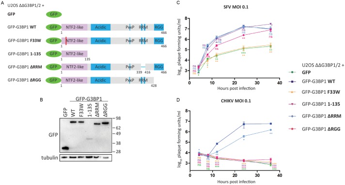
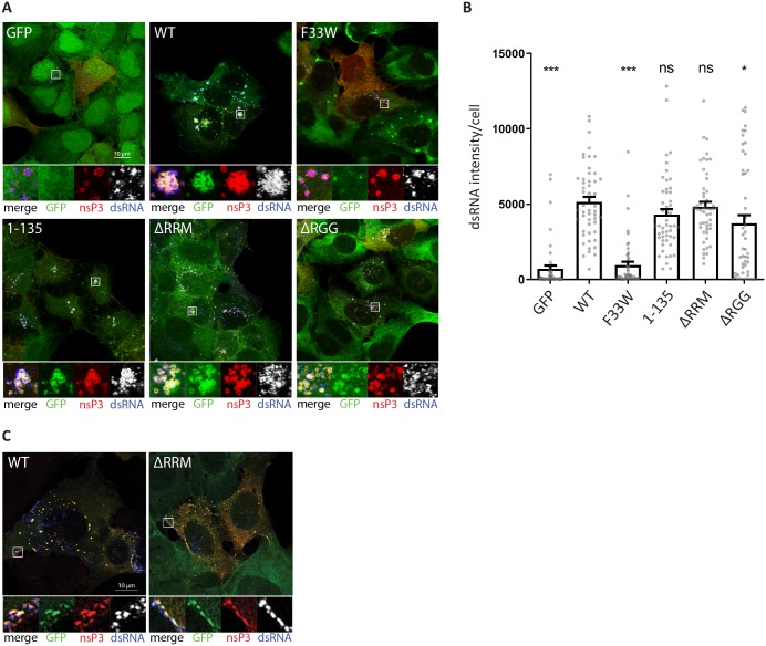
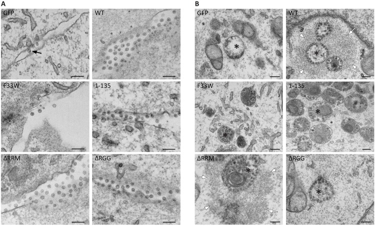
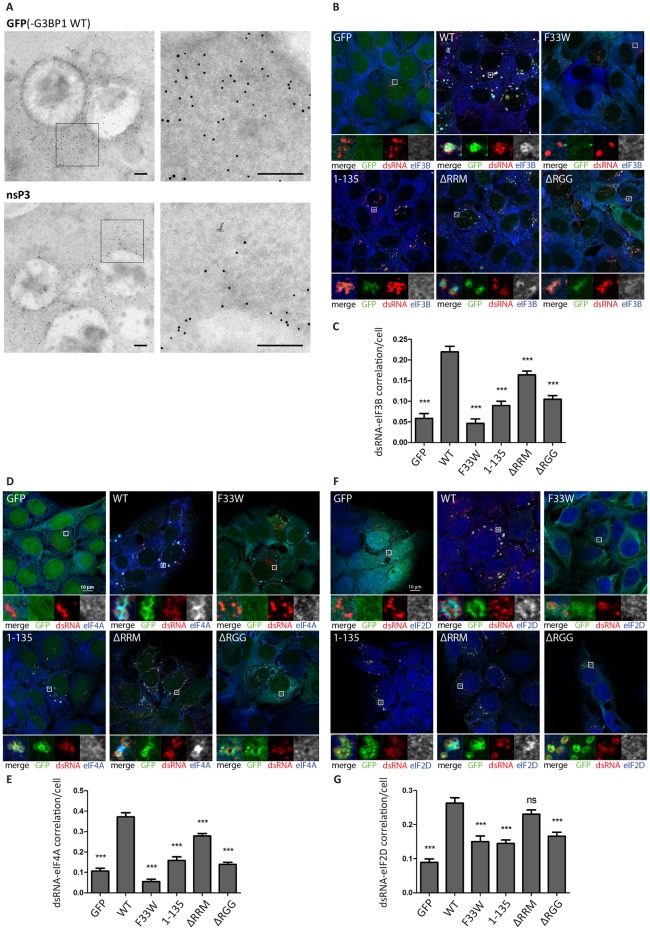
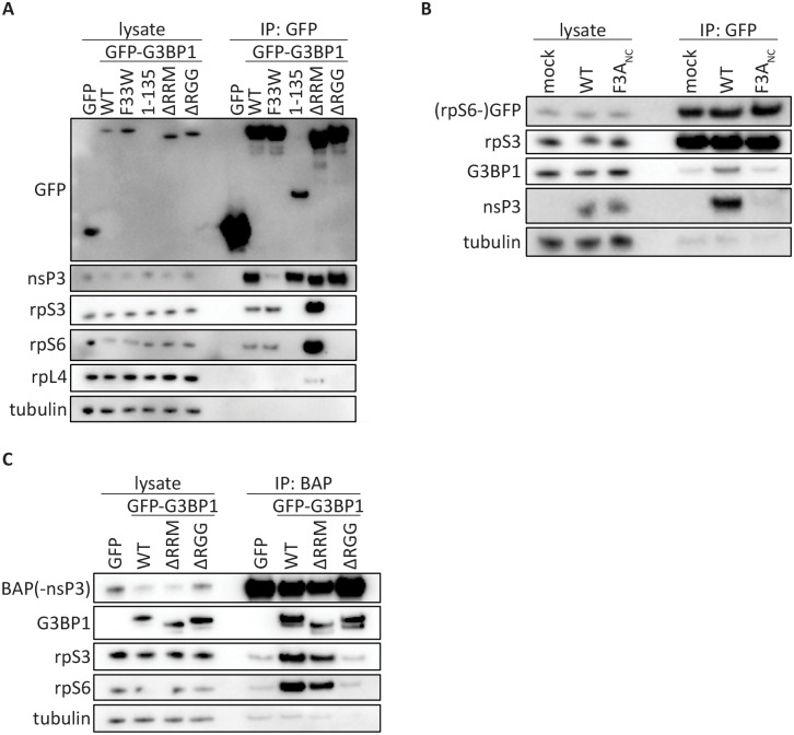
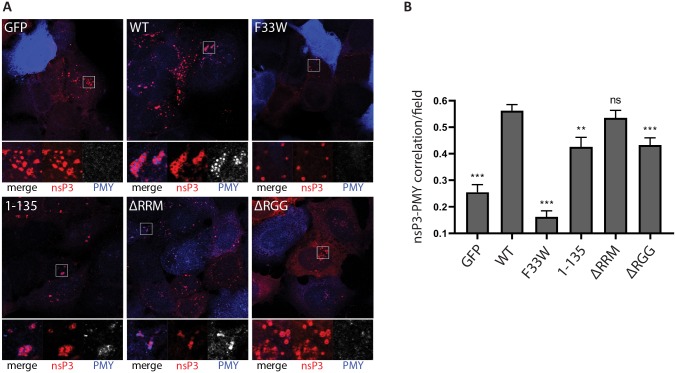
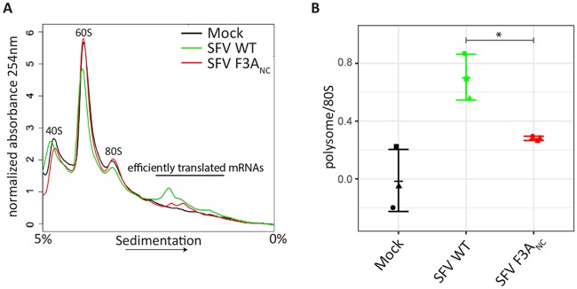
G3BP-1 and -2 (hereafter referred to as G3BP) are multifunctional RNA-binding proteins involved in stress granule (SG) assembly. Viruses from diverse families target G3BP for recruitment to replication or transcription complexes in order to block SG assembly but also to acquire pro-viral effects via other unknown functions of G3BP. The Old World alphaviruses, including Semliki Forest virus (SFV) and chikungunya virus (CHIKV) recruit G3BP into viral replication complexes, via an interaction between FGDF motifs in the C-terminus of the viral non-structural protein 3 (nsP3) and the NTF2-like domain of G3BP. To study potential proviral roles of G3BP, we used human osteosarcoma (U2OS) cell lines lacking endogenous G3BP generated using CRISPR-Cas9 and reconstituted with a panel of G3BP1 mutants and truncation variants. While SFV replicated with varying efficiency in all cell lines, CHIKV could only replicate in cells expressing G3BP1 variants containing both the NTF2-like and the RGG domains. The ability of SFV to replicate in the absence of G3BP allowed us to study effects of different domains of the protein. We used immunoprecipitation to demonstrate that that both NTF2-like and RGG domains are necessary for the formation a complex between nsP3, G3BP1 and the 40S ribosomal subunit. Electron microscopy of SFV-infected cells revealed that formation of nsP3:G3BP1 complexes via the NTF2-like domain was necessary for clustering of cytopathic vacuoles (CPVs) and that the presence of the RGG domain was necessary for accumulation of electron dense material containing G3BP1 and nsP3 surrounding the CPV clusters. Clustered CPVs also exhibited localised high levels of translation of viral mRNAs as detected by ribopuromycylation staining. These data confirm that G3BP is a ribosomal binding protein and reveal that alphaviral nsP3 uses G3BP to concentrate viral replication complexes and to recruit the translation initiation machinery, promoting the efficient translation of viral mRNAs.


Conflict of interest statement

The authors have declared that no competing interests exist.

Figures

Fig 1
Fig 1. The NTF2-like and RGG domains of G3BP are necessary for efficient Old World alphavirus replication.
A. Schematic of GFP-G3BP1 constructs used for reconstitution of G3BP1/2 double KO cell lines. B. Indicated cell lines were lysed and analysed by SDS-PAGE and immunoblotting for GFP and β-tubulin. C. and D. Indicated cells lines were infected with WT SFV (C) or WT CHIKV (D) at a multiplicity of infection (MOI) of 0.1. At 4, 8, 12, 24 and 36 hpi, supernatants were collected, and viral titres were quantified by plaque assay on BHK cells. Data are means of three independent experiments. Error bars indicate standard error of mean (SEM). For statistical analysis samples within each timepoint were compared to the respective titre obtained in U2OS ΔΔG3BP1/2 + GFP-G3BP1 WT cells. ns P>0.05, *P≤0.05, **P≤0.01, ***P≤0.001.
Fig 2
Fig 2. The NTF2-like and RGG domains of G3BP are differently required for efficient formation of SFV and CHIKV dsRNA-positive replication complexes.
A. Indicated cell lines were infected with WT SFV at MOI 10. At 8 hpi cells were fixed and stained for nsP3 (red) and dsRNA (blue). Representative images from 3 independent experiments are shown. B. DsRNA signal intensities from images shown in A. were quantified using CellProfiler software. Bars represent mean + SEM for n = 50 cells per cell line. Individual dsRNA intensities for each analysed cell are shown as grey dots. ns P>0.05, *P≤0.05, **P≤0.01, ***P≤0.001. C. Indicated cell lines were infected with CHIKV at MOI 1. At 8 hpi cells were fixed and stained for nsP3 (red) and dsRNA (blue). Representative images from 3 independent experiments are shown.
Fig 3
Fig 3. Accumulation of electron-dense patches in proximity of SFV-induced cytopathic vacuoles depend on the presence of the RGG domain of G3BP.
Indicated cell lines were infected with WT SFV at MOI 100, fixed at 3 (A) or 8 (B) hpi and analysed by transmission electron microscopy. Representative images from 2 independent experiments are shown. A. Images of cell plasma membranes (PM) show alphavirus spherules (black arrows). B. Endocytosis of PM-associated spherules leads to formation of cytopathic vacuoles (CPV, asterisks) in the perinuclear region. In cells expressing GFP-G3BP1 WT and ΔRRM the CPVs are surrounded by electron-dense material (white arrows). The scale bar is 200 nm.
Fig 4
Fig 4. Accumulation of GFP-G3BP1, nsP3 and translation initiation factors around SFV-induced CPVs.
A. U2OS ΔΔG3BP1/2 + GFP-G3BP1 WT cells were infected with WT SFV at MOI 100 and fixed 8h pi. Samples were prepared for immunogold labeling against GFP(-G3BP1 WT) or nsP3 and analysed by electron microscopy. Representative cytoplasmic regions that stained positive for GFP(-G3BP1 WT) or nsP3 are shown. The scale bar is 200 nm. B. Indicated cell lines were infected with WT SFV at MOI 10. At 8 hpi cells were fixed and stained for dsRNA (red) and B. eIF3B (blue) D. eIF4A (blue) or F. eIF2D (blue). Representative images from 3 independent experiments are shown. Correlations of dsRNA with eIF3B (C.), eIF4A (E.) and eIF2D (G.) were calculated in CellProfiler based on Pearson’s correlation coefficient for n = 50 infected cells per sample. Bars represent mean + SEM. For statistical analysis indicated cell lines were compared to U2OS ΔΔG3BP1/2 + GFP-G3BP1 WT cells. ns P>0.05, *P≤0.05, **P≤0.01, ***P≤0.001.
Fig 5
Fig 5. Old World alphavirus nsP3 forms a complex with 40S ribosomal subunits via G3BP.
A. Indicated cell lines were infected with WT SFV at MOI 10. At 7 hpi, cell lysates were prepared, immunoprecipitated using GFP-Trap (ChromoTek) and separated by SDS–PAGE. Lysates and IPs were probed for GFP, nsP3, rpS3, rpS6, rpL4 or β-tubulin. B. GFP-rpS6 cells were infected with WT-SFV or SFV-F3ANC at MOI 0.5. At 8 hpi, cell lysates were prepared, immunoprecipitated using GFP-Trap (ChromoTek) and separated by SDS–PAGE. Lysates and IPs were probed for GFP, rpS3, G3BP1, nsP3 or β-tubulin. C. Indicated cell lines were transfected with biotinylated CHIKV nsP3 (BAP-nsP3). Cell lysates were prepared 24 h after transfection, precipitated with streptavidin-coated beads, and separated by SDS-PAGE. Lysates and precipitates were probed with antisera for nsP3, GFP, rpS3, rpS6 or β-tubulin. All data in Fig 5 are representative of at least two independent experiments.
Fig 6
Fig 6. Ribopuromycylation of SFV-infected cells: The NTF2-like and RGG domains of G3BP are required for enhanced translational activity.
A. Indicated cell lines were infected with WT SFV at MOI 10. At 8 hpi cells were pulse-labeled with puromycin (PMY) for 5 min, fixed and stained for nsP3 (red) and PMY (blue/white). Nsp3 foci indicate the localization of viral replication complexes. Staining of puromycylated nascent polypeptide chains visualizes translational activity. Representative images from 3 independent experiments are shown. B. Correlations of nsP3 and puromycin were calculated in CellProfiler based on Pearson’s correlation coefficient for n = 20 nsP3-positive fields from ≥10 cells per cell line. Bars represent mean + SEM. For statistical analysis indicated cell lines were compared to U2OS ΔΔG3BP1/2 + GFP-G3BP1 WT cells. ns P>0.05, *P≤0.05, **P≤0.01, ***P≤0.001.
Fig 7
Fig 7. G3BP-binding enhances translation initiation in SFV-infected cells.
A. BHK-21 cells were infected with SFV WT or SFV F3ANC at MOI 10. At 8 hpi cells were treated with cycloheximide and lysates analysed on 5%–50% sucrose gradients. Absorbance at 254 nm is shown as a function of sedimentation and fractions representing 40S, 60S, 80S and polysomes are indicated. A representative figure of three independent experiments is shown. B. The area under the curve for polysomes and the 80S peak were calculated and the ratio is shown. Data are means of three independent experiments. Error bars indicate standard deviation (SD). *P<0.05.

Similar articles

Cited by

References

    1. Tourriere H, Chebli K, Zekri L, Courselaud B, Blanchard JM, Bertrand E, et al. The RasGAP-associated endoribonuclease G3BP assembles stress granules. J Cell Biol. 2003;160(6):823–31. Epub 2003/03/19. 10.1083/jcb.200212128 - DOI - PMC - PubMed
    1. Kedersha N, Panas MD, Achorn CA, Lyons S, Tisdale S, Hickman T, et al. G3BP-Caprin1-USP10 complexes mediate stress granule condensation and associate with 40S subunits. J Cell Biol. 2016;212(7):845–60. Epub 2016/03/30. 10.1083/jcb.201508028 - DOI - PMC - PubMed
    1. Reineke LC, Lloyd RE. Diversion of stress granules and P-bodies during viral infection. Virology. 2013;436(2):255–67. Epub 2013/01/08. 10.1016/j.virol.2012.11.017 - DOI - PMC - PubMed
    1. Panas MD, Schulte T, Thaa B, Sandalova T, Kedersha N, Achour A, et al. Viral and cellular proteins containing FGDF motifs bind G3BP to block stress granule formation. PLoS Pathog. 2015;11(2):e1004659 Epub 2015/02/07. 10.1371/journal.ppat.1004659 - DOI - PMC - PubMed
    1. Panas MD, Varjak M, Lulla A, Eng KE, Merits A, Karlsson Hedestam GB, et al. Sequestration of G3BP coupled with efficient translation inhibits stress granules in Semliki Forest virus infection. Mol Biol Cell. 2012;23(24):4701–12. Epub 2012/10/23. 10.1091/mbc.E12-08-0619 - DOI - PMC - PubMed

Publication types

MeSH terms