. 2019 Jun 14;15(6):e1007879.
doi: 10.1371/journal.ppat.1007879. eCollection 2019 Jun.

Rab32 GTPase, as a direct target of miR-30b/c, controls the intracellular survival of Burkholderia pseudomallei by regulating phagosome maturation

Affiliations

Rab32 GTPase, as a direct target of miR-30b/c, controls the intracellular survival of Burkholderia pseudomallei by regulating phagosome maturation

Zhi-Qiang Hu et al. PLoS Pathog. .

Abstract

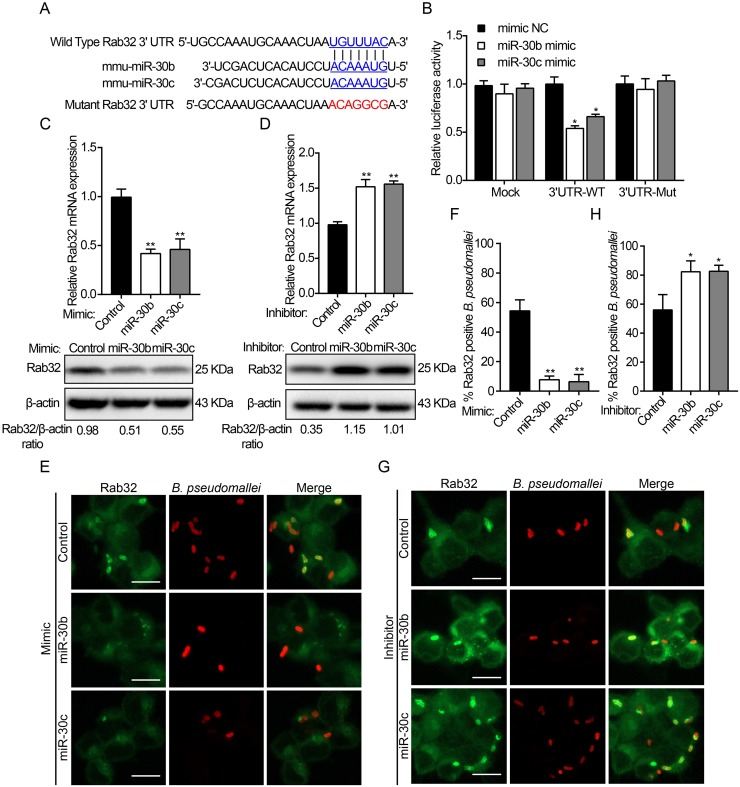
Burkholderia pseudomallei is a gram-negative, facultative intracellular bacterium, which causes a disease known as melioidosis. Professional phagocytes represent a crucial first line of innate defense against invading pathogens. Uptake of pathogens by these cells involves the formation of a phagosome that matures by fusing with early and late endocytic vesicles, resulting in killing of ingested microbes. Host Rab GTPases are central regulators of vesicular trafficking following pathogen phagocytosis. However, it is unclear how Rab GTPases interact with B. pseudomallei to regulate the transport and maturation of bacterial-containing phagosomes. Here, we showed that the host Rab32 plays an important role in mediating antimicrobial activity by promoting phagosome maturation at an early phase of infection with B. pseudomallei. And we demonstrated that the expression level of Rab32 is increased through the downregulation of the synthesis of miR-30b/30c in B. pseudomallei infected macrophages. Subsequently, we showed that B. pseudomallei resides temporarily in Rab32-positive compartments with late endocytic features. And Rab32 enhances phagosome acidification and promotes the fusion of B. pseudomallei-containing phagosomes with lysosomes to activate cathepsin D, resulting in restricted intracellular growth of B. pseudomallei. Additionally, Rab32 mediates phagosome maturation depending on its guanosine triphosphate/guanosine diphosphate (GTP/GDP) binding state. Finally, we report the previously unrecognized role of miR-30b/30c in regulating B. pseudomallei-containing phagosome maturation by targeting Rab32 in macrophages. Altogether, we provide a novel insight into the host immune-regulated cellular pathway against B. pseudomallei infection is partially dependent on Rab32 trafficking pathway, which regulates phagosome maturation and enhances the killing of this bacterium in macrophages.


Conflict of interest statement

The authors have declared that no competing interests exist.

Figures

Fig 1
Fig 1. B. pseudomallei infection increased the expression of Rab32 and its recruitment into B. pseudomallei-containing phagosomes.
(A-D) B. pseudomallei infection increased the expression levels (mRNA and protein) of Rab32 in RAW264.7 cells. RAW264.7 cells were treated with B. pseudomallei (MOI = 10:1) for 0, 1, 2, 4, and 6 h, or at MOI = 0, 1, 10, 20, 50, and 100 for 2 h. (E) RAW264.7 cells were infected with B. pseudomallei (MOI = 10:1) and imaged at the indicated time points: 1 to 6 h and stained with an anti-Rab32 antibody (green) anti-B. pseudomallei antibody (red). Images show maximum-intensity projections of confocal Z-stacks. Scale bar is 5 μm. (F) The percentage of association of live and heat-killed B. pseudomallei phagosomes with Rab32 at each time point is depicted in this panel. Scale bar is 5 μm. ns, no significant difference. Data are representative of at least three independent experiments (*P<0.05, **P<0.01).
Fig 2
Fig 2. MiR-30b and miR-30c are downregulated after B. pseudomallei infection.
(A) Total RNA from RAW264.7 cells were infected with B. pseudomallei for 4 h to perform microarray assay. Hierarchical clustering analysis was performed to show downregulated miRNAs by B. pseudomallei infection. (B) The region of the mouse Rab32 mRNA 3′UTR predicted to be targeted by miR-30b, miR-30c, miR-30d, and miR-30e, respectively (TargetScan 6.2). (C and D) Confirmation of microarray results by qRT-PCR. qRT-PCR analysis of the expression levels of miR-30b, miR-30c, miR-30d, and miR-30e in RAW264.7 cells infected with B. pseudomallei (MOI = 10) for 0, 1, 2, 4, 6, and 8 h, or at MOI = 0, 1, 10, 20, 50, and 100 for 4 h. (E) Expression of miR-30b and miR-30c in RAW264.7 cells infected with B. thailandensis, S. typhimurium and E. coli (MOI = 10). *P<0.05, **P<0.01. Experiments performed in triplicates showed consistent results.
Fig 3
Fig 3. Rab32 is a direct target of miR-30b and miR-30c.
(A) Sequences of miR-30b and miR-30c, and the potential binding site at the 3′UTR of Rab32. Also shown are nucleotides mutated in Rab32-3′UTR mutant. (B) HEK293 cells were co-transfected with luciferase reporter vectors carrying Rab32 WT or mutant constructs (Mut), along with an miR30b or miR-30c mimic or control. Luciferase activity was normalized to the activity of Renilla luciferase. (C and D) RAW264.7 cells were transiently transfected with miR-30b and miR-30c mimic, inhibitor, or control for 24 h. The mRNA and protein levels of Rab32 were determined by qRT-PCR and Western blot, respectively. (E) RAW264.7 cells transfected with miR-30b/30c mimic and control were infected with B. pseudomallei for 2 h, then fixed, and stained with an anti-Rab32 (green) or anti-B. pseudomallei antibody (red). (F) Quantitative analysis of Rab32 associated with B. pseudomallei phagosomes. (G) RAW264.7 cells transfected with miR-30b/30c inhibitor and control were infected with B. pseudomallei for 2 h, and association of Rab32 to B. pseudomallei phagosomes as in E. (H) Quantitative analysis of Rab32 associated with B. pseudomallei phagosomes as indicated in F. Scale bar is 5 μm. Data are representative of at least three independent experiments (*P<0.05, **P<0.01).
Fig 4
Fig 4. B. pseudomallei resides in a Rab32-labeled compartment with late endosomal features.
(A) RAW264.7 cells were infected with B. pseudomallei, at an MOI of 10 for 2 h. Cells were stained with anti-EEA1, anti-Rab5, and anti-Rab7 antibodies (green), or anti-B. pseudomallei antibody (red) and colocalization was determined by confocal microscopy. Scale bar is 5 μm. (B) The percentage of EEA1, Rab5, or Rab7 colocalizing with B. pseudomallei phagosomes was enumerated by fluorescence microscopy. At least 100 bacterial phagosomes were counted for each time point. Results are represented as mean ± S.D. of three independent experiments. (C) RAW264.7 cells expressing EGFP-Rab32 were infected with B. pseudomallei for 2 h, afterwards cells were subjected to immunofluorescence for LAMP1 or LAMP2 (red) and stained with an anti-B. pseudomallei antibody (blue). (D) RAW264.7 cells expressing EGFP-Rab32 were infected with B. pseudomallei, fixed, and immunostained for LAMP1 (red) and B. pseudomallei (blue). B. pseudomallei phagosomes that colocalized with Rab32 and LAMP1 were quantified. Results are the means ± SD of three independent experiments. (E) Quantitative analysis of B. pseudomallei phagosomes that colocalized with Rab32 and LAMP2 as indicated in D. (F) RAW264.7 cells expressing EGFP-Rab32 were incubated with 50 nM Lysotracker (red) for 1 h before infection with B. pseudomallei for 2 h. Cells were stained with anti-B. pseudomallei antibody (blue) and colocalization was determined by confocal microscopy. (G) Quantitative analysis of B. pseudomallei phagosomes that colocalized with Rab32 and Lysotracker as indicated in D. Symbols represent single bacteria or distinct B. pseudomallei groups. Scale bar is 5 μm.
Fig 5
Fig 5. Rab32-positive phagosomes were recruited to limit the growth of B. pseudomallei.
(A) The relative mRNA or protein concentrations of Rab32 were measured in RAW264.7 cells after transfection with 100 nM Rab32 specific siRNA or control RNA for 24 h. (B and C) Representative images of control and Rab32 silenced cells were infected with B. pseudomallei for 2 h, and stained with anti-LAMP1 antibody (red), or anti-B. pseudomallei antibody (green) or DAPI (blue). Quantitative analysis of LAMP1 fluorescence intensity associated with B. pseudomallei phagosomes. Scale bar is 5 μm. (D and E) Association of LAMP2 with B. pseudomallei as in B and a quantitative analysis of LAMP2 association with B. pseudomallei phagosomes as in C. Scale bar is 5 μm. (F) Representative TEM images of control and Rab32 silenced RAW264.7 cells infected with B. pseudomallei (asterisks) for 2 h showing differences in the intracellular location of B. pseudomallei. (white arrowheads = phagosome membrane; N = nucleus). Scale bar is 5 μm. (G) Quantitative analysis of bacteria associated with single-membrane phagosomes. At least 150 bacteria were quantified. (H) Intracellular B. pseudomallei burdens in Rab32 silenced and control RAW264.7 cells at the indicated times after infection. Data is shown as the mean ± SD of three independent experiments. *P<0.05, **P<0.01.
Fig 6
Fig 6. Rab32 increases fusion of B. pseudomallei-containing phagosomes with lysosomes.
(A) RAW264.7 cells expressing the different constructs were incubated with 50 nM Lysotracker for 1 h before infection with B. pseudomallei, and stained for B. pseudomallei (blue). Scale bar is 5 μm. (B) Quantitative analysis of B. pseudomallei phagosomes associated with Lysotracker. Results are represented as mean ± S.D. of three independent experiments. (C) RAW264.7 cells transfected with the indicated constructs were infected with B. pseudomallei, and cells were fixed and stained for cathepsin D (CTSD, red) and B. pseudomallei (blue). Scale bar is 5 μm. (D) Analysis of the number of B. pseudomallei phagosomes positive for CTSD. Results are represented as mean ± S.D. of three independent experiments. (E) Rab32 and its mutants were overexpressed in RAW264.7 cells and cells were infected with B. pseudomallei (MOI = 10) for 2 h, total cellular extracts were prepared and subjected to western blotting using antibody against CTSD. (F) RAW264.7 cells selected for the expression of EGFP, EGFP-Rab32, EGFP-Rab32-T37N, and EGFP-Rab32-Q83L were infected with B. pseudomallei at the indicated time points, cells were lysed, and CFU determined. Results are represented as mean ± SD for at least three separate experiments (*P<0.05, **P<0.01).
Fig 7
Fig 7. MiR-30b and miR-30c affect phagosome maturation and modulate B. pseudomallei intracellular survival in macrophages.
(A) BMDMs transfected with control, miR-30b mimic, and miR-30c mimic were incubated with 50 nM Lysotracker (red) for 1 h before infection with B. pseudomallei for 2 h and fixed, stained with an anti-CTSD (red) or anti-B. pseudomallei (green). Scale bar is 5 μm. (B and C) Percent of B. pseudomallei phagosomes that colocalized with Lysotracker and CTSD at 2 h post-infection as in A. Results represented are of three independent experiments. (D) BMDMs were transfected by control, miR-30b inhibitor or miR-30c inhibitor for 24 h, followed by B. pseudomallei infection for 2 h, and were labeled with Lysotracker (red) or stained with an anti-CTSD (red). Scale bar is 5 μm. (E and F) Quantification showing the percentage of association of the bacteria phagosomes with Lysotracker and CTSD as in D. (G) BMDMs were transfected with control, miR-30b mimic, miR-30c mimic, miR-30b inhibitor, and miR-30c inhibitor for 24 h and then infected with B. pseudomallei. The expression of CTSD was determined by Western blot analysis. (H) BMDMs were transfected with control, miR-30b mimic, and miR-30c mimic at 100 nM for 24 h, and were then infected with B. pseudomallei for different periods of time (0, 1, 2, 4, and 6 h). Intracellular bacterial counts were determined. (I) After BMDMs were transfected with control, miR-30b inhibitor or miR-30c inhibitor for 24 h, intracellular survival of B. pseudomallei was detected as in H. Data are representative of at least three independent experiments (*P<0.05, **P<0.01).
Fig 8
Fig 8. Schematic representation of Rab32 in the control of Burkholderia pseudomallei intracellular growth by macrophages.
After internalization into macrophages, host cell responds to B. pseudomallei infection by downregulating the expression of miR-30b/30c, which results in an increase in Rab32 expression. Subsequently, Rab32 is recruited to the B. pseudomallei-containing phagosomes and promotes the transport to late endocytic compartment. Finally, Rab32 accelerates the B. pseudomallei-containing phagosomes trafficking to the lysosomal compartment with degradative activity. Additionally, a portion of B. pseudomallei can escape from the Rab32-positive phagosome into the cytosol in a process which is largely uncharacterized but likely involves the TTSS.

Similar articles

Cited by

References

    1. Weiss G, Schaible UE. Macrophage defense mechanisms against intracellular bacteria. Immunol Rev. 2015;264(1):182–203. 10.1111/imr.12266 - DOI - PMC - PubMed
    1. Flannagan RS, Cosio G, Grinstein S. Antimicrobial mechanisms of phagocytes and bacterial evasion strategies. Nature reviews Microbiology. 2009;7(5):355–66. 10.1038/nrmicro2128 - DOI - PubMed
    1. Stuart LM, Ezekowitz RA. Phagocytosis: elegant complexity. Immunity. 2005;22(5):539–50. 10.1016/j.immuni.2005.05.002 - DOI - PubMed
    1. Kinchen JM, Ravichandran KS. Phagosome maturation: going through the acid test. Nat Rev Mol Cell Biol. 2008;9(10):781–95. 10.1038/nrm2515 - DOI - PMC - PubMed
    1. Stenmark H. Rab GTPases as coordinators of vesicle traffic. Nat Rev Mol Cell Biol. 2009;10(8):513–25. 10.1038/nrm2728 - DOI - PubMed

Publication types