. 2019 Jun 14;14(6):e0218449.
doi: 10.1371/journal.pone.0218449. eCollection 2019.

Cordycepin kills Mycobacterium tuberculosis through hijacking the bacterial adenosine kinase

Affiliations

Cordycepin kills Mycobacterium tuberculosis through hijacking the bacterial adenosine kinase

Feng Huang et al. PLoS One. .

Abstract

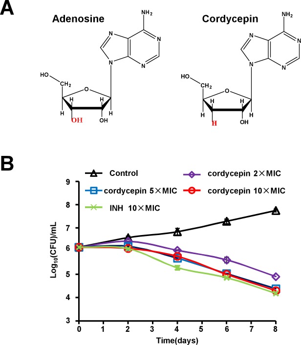
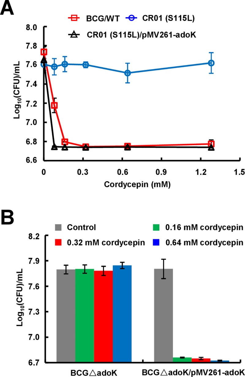
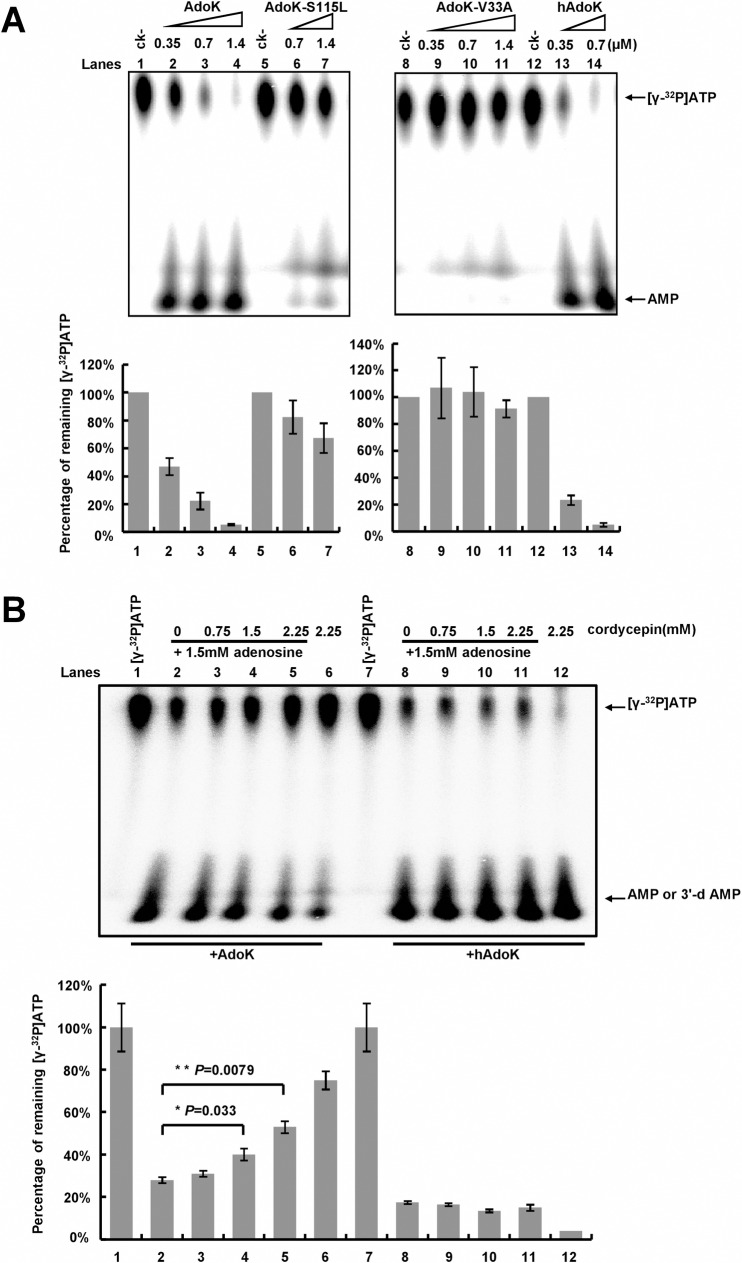
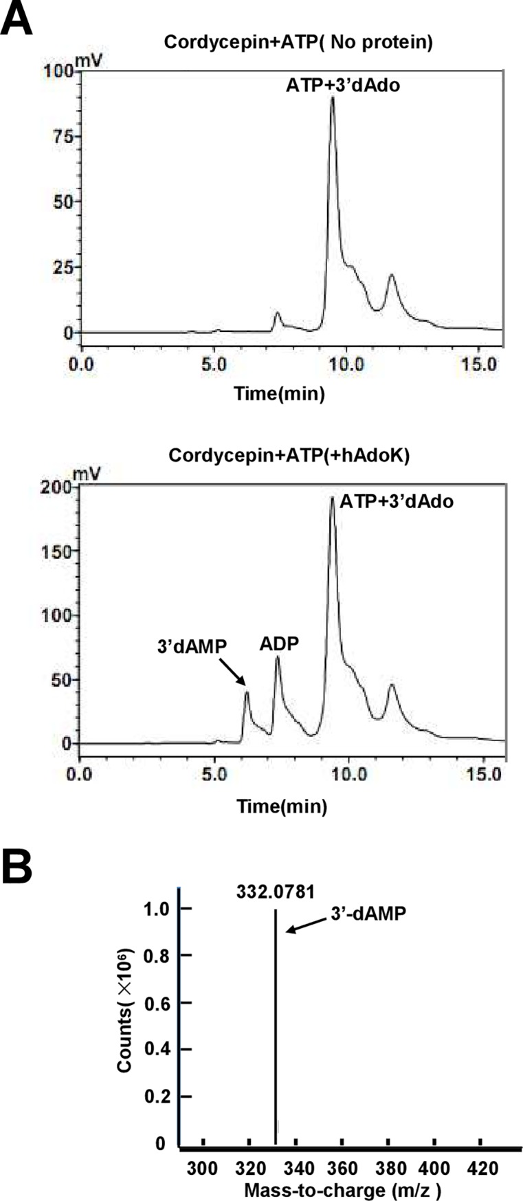
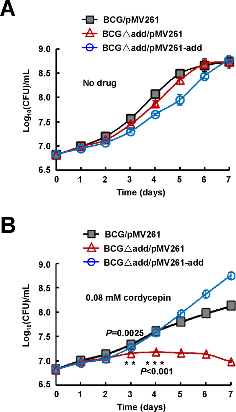
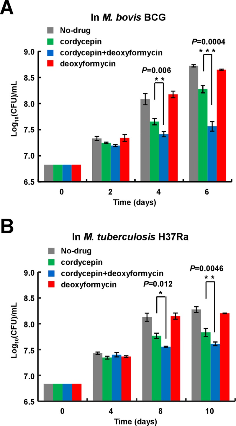
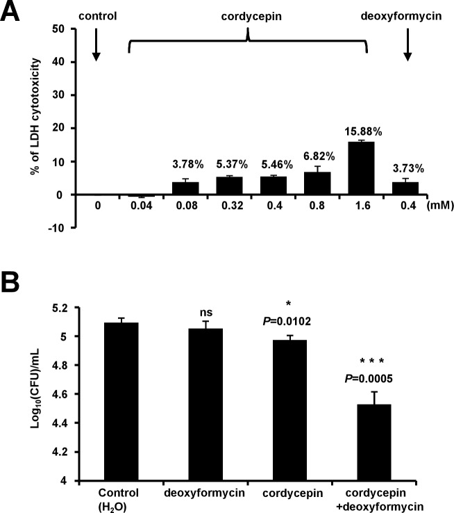
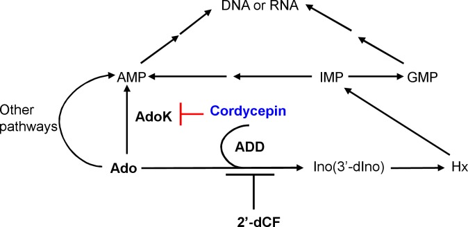
Cordycepin is an efficient component of Cordyceps spp, a traditional Chinese medicine widely used for healthcare in China, and has been recently acted as a strong anticancer agent for clinic. However, whether and how it may play a role in combating tuberculosis, caused by Mycobacterium tuberculosis, remains unknown. Here we report that cordycepin can kill Mycobacterium by hijacking the bacterial adenosine kinase (AdoK), a purine salvage enzyme responsible for the phosphorylation of adenosine (Ado) to adenosine monophosphate (AMP). We show that cordycepin is a poor AdoK substrate but it competitively inhibits the catalytic activity of AdoK for adenosine phosphorylation. Cordycepin does not affect the activity of the human adenosine kinase (hAdoK), whereas hAdoK phosphorylates cordycepin to produce a new monophosphate derivative. Co-use of cordycepin and deoxycoformycin, an inhibitor of adenosine deaminase (ADD), more efficiently kills M. bovis and M. tuberculosis. The add-deleted mycobacterium is more sensitive to cordycepin. This study characterized cordycepin as a new mycobactericidal compound and also uncovered a potential anti-mycobacterial mechanism.


Conflict of interest statement

The authors have declared that no competing interests exist.

Figures

Fig 1
Fig 1. Cordycepin are active against Mycobacterium bovis BCG in vitro.
(A) Structures of adenosine (left panel) and cordycepin (right panel). (B) Kill kinetics of cordycepin for M. bovis BCG over a period of 8 days.
Fig 2
Fig 2. AdoK is responsible for mycobacterial sensitivity to cordycepin.
(A) Mutant strain CR01-S115L is resistant to cordycepin. Mycobacterial strains were grown in 7H9 medium containing 0, 0.08, 0.16, 0.32, 0.64, or 1.28 mM cordycepin at 37°C for 9 days. Their CFUs were then determined and indicated in the figure. Both wildtype BCG/WT and the complemented strain CR01-S115L/pMV261-adoK are sensitive to cordycepin. (B) Assays for the sensitivities of adoK-deleted and its complemented strains to cordycepin. The mycobacterial strains were grown in 7H9 medium containing 0 (control), 0.16, 0.32, and 0.64 mM cordycepin at 37°C for 9 days, and CFUs were measured. All error bars in the figures represent the standard deviations (SD) of the data derived from three biological replicates.
Fig 3
Fig 3. Thin-layer chromatography assays for the adenosine kinase activity and the inhibitory effect of cordycepin.
(A) Assays for the kinase activities of wildtype and mutant AdoK proteins. Radioactive-labeled ATP and AMP are indicated by arrows on the right of the figure. The protein concentrations are indicated on top of the panels. Quantification assays for the percentage of remaining radioactive ATP in the reactions (upper panels) were performed and correspondingly shown as the lower panels. (B) Competitive TLC assays for the inhibitory effect of cordycepin on the activity of AdoK. An increasing concentration of cordycepin gradually inhibited the activity of AdoK, but did not for hAdoK, and the remaining ATP correspondingly increased. Relative percentages of remaining [γ-32P] ATP in the reaction mixtures were quantified, and the mean values of three independent experiments along with error bars (SD) are shown. The P-values were calculated by unpaired two-tailed Student’s t-test using GraphPad Prism 5. The P-values are indicated on top of the columns.
Fig 4
Fig 4. Assay for the new compound produced from cordycepin by human kinase.
(A) HPLC separation of the new products from cordycepin. ATP and cordycepin were co-incubated with (lower panel) or without (upper panel) hAdoK. A new peak for the product on the left of ADP peak is indicated by an arrow. (B) LC-MS characterization of the new product. The mass spectra peak at m/z 332.0781 corresponds to 3′-deoxyadenosine monophosphate (3′-dAMP) in methanol and is indicated.
Fig 5
Fig 5. Effects of adenosine deaminase shunt on the mycobactericidal activity of cordycepin.
Mycobacterial strains were grown in 7H9 medium without (A) or with 0.08 mM cordycepin (B). The add-deleted BCG strain is more sensitive to cordycepin than wildtype strain. Asterisks (*) denote a significant difference between the add-deleted and wildtype strain, and the P-values are indicated. The P-values were calculated by unpaired two-tailed Student’s t-test using GraphPad Prism 5.
Fig 6
Fig 6. Effect of deoxycoformycin on the bactericidal role of cordycepin.
M. bovis BCG (A) and M. tuberculosis H37Ra (B) strains were grown in 7H9 medium in the presence of either cordycepin or deoxycoformycin, or both drugs, and the CFUs were measured, respectively. All experiments were repeated three times. Error bars represent the SD in the figure. Asterisks indicate P-values (*, P<0.05; **, P<0.01; ***, P<0.001) from the unpaired two-tailed Student’s t-test using GraphPad Prism 5.
Fig 7
Fig 7. Effects of cordycepin and deoxyformycin on host cell and intracellular Mycobacterium bovis BCG.
(A) Determination of LDH cytotoxicity of cordycepin or deoxyformycin in BMDMs. BMDMs (5×10⁴ cells per well) were plated in a 96-well plate and incubated overnight in an incubator at 37°C, 5% CO2. And on the next day, different concentrations of cordycepin or deoxyformycin were added to the culture media and incubated for 18 hours. LDH cytotoxicity was measured using the Pierce LDH Cytotoxicity Assay Kit. All experiments were repeated three times. Error bars are standard deviations. (B) BMDMs were infected with Mycobacterium bovis BCG (MOI = 1) and treated with 0.4 mM cordycepin or/and 0.4 mM deoxyformycin for 18 h, and the mycobacterial survival was assayed by determining colony-forming units (CFUs). All experiments were repeated three times. Error bars represent the SD in the figure. Asterisks indicate P-values (*, P<0.05; ***, P<0.001) from the unpaired two-tailed Student’s t-test using GraphPad Prism 5.
Fig 8
Fig 8. Metabolic pathways of Ado in M. tuberculosis and mycobactericidal mechanism of cordycepin.
Ado is converted into AMP by adenosine kinase (AdoK) or inosine (Ino) by adenosine deaminase (ADD) in M. tuberculosis. Cordycepin targets AdoK to form a lethal intermediate structure. Deoxycoformycin (2′-dCF) inhibits the activity of ADD to enhance the mycobactericidal role of cordycepin.

Similar articles

Cited by

References

    1. World Health Organization. Global Tuberculosis Report 2018. WHO; 2017.
    1. Cunningham KG, Manson W, Spring FS, Hutchinson SA. Cordycepin, a metabolic product isolated from cultures of Cordyceps militaris (Linn.) Link. Nature 1950;166:949. - PubMed
    1. Zhou XX, Meyer CU, Schmidtke P, Zepp F. Effect of cordycepin on interleukin-10 production of human peripheral blood mononuclear cells. Eur J Pharmacol 2002;453:309–317. - PubMed
    1. Sugar AM, McCaffrey RP. Antifungal activity of 3’-deoxyadenosine (cordycepin). Antimicrob Agents Ch 1998;42:1424–1427. - PMC - PubMed
    1. Nakamura K, Yoshikawa N, Yamaguchi Y, Kagota S, Shinozuka K, Kunitomo M. Antitumor effect of cordycepin (3’-deoxyadenosine) on mouse melanoma and lung carcinoma cells involves adenosine A3 receptor stimulation. Anticancer Res 2006;26:43–47. - PubMed

Publication types

MeSH terms