. 2019 Oct:157:107666.
doi: 10.1016/j.neuropharm.2019.107666. Epub 2019 Jun 12.

The role of the neuropeptide PEN receptor, GPR83, in the reward pathway: Relationship to sex-differences

Affiliations

The role of the neuropeptide PEN receptor, GPR83, in the reward pathway: Relationship to sex-differences

Amanda K Fakira et al. Neuropharmacology. 2019 Oct.

Abstract

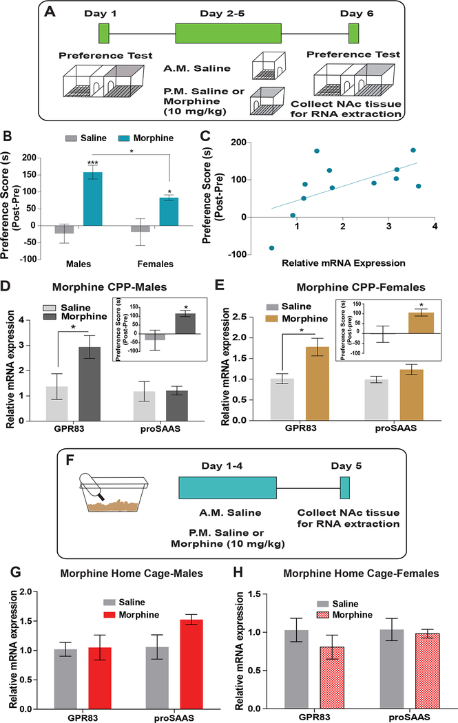
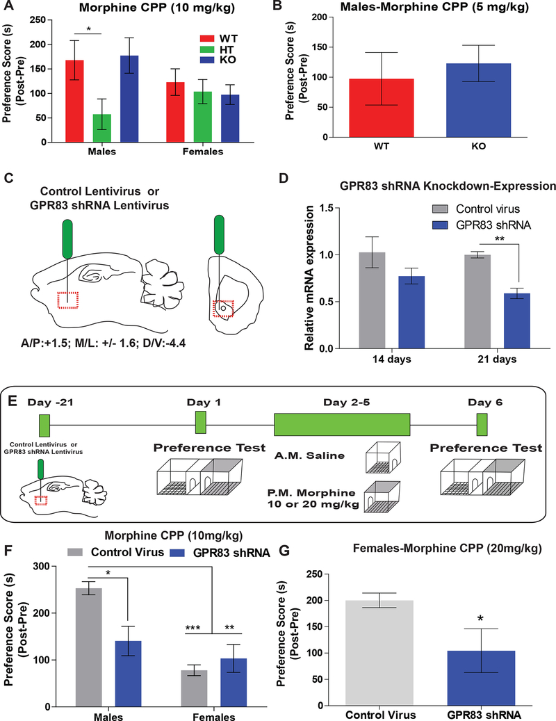
GPR83, the receptor for the neuropeptide PEN, exhibits high expression in the nucleus accumbens of the human and rodent brain, suggesting that it plays a role in modulating the mesolimbic reward pathway. However, the cell-type specific expression of GPR83, its functional impact in the reward pathway, and in drug reward-learning has not been fully explored. Using GPR83/eGFP mice, we show high GPR83 expression on cholinergic interneurons in the nucleus accumbens and moderate expression on ventral tegmental area dopamine neurons. In GPR83 knockout mice, baseline dopamine release in the nucleus accumbens is enhanced which disrupts the ratio of tonic vs phasic release. Additionally, GPR83 knockout leads to changes in the expression of dopamine-related genes. Using the morphine conditioned place preference model, we identify sex differences in morphine reward-learning, show that GPR83 is upregulated in the nucleus accumbens following morphine conditioned place preference, and show that shRNA-mediated knockdown of GPR83 in the nucleus accumbens leads to attenuation morphine reward. Together, these findings detect GPR83 expression in the reward-pathway, and show its involvement in dopamine release and morphine reward-learning.

Keywords: Cholinergic interneurons; Dopamine; GIR; Morphine; Voltammetry; proSAAS.


Figures

Figure 1:
Figure 1:. GPR83 expression in the NAc.
A) In situ hybridization (ISH) image from the Allen Brain Developing Mouse Atlas of GPR83 (Left) and corresponding brain atlas image (right) of a striatal brain section. B) Enlarged image of area within boxed image in A) demonstrating the pattern of GPR83 expression within the NAc. C) Low magnification image of GPR83 (green) expression in the NAc using GPR83 reporter mice from Gensat. D) and E) Higher magnification images showing that GPR83 is expressed in neurons in the NAc. RNAscope ISH probes for F) eGFP (green) and G) GPR83 (red) show that these markers colocalize H) in the same cells in GPR83/eGFP mice.
Figure 2:
Figure 2:. Cell-type expression of GPR83 in the NAc.
A) GPR83 (green) and B) DARPP-32 (red) expression in the NAc shows that GPR83 is not C) co-localized in medium spiny neurons. Region to the right of the dashed lines is the lateral septum (LS). D) GPR83 (green) and E) choline acetyltransferase (ChAT; red) expression in the NAc showing that GPR83 is F) co-localized in cholinergic interneurons (yellow). The regions outlined by dashed lines are the anterior commissure (ac) and lateral ventricles (VL). Higher magnification images of GPR83, DARPP-32 (G-I) and ChAT (J-L) expression. M) Quantification of percentage of GPR83 expressing cells that co-express ChAT and DARPP-32. N) GPR83 (green) and O) ChAT (red) are only co-expressed P) in neurons in the NAc and olfactory tubercle, outlined by dashed lines. ChAT positive neurons in the Diagonal Band Nucleus do not express GPR83 O) and P). Co-expressing neurons are indicated by a star. The data represents mean ± SEM. Representative images of 15 sections from 4 mice for ChAT staining and 10 sections from 4 mice for DARPP-32 staining are shown.
Figure 3:
Figure 3:. GPR83 expression within the VTA.
A) ISH image from the Allen Brain Developing Mouse Atlas of GPR83 (Left) and corresponding brain atlas image (right) of a brain section containing the VTA. B1) Enlarged 26 image of area within boxed outline in A) demonstrating the pattern of GPR83 expression within the VTA. B2) Enlarged image of area within the circled outline in A) demonstrating a brain region (hypothalamic mammillary bodies (MB)) which has high GPR83 expression compared to VTA. C) Brain section from GPR83/eGFP mice containing the VTA and mammillary bodies stained with TH and GFP. D) Brain section from GPR83/eGFP mice containing the VTA and mammillary bodies stained with TH and GFP. Boxed region 3) contains VTA dopamine neurons and boxed region 4) hypothalamic mammillary bodies. 3a) Enlarged image of TH staining from boxed region (3) in D) and 3b) is the same region with staining for eGFP (GPR83). 3c) Merged image of 3a and 3b. 4a) Enlarged image of TH staining from boxed (4) region in D) and 4b) is the same region with staining for GFP (GPR83). 4c) Merged image of 4a and 4b. Representative images of 8 sections from 2 mice are shown. E) Quantification of percentage of GPR83 expressing cells that co-express TH and percentage of TH expressing cells that do not co-express GPR83. Representative images of 7 sections from 2 mice.
Figure 4:
Figure 4:. Analysis of dopamine responses in the NAc of GPR83 KO mice.
Fast scanning cyclic voltammetry in the NAc of GPR83 WT and KO mice was used to examine differences in dopamine release. A) Current versus time plots (left) and color plots (right) showing dopamine responses following single pulse stimulations. B) Grouped data from A) showing enhanced dopamine release in GPR83 KO mice compared to WT (Unpaired t-test; t(8)=3.794, p<0.01, n=4–6). C) Aligned current versus time plots for measurement of dopamine uptake. Grouped data for D) maximal dopamine uptake (Vmax, Unpaired t-test, t(8)=1.375, p=0.2110) and E) dopamine clearance rate (tau, Unpaired t-test, t(8)=1.349, p=0.2141) indicating no significant differences. F) Phasic stimulation (5 pulses at 5, 10 and 20 Hz) current versus time plots (top) and color plots (bottom) showing enhanced dopamine release in response to increasing frequency of five pulse stimulations. G) Summary of dopamine release in response to phasic stimulation in GPR83 KO mice compared to WT (Two-way ANOVA Interaction F(3,24)= 1.175, p=0.3399, Frequency F(3,24)=22.2, ***p<0.0001, Genotype F(1,8)=9.098, **p<0.01, Holm-Sidak’s multiple comparisons test, *p<0.05, **p<0.01, n=4–6mice/gp). H) Current versus time plots of dopamine release represented as a percent of one pulse (tonic) release. I) Summary of normalized dopamine 27 release in response to phasic stimulation showing that the percent increase in dopamine release issignificantly blocked in GPR83 KO mice compared to WT. (Two-way ANOVA Interaction F(2,18)= 6.460, ** p<0.01, Frequency F(2,18)=26.27, ***p<0.0001, Genotype F(1,9)=4.046, p=0.751,n=4–6 mice/gr). J) Dopamine release measured before and after treatment with nicotinic acetylcholine receptor antagonist mecamylamine (2μM, MEC) in GPR83 WT (red) and GPR83 KO (blue). Data are represented as percent 1 pulse stimulation. (WT; Two-way ANOVA Interaction F(5,48)= 2.44, * p<0.05, MEC F(1,48)=5.41,*p<0.05, Frequency F(5,48)=5.48, ***p<0.001, KO; Two-way ANOVA Interaction F(5,30)= 10.41, *** p<0.0001, MEC F(1,30)=2.51,p=0.1645, Frequency F(5,30)=11.39, ***p<0.001, Bonferroni post-hoc analysis, *p<0.05, **p<0.01, n=4–5 mice/gr). The data represents mean ± SEM.
Figure 5:
Figure 5:. Analysis of dopamine related proteins in GPR83 KO mice.
VTA punches from GPR83 WT and KO mice were used to determine phosphorylation of tyrosine hydroxylase (TH). A) Western blot images of TH and pS40 pTH in the VTA of GPR83 WT and KO mice. B) Summary graphs of pTH (left) and total TH levels (right) in GPR83 WT versus KO mice (Unpaired t-test, pTH/TH, *p<0.05, t(10)=2.64, n=8–9mice/gr; TH/actin t(10)=0.09794, n=5–6 mice/gr). C) qPCR analysis of dopamine related proteins in the NAc of male and female GPR83 KO mice compared to WT. qPCR data are normalized to GPR83 WT (Unpaired t-tests, see Supplemental Table 2 for details, *p<0.05, n-=4–5 mice/gr). The data represents mean ± SEM.
Figure 6:
Figure 6:. Regulation of GPR83 expression by morphine reward-learning.
A) Schematic of experimental design for morphine CPP (10mg/kg, i.p.; 4 consecutive days) and collection of tissue for qPCR analysis of GPR83 and proSAAS. B) In male and female mice, morphine CPP results in preference for the morphine paired chamber compared to saline controls. Females have a lower preference than male mice. (Two-way ANOVA Interaction F(1,90)= 3.42, p=0.0675, Morphine F(1,90)=38.98,***p<0.0001, Sex F(1,90)=1.98, p=0.1631, Bonferroni post-hoc analysis, *p<0.05, ***p<0.0001, n= 8–29 mice saline groups, n=24–36 mice morphine groups). C) Correlation analysis of GPR83 expression in the NAc and morphine preference score (r=0.3572; p=0.04, n=12 mice). D and E) A select group of male and female mice exposed to morphine CPP with similar morphine paired scores (inset; average males 116±17.5s, n=5; Average females 106.2±43.69 s, n= 6). (Males: Unpaired t-test *p<0.05, t(8)=2.523, n=5/gr; Females: Unpaired t-test *p<0.05, t(11)=2.85, n=6–7/gr). Morphine CPP increases expression of GPR83 but not proSAAS in the NAc in both male and female mice. (Unpaired t-test, *p<0.05, GPR83, t(6)=2.312, proSAAS, t(7)=0.081, n=4–5 mice/gr; Females: Unpaired t-test, GPR83 *p<0.05, t(10)=2.91, proSAAS, p=0.11, t(10)=1.74, n=6 mice/gr). F) Schematic of experimental design for morphine home cage treatment and collection of tissue for qPCR analysis of GPR83 and proSAAS. G and H) Morphine administered in the home cage (same dose and schedule) does not increase expression of GPR83 or proSAAS in the NAc of male or female mice. (Males: Unpaired t-test, GPR83, p=0.91, t(7)=0.123, proSAAS, p=0.082 t(6)=2.088, n=4 mice/gr; Females: Unpaired t-test, GPR83, p=0.35, t(6)=1.01, proSAAS, p=0.74, t(6)=0.35, n=4 mice/gr). The data represents mean ± SEM.
Figure 7:
Figure 7:. The effect of GPR83 knockout and knockdown in the NAc on morphine reward.
A) Morphine CPP (10 mg/kg) in male and female GPR83 WT, HT and KO mice. (Two-way ANOVA Interaction F(2,51)= 1.93, p=0.1550, Genotype F(2,51)=2.38, p=0.1032, Sex F(1,51)=0.94, p=0.3377, Bonferroni post-hoc analysis, *p<0.05, n= 7–12 mice/gr. B) Morphine CPP (5 mg/kg) in male GPR83 WT and KO mice. (Unpaired t-test, p=0.6315, t(21)=0.4868, n=11–12 mice/gr. C)Schematic of control and GPR83 shRNA lentiviral injections into the NAc of male and female mice. C) Two and three weeks later punches of NAc were collected for qPCR analysis (Twoway ANOVA, Interaction F(1,8)=0.65, p=0.44; GPR83 KD F(1,8)= 11.46, p<0.01; Time F(1,8)=1.15, p=0.32; Bonferroni post-hoc tests, Control Virus vs GPR83 shRNA, at 21 days, p<0.05, n=3/gr). C) Schematic of experiments using lentiviral knockdown of GPR83 in the NAc followed by morphine CPP training. D) Knockdown of GPR83 in the NAc decreases morphine preference in male but not female mice using a 10 mg/kg dose (Two-way ANOVA, Interaction, F(1,25)=8.16, p<0.01, GPR83 shRNA, F(1,25)=3.28, p=0.08, Sex F(1,25)=19.32, p<0.0001; Bonferroni post-hoc tests Control virus vs GPR83 shRNA males **p<0.01, n=6–8 mice/gr). E) In female mice morphine CPP using a 20 mg/kg dose results in a higher preference than the 10 mg/kg dose; this is attenuated by GPR83 knockdown in the NAc. Unpaired t-test *p<0.05, t=2.414, df=14, n=7–9/gp. The data represents mean ± SEM.

Similar articles

Cited by

References

    1. Abad ATK, Miladi-Gorji H, Bigdeli I, 2016. Effects of swimming exercise on morphine-induced reward and behavioral sensitization in maternally-separated rat pups in the conditioned place preference procedure. Neurosci. Lett 10.1016/j.neulet.2016.08.011 - DOI - PubMed
    1. Adams F, Grassie M, Shahid M, Hill DR, Henry B, 2003. Acute oral dexamethasone administration reduces levels of orphan GPCR glucocorticoid-induced receptor (GIR) mRNA in rodent brain: Potential role in HPA-axis function. Mol. Brain Res 117, 39–46. 10.1016/S0169-328X(03)00280-8 - DOI - PubMed
    1. Alexander BK, Coambs RB, Hadaway PF, 1978. The effect of housing and gender on morphine self-administration in rats. Psychopharmacology (Berl) 10.1007/BF00426903 - DOI - PubMed
    1. Aosaki T, Kimura M, Graybiel AM, 1995. Temporal and spatial characteristics of tonically active neurons of the primate’s striatum. J. Neurophysiol 10.1152/jn.1995.73.3.1234 - DOI - PubMed
    1. Aosaki T, Tsubokawa H, Ishida A, Watanabe K, Graybiel AM, Kimura M, 1994. Responses of tonically active neurons in the primate’s striatum undergo systematic changes during behavioral sensorimotor conditioning. J. Neurosci - PMC - PubMed

Publication types

MeSH terms